|
|
|
Overcoming oral insulin delivery barriers: application of cell penetrating peptide and silica-based nanoporous composites |
Huining HE1,4,5, Junxiao YE1, Jianyong SHENG2, Jianxin WANG2, Yongzhuo HUANG2,3, Guanyi CHEN4, Jingkang WANG1( ), Victor C YANG5,6() ), Victor C YANG5,6() |
| 1. School of Chemical Engineering and Technology, Tianjin University, Tianjin 300072, China; 2. Department of Pharmaceutics, School of Pharmacy, Fudan University; Key Laboratory of Smart Drug Delivery, Ministry of Education & PLA, Shanghai 201203, China; 3. Shanghai Institute of Materia Medica, Chinese Academy of Sciences, Shanghai 201203, China; 4. School of Environmental Science and Engineering, State Key Laboratory of Engines, Tianjin University, Tianjin 300072, China; 5. Tianjin Key Laboratory on Technologies Enabling Development of Clinical Therapeutics and Diagnostics, School of Pharmacy, Tianjin Medical University, Tianjin 300070, China; 6. Department of Pharmaceutical Sciences, College of Pharmacy, University of Michigan, Michigan 48109-1065, USA |
|
|
|
|
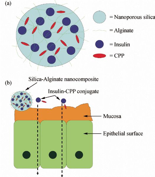
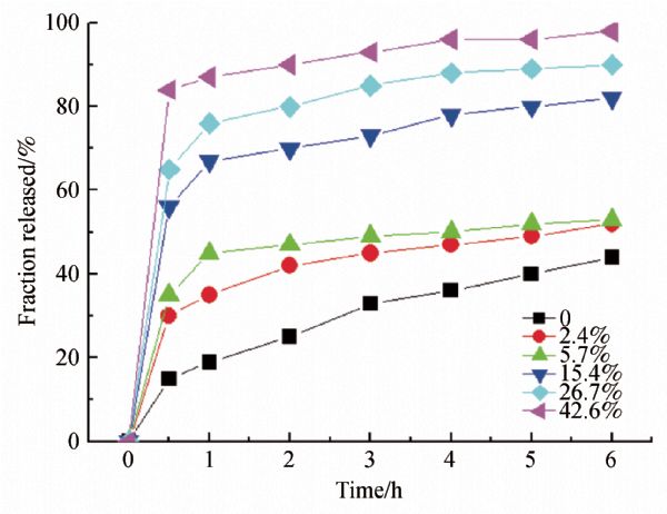
Abstract Oral insulin delivery has received the most attention in insulin formulations due to its high patient compliance and, more importantly, to its potential to mimic the physiologic insulin secretion seen in non-diabetic individuals. However, oral insulin delivery has two major limitations: the enzymatic barrier that leads to rapid insulin degradation, and the mucosal barrier that limits insulin’s bioavailability. Several approaches have been actively pursued to circumvent the enzyme barrier, with some of them receiving promising results. Yet, thus far there has been no major success in overcoming the mucosal barrier, which is the main cause in undercutting insulin’s oral bioavailability. In this review of our group’s research, an innovative silica-based, mucoadhesive oral insulin formulation with encapsulated-insulin/cell penetrating peptide (CPP) to overcome both enzyme and mucosal barriers is discussed, and the preliminary and convincing results to confirm the plausibility of this oral insulin delivery system are reviewed. In vitro studies demonstrated that the CPP-insulin conjugates could facilitate cellular uptake of insulin while keeping insulin’s biologic functions intact. It was also confirmed that low molecular weight protamine (LMWP) behaves like a CPP peptide, with a cell translocation potency equivalent to that of the widely studied TAT. The mucoadhesive properties of the produced silica-chitosan composites could be controlled by varying both the pH and composition; the composite consisting of chitosan (25 wt-%) and silica (75 wt-%) exhibited the greatest mucoadhesion at gastric pH. Furthermore, drug release from the composite network could also be regulated by altering the chitosan content. Overall, the universal applicability of those technologies could lead to development of a generic platform for oral delivery of many other bioactive compounds, especially for peptide or protein drugs which inevitably encounter the poor bioavailability issues.
|
| Keywords
insulin
cell penetrating peptide
mucoadhesive composites
oral delivery
|
|
Corresponding Author(s):
WANG Jingkang,Email:wangjkch@tju.edu.cn; YANG Victor C,Email:vcyang@umich.edu
|
|
Issue Date: 05 March 2013
|
|
| 1 |
Capaldi B. Treatments and devices for future diabetes management. Nursing Times , 2005, 101(18): 30–32
|
| 2 |
Cobble M E. Initiating and intensifying insulin therapy for type 2 diabetes: why, when, and how. American Journal of Therapeutics , 2009, 16(1): 56–64
doi: 10.1097/MJT.0b013e3181966bf0
|
| 3 |
Prevention Cf DCa. National diabetes fact sheet general information and national estimates on diabetes in the United States. Centers for Disease Control and Prevention , 2003
|
| 4 |
Heinemann L. New ways of insulin delivery. International Journal of Clinical Practice. Supplement , 2011, 65(170): 31–46
doi: 10.1111/j.1742-1241.2010.02577.x
|
| 5 |
Gordon Still J. Development of oral insulin: progress and current status. Diabetes/Metabolism Research and Reviews , 2002, 18(S1): S29–S37
doi: 10.1002/dmrr.207
|
| 6 |
Heinemann L. New ways of insulin delivery. International Journal of Clinical Practice. Supplement , 2010, 64: 29–40
doi: 10.1111/j.1742-1241.2009.02276.x
|
| 7 |
Reis C P, Damge C. Nanotechnology as a promising strategy for alternative routes of insulin delivery. Methods in Enzymology , 2012, 508: 271–294
doi: 10.1016/B978-0-12-391860-4.00014-8
|
| 8 |
Fonte P, Andrade F, Araujo F, Andrade C, Neves J, Sarmento B. Chitosan-coated solid lipid nanoparticles for insulin delivery. Methods in Enzymology , 2012, 508: 295–314
doi: 10.1016/B978-0-12-391860-4.00015-X
|
| 9 |
Card J W, Magnuson B A. A review of the efficacy and safety of nanoparticle-based oral insulin delivery systems. American Journal of Physiology. Gastrointestinal and Liver Physiology , 2011, 301(6): G956–G967
doi: 10.1152/ajpgi.00107.2011
|
| 10 |
He P, Tang Z, Lin L, Deng M, Pang X, Zhuang X, Chen X. Novel biodegradable and pH-sensitive poly(ester amide) microspheres for oral insulin delivery. Macromolecular Bioscience , 2012, 12(4): 547–556
doi: 10.1002/mabi.201100358
|
| 11 |
Cefalu W T. Concept, strategies, and feasibility of noninvasive insulin delivery. Diabetes Care , 2004, 27(1): 239–246
doi: 10.2337/diacare.27.1.239
|
| 12 |
Krishnankutty R K, Mathew A, Sedimbi S K, Suryanarayan S, Sanjeevi C B. Alternative routes of insulin delivery. Zhong Nan Da Xue Xue Bao. Yi Xue Ban , 2009, 34(10): 933–948
|
| 13 |
Bellary S, Barnett A H. Inhaled insulin: new technology, new possibilities. International Journal of Clinical Practice , 2006, 60(6): 728–734
doi: 10.1111/j.1742-1241.2006.00976.x
|
| 14 |
Cefalu W T. Evolving strategies for insulin delivery and therapy. Drugs , 2004, 64(11): 1149–1161
doi: 10.2165/00003495-200464110-00001
|
| 15 |
Sajeesh S, Bouchemal K, Marsaud V, Vauthier C, Sharma C P. Cyclodextrin complexed insulin encapsulated hydrogel microparticles: An oral delivery system for insulin. Journal of Controlled Release , 2010, 147(3): 377–384
doi: 10.1016/j.jconrel.2010.08.007
|
| 16 |
Yadav N, Morris G, Harding S E, Ang S, Adams G G. Various non-injectable delivery systems for the treatment of diabetes mellitus. Endocrine, Metabolic & Immune Disorders Drug Targets , 2009, 9(1): 1–13
doi: 10.2174/187153009787582405
|
| 17 |
Banting F G, Best C H, Collip J B, Campbell W R, Fletcher A A. Pancreatic extracts in the treatment of diabetes mellitus. Canadian Medical Association Journal , 1922, 7: 6
|
| 18 |
Del Curto M D, Maroni A, Palugan L, Zema L, Gazzaniga A, Sangalli M E. Oral delivery system for two-pulse colonic release of protein drugs and protease inhibitor/absorption enhancer compounds. Journal of Pharmaceutical Sciences , 2011, 100(8): 3251–3259
doi: 10.1002/jps.22560
|
| 19 |
Jelvehgari M, Milani P Z, Siahi-Shadbad M R, Monajjemzadeh F, Nokhodchi A, Azari Z, Valizadeh H. In vitro and in vivo evaluation of insulin microspheres containing protease inhibitor. Arzneimittel-Forschung , 2011, 61(1): 14–22
doi: 10.1055/s-0031-1296163
|
| 20 |
Su F Y, Lin K J, Sonaje K, Wey S P, Yen T C, Ho Y C, Panda N, Chuang E Y, Maiti B, Sung H W. Protease inhibition and absorption enhancement by functional nanoparticles for effective oral insulin delivery. Biomaterials , 2012, 33(9): 2801–2811
doi: 10.1016/j.biomaterials.2011.12.038
|
| 21 |
Marschutz M K, Bernkop-Schnurch A. Oral peptide drug delivery: polymer-inhibitor conjugates protecting insulin from enzymatic degradation in vitro. Biomaterials , 2000, 21(14): 1499–1507
doi: 10.1016/S0142-9612(00)00039-9
|
| 22 |
Saudek C D. Novel forms of insulin delivery. Endocrinology and Metabolism Clinics of North America , 1997, 26(3): 599–610
doi: 10.1016/S0889-8529(05)70269-3
|
| 23 |
Avadi M R, Sadeghi A M, Mohammadpour N, Abedin S, Atyabi F, Dinarvand R, Rafiee-Tehrani M. Preparation and characterization of insulin nanoparticles using chitosan and Arabic gum with ionic gelation method. Nanomedicine; Nanotechnology, Biology, and Medicine , 2010, 6(1): 58–63
doi: 10.1016/j.nano.2009.04.007
|
| 24 |
Cui F, He C, He M, Tang C, Yin L, Qian F, Yin C. Preparation and evaluation of chitosan-ethylenediaminetetraacetic acid hydrogel films for the mucoadhesive transbuccal delivery of insulin. Journal of Biomedical Materials Research. Part A , 2009, 89A(4): 1063–1071
doi: 10.1002/jbm.a.32071
|
| 25 |
Cui F, Qian F, Zhao Z, Yin L, Tang C, Yin C. Preparation, characterization, and oral delivery of insulin loaded carboxylated chitosan grafted poly(methyl methacrylate) nanoparticles. Biomacromolecules , 2009, 10(5): 1253–1258
doi: 10.1021/bm900035u
|
| 26 |
Schilling R, Mitra A. Degradation of insulin by trypsin and alpha-chymotrypsin. Pharmaceutical Research , 1991, 8(6): 721–727
doi: 10.1023/A:1015893832222
|
| 27 |
Nishihata T, Rytting J H, Kamada A, Higuchi T. Enhanced intestinal absorption of insulin in rats in the presence of sodium 5-methoxysalicylate. Diabetes , 1981, 30(12): 1065–1067
doi: 10.2337/diabetes.30.12.1065
|
| 28 |
Cui C Y, Lu W L, Xiao L, Zhang S Q, Huang Y B, Li S L, Zhang R J, Wang G L, Zhang X, Zhang Q. Sublingual delivery of insulin: effects of enhancers on the mucosal lipid fluidity and protein conformation, transport, and in vivo hypoglycemic activity. Biological & Pharmaceutical Bulletin , 2005, 28(12): 2279–2288
doi: 10.1248/bpb.28.2279
|
| 29 |
Muranishi S. Delivery system design for improvement of intestinal absorption of peptide drugs. Yakugaku Zasshi , 1997, 117(7): 394–414
|
| 30 |
Chung S W, Hil-lal T A, Byun Y. Strategies for non-invasive delivery of biologics. Journal of Drug Targeting , 2012, 20(6): 481–501
doi: 10.3109/1061186X.2012.693499
|
| 31 |
Schwarze S R, Ho A, Vocero-Akbani A, Dowdy S F. In vivo protein transduction: delivery of a biologically active protein into the mouse. Science , 1999, 285(5433): 1569–1572
doi: 10.1126/science.285.5433.1569
|
| 32 |
Schwarze S R, Dowdy S F. In vivo protein transduction: intracellular delivery of biologically active proteins, compounds and DNA. Trends in Pharmacological Sciences , 2000, 21(2): 45–48
doi: 10.1016/S0165-6147(99)01429-7
|
| 33 |
Cooper I, Sasson K, Teichberg V I, Schnaider-Beeri M, Fridkin M, Shechter Y. Peptide derived from HIV-1 TAT protein, destabilizes a monolayer of endothelial cells in an in vitro model of the blood-brain barrier, and allows permeation of high molecular weight proteins. Journal of Biological Chemistry , 2012, ???:
doi: 10.1074/jbc.M112.395384
|
| 34 |
Yu R, Zeng Z, Guo X, Zhang H, Liu X, Ding Y, Chen J. The TAT peptide endows PACAP with an enhanced ability to traverse bio-barriers. Neuroscience Letters , 2012, 527(1): 1–5
doi: 10.1016/j.neulet.2012.08.005
|
| 35 |
Elliott G, O'Hare P. Intercellular trafficking and protein delivery by a herpesvirus structural protein. Cell , 1997, 88(2): 223–233
doi: 10.1016/S0092-8674(00)81843-7
|
| 36 |
Min S H, Kim D M, Kim M N, Ge J, Lee D C, Park I Y, Park K C, Hwang J S, Cho C W, Yeom Y I. Gene delivery using a derivative of the protein transduction domain peptide, K-Antp. Biomaterials , 2010, 31(7): 1858–1864
doi: 10.1016/j.biomaterials.2009.11.019
|
| 37 |
Derossi D, Joliot A H, Chassaing G, Prochiantz A. The third helix of the antennapedia homeodomain translocates through biological membranes. Journal of Biological Chemistry , 1994, 269(14): 10444–10450
|
| 38 |
Jin G S, Zhu G D, Zhao Z G, Liu F S. VP22 enhances the expression of glucocerebrosidase in human Gaucher II fibroblast cells mediated by lentiviral vectors. Clinical and Experimental Medicine , 2012, 12(3): 135–143
doi: 10.1007/s10238-011-0152-7
|
| 39 |
Tanaka M, Kato A, Satoh Y, Ide T, Sagou K, Kimura K, Hasegawa H, Kawaguchi Y. Herpes simplex virus 1 VP22 regulates translocation of multiple viral and cellular proteins and promotes neurovirulence. Journal of Virology , 2012, 86(9): 5264–5277
doi: 10.1128/JVI.06913-11
|
| 40 |
Chang L C, Lee H F, Yang Z, Yang V. Low molecular weight protamine (LMWP) as nontoxic heparin/low molecular weight heparin antidote (I): Preparation and characterization. AAPS PharmSci , 2001, 3(3): 7–14
doi: 10.1208/ps030317
|
| 41 |
Chang L C, Liang J, Lee H F, Lee L, Yang V. Low molecular weight protamine (LMWP) as nontoxic heparin/low molecular weight heparin antidote (II): In vitro evaluation of efficacy and toxicity. AAPS PharmSci , 2001, 3(3): 15–23
doi: 10.1208/ps030318
|
| 42 |
Chang L C, Wrobleski S, Wakefield T, Lee L, Yang V. Low molecular weight protamine as nontoxic heparin/low molecular weight heparin antidote (III): Preliminary in vivo evaluation of efficacy and toxicity using a canine model. AAPS PharmSci , 2001, 3(3): 24–31
doi: 10.1208/ps030319
|
| 43 |
Park Y J, Chang L C, Liang J F, Moon C, Chung C P, Yang V C. Nontoxic membrane translocation peptide from protamine, low molecular weight protamine (LMWP), for enhanced intracellular protein delivery: in vitro and in vivo study. FASEB Journal , 2005, 19(11): 1555–1557
|
| 44 |
Xia H, Gao X, Gu G, Liu Z, Zeng N, Hu Q, Song Q, Yao L, Pang Z, Jiang X, Chen J, Chen H. Low molecular weight protamine-functionalized nanoparticles for drug delivery to the brain after intranasal administration. Biomaterials , 2011, 32(36): 9888–9898
doi: 10.1016/j.biomaterials.2011.09.004
|
| 45 |
Ramadas M W P, Dileep K J, Ramadas M, Anitha Y, Sharma C P, 0. Lipoinsulin encapsulated alginate-chitosan capsules: intestinal delivery in diabetic rats. Journal of Microencapsulation , 2000, 17(4): 405–411
doi: 10.1080/026520400405660
|
| 46 |
Kimura T, Sato K, Sugimoto K, Tao R, Murakami T, Kurosaki Y, Nakayama T. Oral administration of insulin as poly(vinyl alcohol)-gel spheres in diabetic rats. Biological & Pharmaceutical Bulletin , 1996, 19(6): 897–900
doi: 10.1248/bpb.19.897
|
| 47 |
Mitchell D J, Steinman L, Kim D T, Fathman C G, Rothbard J B. Polyarginine enters cells more efficiently than other polycationic homopolymers. Journal of Peptide Research , 2000, 56(5): 318–325
doi: 10.1034/j.1399-3011.2000.00723.x
|
| 48 |
Futaki S, Nakase I, Suzuki T, Zhang, Sugiura Y. Translocation of branched-chain arginine peptides through cell membranes: flexibility in the spatial disposition of positive charges in membrane-permeable peptides. Biochemistry , 2002, 41(25): 7925–7930
doi: 10.1021/bi0256173
|
| 49 |
Wong T W. Chitosan and its use in design of insulin delivery system. Recent Patents on Drug Delivery & Formulation , 2009, 3(1): 8–25
doi: 10.2174/187221109787158346
|
| 50 |
Damge C, Maincent P, Ubrich N. Oral delivery of insulin associated to polymeric nanoparticles in diabetic rats. Journal of Controlled Release , 2007, 117(2): 163–170
doi: 10.1016/j.jconrel.2006.10.023
|
| 51 |
Sarmento B, Ribeiro A, Veiga F, Sampaio P, Neufeld R, Ferreira D. Alginate/chitosan nanoparticles are effective for oral insulin delivery. Pharmaceutical Research , 2007, 24(12): 2198–2206
doi: 10.1007/s11095-007-9367-4
|
| 52 |
Liang J F, Zhen L, Chang L C, Yang V C. A less toxic heparin antagonist—low molecular weight protamine. Biochemistry. Biokhimiia , 2003, 68(1): 116–120
doi: 10.1023/A:1022109905487
|
| 53 |
Tsui B, Singh V K, Liang J F, Yang V C. Reduced reactivity towards anti-protamine antibodies of a low molecular weight protamine analogue. Thrombosis Research , 2001, 101(5): 417–420
doi: 10.1016/S0049-3848(00)00427-8
|
| 54 |
Carlsson J, Drevin H, Axén R. Protein thiolation and reversible protein-protein conjugation.N-Succinimidyl 3-(2-pyridyldithio)propionate, a new heterobifunctional reagent. Biochemical Journal , 1978, 173(3): 723–737
|
| 55 |
Chickering D E, Mathiowitz E. Bioadhesive microspheres I. A novel electrobalance-based method to study adhesive interactions between individual microspheres and intestinal mucosa. Journal of Controlled Release , 1995, 34(3): 251–262
doi: 10.1016/0168-3659(95)00011-V
|
| 56 |
Sudhakar Y, Kuotsu K, Bandyopadhyay A K. Buccal bioadhesive drug delivery—a promising option for orally less efficient drugs. Journal of Controlled Release , 2006, 114(1): 15–40
doi: 10.1016/j.jconrel.2006.04.012
|
|
Viewed |
|
|
|
Full text
|
|
|
|
|
Abstract
|
|
|
|
|
Cited |
|
|
|
|
| |
Shared |
|
|
|
|
| |
Discussed |
|
|
|
|