|
|
|
Cystine oligomers successfully attached to peptide cysteine-rich fibrils |
Christian Bortolini,Mingdong Dong( ) ) |
| Interdisciplinary Nanoscience Center (iNANO), Aarhus University, Aarhus C 8000, Denmark |
|
|
|
|
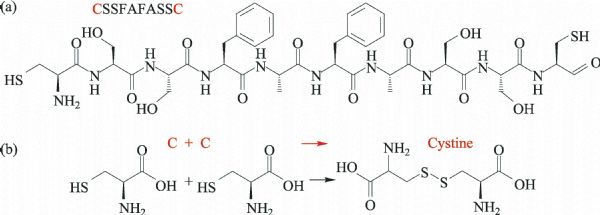
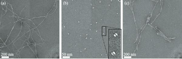
Abstract Amyloid peptides are renowned to be related to neurodegenerative diseases, however, a fruitful avenue is to employ them as high-performance nanomaterials. These materials benefit from the intrinsic outstanding mechanical robustness of the amyloid backbone made of b-strands. In this work, we exploited amyloid-like fibrils as functional material to attach pristine L-cysteine aggregates (cystine oligomers) and gold nanoparticles, without the need of templating compounds. This work will open new avenues on functional materials design and their realisation.
|
| Keywords
cysteine
peptide fibrils
gold nanoparticles
amyloids
oligomers
nanomaterials
|
|
Corresponding Author(s):
Mingdong Dong
|
|
Online First Date: 25 January 2016
Issue Date: 29 February 2016
|
|
| 1 |
Bortolini C, Liu L, Gronewold T M A, Wang C, Besenbacher F, Dong M D. The position of hydrophobic residues tunes peptide self-assembly. Soft Matter, 2014, 10(31): 5656–5661
https://doi.org/10.1039/C4SM01065E
|
| 2 |
Paramonov S E, Jun H W, Hartgerink J D. Self-assembly of peptide-amphiphile nanofibers: The roles of hydrogen bonding and amphiphilic packing. Journal of the American Chemical Society, 2006, 128(22): 7291–7298
https://doi.org/10.1021/ja060573x
|
| 3 |
Liu L, Busuttil K, Zhang S, Yang Y L, Wang C, Besenbacher F, Dong M D. The role of self-assembling polypeptides in building nanomaterials. Physical Chemistry Chemical Physics, 2011, 13(39): 17435–17444
https://doi.org/10.1039/c1cp21338e
|
| 4 |
Huang J F, Sun I W. Fabrication and surface functionalization of nanoporous gold by electrochemical alloying/dealloying of Au-Zn in an ionic liquid, and the self-assembly of L-cysteine monolayers. Advanced Functional Materials, 2005, 15(6): 989–994
https://doi.org/10.1002/adfm.200400382
|
| 5 |
Bortolini C, Liu L, Li Z S, Thomsen K, Wang C, Besenbacher F, Dong M D. Identification of cysteine-rich peptide-fiber by specific cysteine-Au nanoparticles binding on fiber surface. Advanced Materials Interfaces, 2014, 9: 1400133
https://doi.org/10.1002/admi.201400133
|
| 6 |
Djalali R, Chen Y, Matsui H. Au nanowire fabrication from sequenced histidine-rich peptide. Journal of the American Chemical Society, 2002, 124(46): 13660–13661
https://doi.org/10.1021/ja028261r
|
| 7 |
Banerjee I A, Yu L T, Matsui H. Cu nanocrystal growth onpeptide nanotubes by biomineralization: Size control of cunanocrystals by tuning peptide conformation. Proceedings of the National Academy of Sciences of the United States of America, 2003, 100(25): 14678–14682
https://doi.org/10.1073/pnas.2433456100
|
| 8 |
Kasotakis E, Mossou E, Adler-Abramovich L, Mitchell E P, Forsyth V T, Gazit E, Mitraki A. Design of metal-binding sites onto self-assembled peptide fibrils. Biopolymers, 2009, 92(3): 164–172
https://doi.org/10.1002/bip.21163
|
| 9 |
Lindgren M, Hallbrink M, Prochiantz A, Langel U. Cell-penetrating peptides. Trends in Pharmacological Sciences, 2000, 21(3): 99–103
https://doi.org/10.1016/S0165-6147(00)01447-4
|
| 10 |
Richard J P, Melikov K, Vives E, Ramos C, Verbeure B, Gait M J, Chernomordik L V, Lebleu B. Cell-penetrating peptides. A reevaluation of the mechanism of cellular uptake. Journal of Biological Chemistry, 2002, 2 78(1): 585–590
https://doi.org/10.1074/jbc.M209548200
|
| 11 |
Walker L C, Jucker M. Amyloid by default. Nature Neuroscience, 2011, 14(6): 669–670
https://doi.org/10.1038/nn.2853
|
| 12 |
Dobson C M. Protein folding and misfolding. Nature, 2003, 426(6968): 884–890
https://doi.org/10.1038/nature02261
|
| 13 |
Knowles T P J, Buehler M J. Nanomechanics of functional and pathological amyloid materials. Nature Nanotechnology, 2011, 6(8): 469–479
https://doi.org/10.1038/nnano.2011.102
|
| 14 |
Shorter J, Lindquist S. Prions as adaptive conduits of memory and inheritance. Nature Reviews. Genetics, 2005, 6(6): 435–450
https://doi.org/10.1038/nrg1616
|
| 15 |
Hauser C A E, Maurer-Stroh S, Martins I C. Amyloid-based nanosensors and nanodevices. Chemical Society Reviews, 2014, 43(15): 5326–5345
https://doi.org/10.1039/C4CS00082J
|
| 16 |
Knowles T P, Fitzpatrick A W, Meehan S, Mott H R, Vendruscolo M, Dobson C M, Welland M E. Role of intermolecular forces in defining material properties of protein nanofibrils. Science, 2007, 318(5858): 1900–1903
https://doi.org/10.1126/science.1150057
|
| 17 |
Luhrs T, Ritter C, Adrian M, Riek-Loher D, Bohrmann B, Doeli H, Schubert D, Riek R. 3D structure of Alzheimer’s amyloid-β(1-42) fibrils. Proceedings of the National Academy of Sciences of the United States of America, 2005, 102(48): 17342–17347
https://doi.org/10.1073/pnas.0506723102
|
| 18 |
Bortolini C, Jones N C, Hoffmann S V, Wang C, Besenbacher F, Dong M D. Mechanical properties of amyloid-like fibrils defined by secondary structures. Nanoscale, 2015, 7(17): 7745–7752
https://doi.org/10.1039/C4NR05109B
|
| 19 |
Scheibel T, Parthasarathy R, Sawicki G, Lin X M, Jaeger H, Lindquist S L. Conducting nanowires built by controlled self-assembly of amyloid fibers and selective metal deposition. Proceedings of the National Academy of Sciences of the United States of America, 2003, 100(8): 4527–4532
https://doi.org/10.1073/pnas.0431081100
|
| 20 |
Miles A J, Janes R W, Brown A, Clarke D T, Sutherland J C, Tao Y, Wallace B A, Hoffmann S V. Light flux density threshold at which protein denaturation isinduced by synchrotron radiation circular dichroismbeamlines. Journal of Synchrotron Radiation, 2008, 15(4): 420–422
https://doi.org/10.1107/S0909049508009606
|
| 21 |
Miles A J, Hoffmann S V, Tao Y, Janes R W, Wallace B A. Synchrotron radiation circular dichroism (SRCD) spectroscopy: New beamlines and new applications in biology. Spectroscopy-an International Journal, 2007, 21(5-6): 245–255
https://doi.org/10.1155/2007/282713
|
| 22 |
Whitmore L, Wallace B A. DICHROWEB, an online server for protein secondary structure analyses from circular dichroism spectroscopic data. Nucleic Acids Research, 2004, 32: 668–673
https://doi.org/10.1093/nar/gkh371
|
| 23 |
Whitmore L, Wallace B A. Protein secondary structure analyses from circular dichroism spectroscopy: Methods and reference databases. Biopolymers, 2008, 89(5): 392–400
https://doi.org/10.1002/bip.20853
|
|
Viewed |
|
|
|
Full text
|
|
|
|
|
Abstract
|
|
|
|
|
Cited |
|
|
|
|
| |
Shared |
|
|
|
|
| |
Discussed |
|
|
|
|