|
|
|
Plasma-enabled healing of graphene nano-platelets layer |
Xiuqi Fang1, Carles Corbella1( ), Denis B. Zolotukhin1,2, Michael Keidar1 ), Denis B. Zolotukhin1,2, Michael Keidar1 |
1. Department of Mechanical & Aerospace Engineering, George Washington University, Washington, DC 20052, USA
2. Department of Physics, Tomsk State University of Control Systems and Radioelectronics, Tomsk 634050, Russia |
|
|
|
|
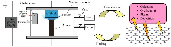
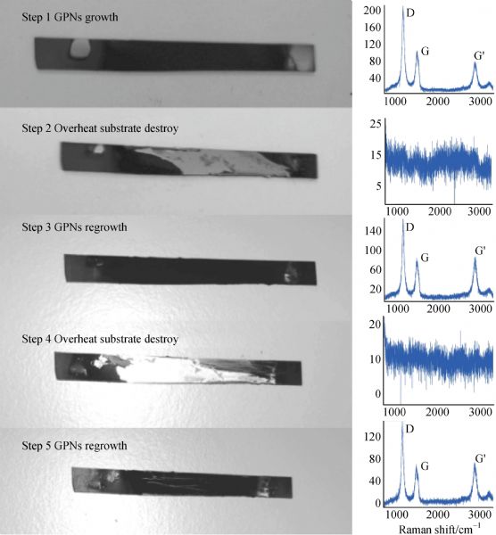
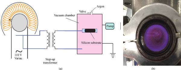
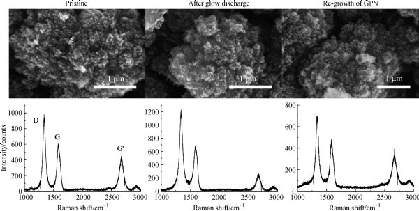
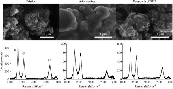
Abstract Graphene platelet networks (GPNs) were deposited onto silicon substrates by means of anodic arc discharge ignited between two graphite electrodes. Substrate temperature and pressure of helium atmosphere were optimized for the production of the carbon nanomaterials. The samples were modified or destroyed with different methods to mimic typical environments responsible of severe surface degradation. The emulated conditions were performed by four surface treatments, namely thermal oxidation, substrate overheating, exposition to glow discharge, and metal coating due to arc plasma. In the next step, the samples were regenerated on the same substrates with identical deposition technique. Damaging and re-growth of GPN samples were systematically characterized by scanning electron microscopy and Raman spectroscopy. The full regeneration of the structural and morphological properties of the samples has proven that this healing method by arc plasma is adequate for restoring the functionality of 2D nanostructures exposed to harsh environments.
|
| Keywords
graphene platelet networks
anodic arc discharge
plasma healing
scanning electron microscopy
Raman spectroscopy
|
|
Corresponding Author(s):
Carles Corbella
|
|
Online First Date: 22 March 2019
Issue Date: 22 May 2019
|
|
| 1 |
MOhring. Materials Science of Thin Films. 2nd ed. San Diego: Academic Press, 2002
|
| 2 |
F JGordillo-Vázquez, V JHerrero, ITanarro. From carbon nanostructures to new photoluminescence sources: An overview of new perspectives and emerging applications of low-pressure PECVD. Chemical Vapor Deposition, 2007, 13(6-7): 267–279
https://doi.org/10.1002/cvde.200604034
|
| 3 |
MKeidar, I I Beilis. Plasma Engineering: Applications from Aerospace to Bio- and Nanotechnology. London: Academic Press, 2013
|
| 4 |
IAdamovich, S D Baalrud, A Bogaerts, P JBruggeman, MCappelli, VColombo, UCzarnetzki, UEbert, J GEden, PFavia, et al. The 2017 plasma roadmap: Low temperature plasma science and technology. Journal of Physics. D, Applied Physics, 2017, 50(32): 323001
https://doi.org/10.1088/1361-6463/aa76f5
|
| 5 |
UCvelbar, J L Walsh, M Černák, H Wde Vries, SReuter, TBelmonte, CCorbella, CMiron, NHojnik, AJurov, et al. White paper on the future of plasma science and technology in plastics and textiles. Plasma Processes and Polymers, 2019, 16(1): e1700228
https://doi.org/10.1002/ppap.201700228
|
| 6 |
AFridman, G Friedman. Plasma Medicine. Weinheim: Wiley, 2013
|
| 7 |
D BGraves. Low temperature plasma biomedicine: A tutorial review. Physics of Plasmas, 2014, 21(8): 080901
https://doi.org/10.1063/1.4892534
|
| 8 |
MKeidar, D Yan, I IBeilis, BTrink, J HSherman. Plasmas for treating cancer: Opportunities for adaptive and self-adaptive approaches. Trends in Biotechnology, 2018, 36(6): 586–593
https://doi.org/10.1016/j.tibtech.2017.06.013
|
| 9 |
SBekeschus, P Favia, ERobert, Tvon Woedtke. White paper on plasma for medicine and hygiene: Future in plasma health sciences. Plasma Processes and Polymers, 2019, 16(1): e1800033
https://doi.org/10.1002/ppap.201800033
|
| 10 |
N AAzarenkov, I BDenisenko, K NOstrikov. A model of a large-area planar plasma producer based on surface wave propagation in a plasma-metal structure with a dielectric sheath. Journal of Physics. D, Applied Physics, 1995, 28(12): 2465–2469
https://doi.org/10.1088/0022-3727/28/12/011
|
| 11 |
QCheng, S Xu, K KOstrikov. Single-step, rapid low-temperature synthesis of Si quantum dots embedded in an amorphous SiC matrix in high-density reactive plasmas. Acta Materialia, 2010, 58(2): 560–569
https://doi.org/10.1016/j.actamat.2009.09.034
|
| 12 |
OVolotskova, J A Fagan, J Y Huh, F R Phelan Jr, A Shashurin, MKeidar. Tailored distribution of single-wall carbon nanotubes from Arc plasma synthesis using magnetic fields. ACS Nano, 2010, 4(9): 5187–5192
https://doi.org/10.1021/nn101279r
|
| 13 |
MKeidar, A Shashurin, OVolotskova, YRaitses, I IBeilis. Mechanism of carbon nanostructure synthesis in arc plasma. Physics of Plasmas, 2010, 17(5): 057101
https://doi.org/10.1063/1.3312879
|
| 14 |
S RZavada, N R McHardy, K L Gordon, T F Scott. Rapid, puncture-initiated healing via oxygen-mediated polymerization. ACS Macro Letters, 2015, 4(8): 819–824
https://doi.org/10.1021/acsmacrolett.5b00315
|
| 15 |
ILevchenko, S Xu, GTeel, DMariotti, M L RWalker, MKeidar. Recent progress and perspectives of space electric propulsion systems based on smart nanomaterials. Nature Communications, 2018, 9(1): 879
https://doi.org/10.1038/s41467-017-02269-7
|
| 16 |
DGolberg, X D Bai, M Mitome, C CTang, C YZhi, YBando. Structural peculiarities of in situ deformation of a multiwalled BN nanotube inside a high-resolution analytical transmission electron microscope. Acta Materialia, 2007, 55(4): 1293–1298
https://doi.org/10.1016/j.actamat.2006.09.034
|
| 17 |
SIijima. Helical microtubules of graphitic carbon. Nature, 1991, 354(6348): 56–58
https://doi.org/10.1038/354056a0
|
| 18 |
AShashurin, M Keidar. Synthesis of 2D materials in arc plasmas. Journal of Physics. D, Applied Physics, 2015, 48(31): 314007
https://doi.org/10.1088/0022-3727/48/31/314007
|
| 19 |
XFang, A Shashurin, GTeel, MKeidar. Determining synthesis region of the single wall carbon nanotubes in arc plasma volume. Carbon, 2016, 107: 273–280
https://doi.org/10.1016/j.carbon.2016.05.061
|
| 20 |
XFang, J Donahue, AShashurin, MKeidar. Plasma-based graphene functionalization in glow discharge. Graphene, 2015, 4(1): 1–6
https://doi.org/10.4236/graphene.2015.41001
|
| 21 |
A CFerrari, J Robertson. Interpretation of Raman spectra of disordered and amorphous carbon. Physical Review. B, 2000, 61(20): 14095–14107
https://doi.org/10.1103/PhysRevB.61.14095
|
| 22 |
A CFerrari, J C Meyer, V Scardaci, CCasiraghi, MLazzeri, FMauri, SPiscanec, DJiang, K SNovoselov, SRoth, et al. Raman Spectrum of graphene and graphene layers. Physical Review Letters, 2006, 97(18): 187401
https://doi.org/10.1103/PhysRevLett.97.187401
|
| 23 |
A CFerrari, D M Basko. Raman spectroscopy as a versatile tool for studying the properties of graphene. Nature Nanotechnology, 2013, 8(4): 235–246
https://doi.org/10.1038/nnano.2013.46
|
| 24 |
M ALieberman, J ALichtenberg. Principles of Plasma Discharges and Material Processing. 2nd ed. Hoboken: Wiley, 2005
|
| 25 |
D BZolotukhin, MKeidar. Optimization of discharge triggering in micro-cathode vacuum arc thruster for CubeSats. Plasma Sources Science & Technology, 2018, 27(7): 074001
https://doi.org/10.1088/1361-6595/aacdb0
|
|
Viewed |
|
|
|
Full text
|
|
|
|
|
Abstract
|
|
|
|
|
Cited |
|
|
|
|
| |
Shared |
|
|
|
|
| |
Discussed |
|
|
|
|