|
|
|
MiRNA-451 is a potential biomarker for estrogenicity in mouse uterus |
Lingyan HOU1, Yun LU2( ), Ying LI2, Li LI1 ), Ying LI2, Li LI1 |
| 1. College of Science, Beijing Forestry University, Beijing 100083, China; 2. State Key Joint Laboratory of Environment Simulation and Pollution Control, School of Environment, Tsinghua University, Beijing 100084, China |
|
|
|
|
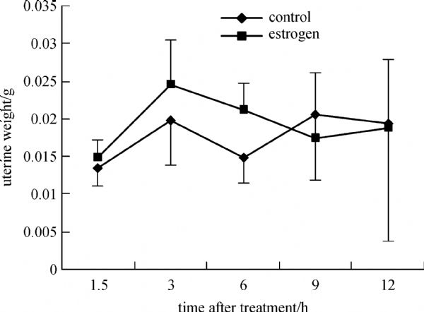
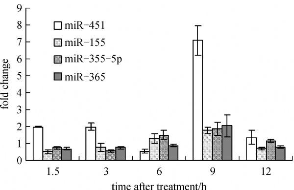
Abstract The uterotrophic assay has been commonly used to test environmental estrogens in vivo, however, it is often not sensitive enough sometimes. An alternative way is to evaluate estrogenicity through biomarker genes. MicroRNA (miRNA) is a class of regulatory gene, which has been shown to be a good biomarker for many diseases and toxicological effects in recent years, and some evidences showed that estrogen induced response was partially mediated by miRNAs. In this study, two types of microarrays were used to test the 17β-estradiol (E2) induced miRNA expression profile at different time points in the immature mouse uterus. Statistical analysis showed the aldehyde slide based array had less variation than the amino slide based array, and 11 dysregulated miRNAs were screened out for significant fold change. Real-time PCR was performed to further confirm that 4 out of 7 selected miRNAs, namely miR-451, miR-155, miR-335-5p, and miR-365, are E2 regulated miRNAs in the uterus. The function of the predicted targets of these miRNAs is involved in cell grow control, which is consistent with the main E2 function in the uterus. MiR-451 had similar strong responses to E2 in the uterus of both immature and overiectomized mice, and could be a potential biomarker for estrogenicity in the uterus.
|
| Keywords
estrogen
microRNA (miRNA)
microarray
biomarker
|
|
Corresponding Author(s):
LU Yun,Email:luyun@tsinghua.edu.cn
|
|
Issue Date: 01 February 2014
|
|
| 1 |
Dechering K, Boersma C, Mosselman S. Estrogen receptors α and β: two receptors of a kind? Current Medicinal Chemistry , 2000, 7(8): 561–576
|
| 2 |
Nilsson S, Gustafsson J A. Biological role of estrogen and estrogen receptors. Critical Reviews in Biochemistry and Molecular Biology , 2002, 37(1): 1–28
doi: 10.1080/10409230290771438
|
| 3 |
Gruber C J, Tschugguel W, Schneeberger C, Huber J C. Production and actions of estrogens. The New England Journal of Medicine , 2002, 346(5): 340–352
doi: 10.1056/NEJMra000471 pmid:11821512
|
| 4 |
Erickson B E. Next-generation risk assessment. Chemical and Engineering News , 2009, 87(25): 30–33
doi: 10.1021/cen-v087n025.p030
|
| 5 |
Moggs J G. Molecular responses to xenoestrogens: mechanistic insights from toxicogenomics. Toxicology , 2005, 213(3): 177–193
doi: 10.1016/j.tox.2005.05.020 pmid:15996808
|
| 6 |
Sharpe R M. The ‘oestrogen hypothesis’–where do we stand now? International Journal of Andrology , 2003, 26(1): 2–15
doi: 10.1046/j.1365-2605.2003.00367.x pmid:12534932
|
| 7 |
Sikka S C, Wang R. Endocrine disruptors and estrogenic effects on male reproductive axis. Asian Journal of Andrology , 2008, 10(1): 134–145
doi: 10.1111/j.1745-7262.2008.00370.x pmid:18087652
|
| 8 |
Markey C M, Michaelson C L, Veson E C, Sonnenschein C, Soto A M. The mouse uterotrophic assay: a reevaluation of its validity in assessing the estrogenicity of bisphenol A. Environmental Health Perspectives , 2001, 109(1): 55–60
doi: 10.1289/ehp.0110955 pmid:11171525
|
| 9 |
Choi K C, Jeung E B. The biomarker and endocrine disruptors in mammals. Journal of Reproduction and Development , 2003, 49(5): 337–345
doi: 10.1262/jrd.49.337 pmid:14967909
|
| 10 |
Heppell S A, Denslow N D, Folmar L C, Sullivan C V. Universal assay of vitellogenin as a biomarker for environmental estrogens. Environmental Health Perspectives , 1995, 103(Suppl 7): 9–15
doi: 10.1289/ehp.95103s79 pmid:8593883
|
| 11 |
An B S, Choi K C, Kang S K. Novel Calbindin-D(9K) protein as a useful biomarker for environmental estrogenic compounds in the uterus of immature rats. Reproductive Toxicology (Elmsford, N.Y.) , 2003, 17(3): 311–319
doi: 10.1016/S0890-6238(03)00003-0 pmid:12759100
|
| 12 |
Jung Y W, Hong E J, Choi K C, Jeung E B. Novel progestogenic activity of environmental endocrine disruptors in the upregulation of calbindin-D9k in an immature mouse model. Toxicological sciences , 2005, 83(1): 78–88
doi: 10.1093/toxsci/kfi015 pmid:15509668
|
| 13 |
An B S, Kang S K, Shin J H, Jeung E B. Stimulation of calbindin- D(9k) mRNA expression in the rat uterus by octyl-phenol, nonylphenol and bisphenol. Molecular and Cellular Endocrinology , 2002, 191(2): 177–186
doi: 10.1016/S0303-7207(02)00042-4 pmid:12062901
|
| 14 |
Ciesla M, Skrzypek K, Kozakowska M, Loboda A, Jozkowicz A, Dulak J. MicroRNAs as biomarkers of disease onset. Analytical and Bioanalytical Chemistry , 2011, 401(7): 2051–2061
doi: 10.1007/s00216-011-5001-8 pmid:21544542
|
| 15 |
Wang B, Majumder S, Nuovo G, Kutay H, Volinia S, Patel T, Schmittgen T D, Croce C, Ghoshal K, Jacob S T. Role of microRNA-155 at early stages of hepatocarcinogenesis induced by choline-deficient and amino acid-defined diet in c57BL/6 mice. Hepatology (Baltimore, Md.) , 2009, 50(4): 1152–1161
doi: 10.1002/hep.23100 pmid:19711427
|
| 16 |
Wang W X, Rajeev B W, Stromberg A J, Ren N, Tang G L, Huang Q W, Rigoutsos I, Nelson P T. The expression of microRNA miR-107 decreases early in Alzheimer’s disease and may accelerate disease progression through regulation of beta-site amyloid precursor protein-cleaving enzyme 1. The Journal of Neuroscience , 2008, 28(5): 1213–1223
doi: 10.1523/JNEUROSCI.5065-07.2008 pmid:18234899
|
| 17 |
Wang K, Marzolf B, Troisch P, Brightman A, Hu Z Y, Hood L E, Zhang S L, Galas D J. Circulating microRNAs, potential biomarkers for drug-induced liver injury. In: Proceedings of the National Academy of Sciences of the United , 2009, 106(11): 4402–4407
|
| 18 |
Ambros V. The functions of animal microRNAs. Nature , 2004, 431(7006): 350–355
doi: 10.1038/nature02871 pmid:15372042
|
| 19 |
Bartel D P. MicroRNAs: genomics, biogenesis, mechanism, and function. Cell , 2004, 116(2): 281–297
doi: 10.1016/S0092-8674(04)00045-5 pmid:14744438
|
| 20 |
Griffiths-Jones S,Saini H K, Dongen S, Enright A J. MiRbase: tools for microRNA genomics. Nucleic Acids Receaprch , 2008, 36(S1): 1540–158
|
| 21 |
Yokoi T, Nakajima M. Toxicological implications of modulation of gene expression by microRNAs. Toxicological Sciences , 2011, 123(1): 1–14
doi: 10.1093/toxsci/kfr168 pmid:21715665
|
| 22 |
Couzin J. Genetics-Erasing microRNAs reveals their powerful punch. Science , 2007, 316(5824): 530–530
doi: 10.1126/science.316.5824.530 pmid:17463259
|
| 23 |
Kovalchuk O, Tryndyak V P, Montgomery B, Boyko A, Kutanzi K, Zemp F, Warbritton A R, Latendresse J R, Kovalchuk I, Beland F A, Pogribny I P. Estrogen-induced rat breast carcinogenesis is characterized by alterations in DNA methylation, histone modifications and aberrant microRNA expression. Cell Cycle (Georgetown, Tex.) , 2007, 6(16): 2010–2018
doi: 10.4161/cc.6.16.4549 pmid:17700064
|
| 24 |
Dai R, Phillips R A, Zhang Y, Khan D, Crasta O, Ahmed S A. Suppression of LPS-induced Interferon-gamma and nitric oxide in splenic lymphocytes by select estrogen-regulated microRNAs: a novel mechanism of immune modulation. Blood , 2008, 112(12): 4591–4597
doi: 10.1182/blood-2008-04-152488 pmid:18791161
|
| 25 |
Nothnick W B, Healy C. Estrogen induces distinct patterns of microRNA expression within the mouse uterus. Reproductive Sciences (Thousand Oaks, Calif.) , 2010, 17(11): 987–994
doi: 10.1177/1933719110377472 pmid:20720260
|
| 26 |
Lewis B P, Shih I H, Jones-Rhoades M W, Bartel D P, Burge C B. Prediction of mammalian microRNA targets. Cell , 2003, 115(7): 787–798
doi: 10.1016/S0092-8674(03)01018-3 pmid:14697198
|
| 27 |
Krek A, Grün D, Poy M N, Wolf R, Rosenberg L, Epstein E J, MacMenamin P, da Piedade I, Gunsalus K C, Stoffel M, Rajewsky N. Combinatorial microRNA target predictions. Nature Genetics , 2005, 37(5): 495–500
doi: 10.1038/ng1536 pmid:15806104
|
| 28 |
John B, Enright A J, Aravin A, Tuschl T, Sander C, Marks D S. Human microRNA targets. PLoS Biology , 2004, 2(11): 1862–1879
doi: 10.1371/journal.pbio.0020363 pmid:15502875
|
| 29 |
Moggs J G, Tinwell H, Spurway T, Chang H S, Pate I, Lim F L, Moore D J, Soames A, Stuckey R, Currie R, Zhu T, Kimber I, Ashby J, Orphanides G. Phenotypic anchoring of gene expression changes during estrogen-induced uterine growth. Environmental Health Perspectives , 2004, 112(16): 1589–1606
doi: 10.1289/ehp.7345 pmid:15598610
|
| 30 |
Klinge C M. Estrogen regulation of microRNA expression. Current Genomics , 2009, 10(3): 169–183
doi: 10.2174/138920209788185289 pmid:19881910
|
| 31 |
Hewitt S C, Deroo B J, Hansen K, Collins J, Grissom S, Afshari C A, Korach K S. Estrogen receptor-dependent genomic responses in the uterus mirror the biphasic physiological response to estrogen. Molecular Endocrinology (Baltimore, Md.) , 2003, 17(10): 2070–2083
doi: 10.1210/me.2003-0146 pmid:12893882
|
| 32 |
Hong S H, Nah H Y, Lee J Y, Gye M C, Kim C H, Kim M K. Analysis of estrogen-regulated genes in mouse uterus using cDNA microarray and laser capture microdissection. Journal of Endocrinology , 2004, 181(1): 157–167
doi: 10.1677/joe.0.1810157 pmid:15072576
|
| 33 |
Watanabe H, Suzuki A, Kobayashi M, Takahashi E, Itamoto M, Lubahn D B, Handa H, Iguchi T. Analysis of temporal changes in the expression of estrogen-regulated genes in the uterus. The Journal of Molecular Endocrinology , 2003, 30(3): 347–358
doi: 10.1677/jme.0.0300347 pmid:12790804
|
| 34 |
Naciff J M, Overmann G J, Torontali S M, Carr G J, Tiesman J P, Richardson B D, Daston G P. Gene expression profile induced by 17 alpha-ethynyl estradiol in the prepubertal female reproductive system of the rat. Toxicological Sciences , 2003, 72(2): 314–330
doi: 10.1093/toxsci/kfg037 pmid:12655037
|
| 35 |
Wu X, Pang S T, Sahlin L, Blanck A, Norstedt G, Flores-Morales A. Gene expression profiling of the effects of castration and estrogen treatment in the rat uterus. Biology of Reproduction , 2003, 69(4): 1308–1317
doi: 10.1095/biolreprod.103.015420 pmid:12801995
|
| 36 |
Kang K S, Kim H S, Ryu D Y, Che J H, Lee Y S. Immature uterotrophic assay is more sensitive than ovariectomized uterotrophic assay for the detection of estrogenicity of p-nonylphenol in Sprague-Dawley rats. Toxicology Letters , 2000, 118(1–2): 109–115
doi: 10.1016/S0378-4274(00)00272-1 pmid:11137316
|
| 39 |
Ferenczy A. Studies on the cytodynamics of human endometrial regeneration. I. Scanning electron microscopy. American Journal of Obstetrics and Gynecology , 1976, 124(1): 64–74
pmid:1244749
|
| 40 |
Punyadeera C, Verbost P, Groothuis P. Oestrogen and progestin responses in human endometrium. The Journal of Steroid Biochemistry and Molecular Biology , 2003, 84(4): 393–410
doi: 10.1016/S0960-0760(03)00061-X pmid:12732285
|
| 41 |
Rosenfeld C S, Roberts R M, Lubahn D B. Estrogen receptor- and aromatase-deficient mice provide insight into the roles of estrogen within the ovary and uterus. Molecular Reproduction and Development , 2001, 59(3): 336–346
doi: 10.1002/mrd.1039 pmid:11424220
|
| 42 |
Honda K, Sawada H, Kihara T, Urushitani M, Nakamizo T, Akaike A, Shimohama S. Phosphatidylinositol 3-kinase mediates neuroprotection by estrogen in cultured cortical neurons. Journal of Neuroscience Research , 2000, 60(3): 321–327
doi: 10.1002/(SICI)1097-4547(20000501)60:3<321::AID-JNR6>3.0.CO;2-T pmid:10797534
|
| 43 |
Ivanova T, Mendez P, Garcia-Segura L M, Beyer C. Rapid stimulation of the PI3-kinase/Akt signalling pathway in developing midbrain neurones by oestrogen. Journal of Neuroendocrinology , 2002, 14(1): 73–79
doi: 10.1046/j.0007-1331.2001.00742.x pmid:11903815
|
| 44 |
Castoria G, Migliaccio A, Bilancio A, Di Domenico M, de Falco A, Lombardi M, Fiorentino R, Varricchio L, Barone M V, Auricchio F. PI3-kinase in concert with Src promotes the S-phase entry of oestradiol-stimulated MCF-7 cells. The EMBO Journal , 2001, 20(21): 6050–6059
doi: 10.1093/emboj/20.21.6050 pmid:11689445
|
| 45 |
Sun M, Paciga J E, Feldman R I, Yuan Z, Coppola D, Lu Y Y, Shelley S A, Nicosia S V, Cheng J Q. Phosphatidylinositol-3-OH kinase (PI3K)/Akt2, activated in breast cancer, regulates and is induced by estrogen receptor alpha (ER alpha) via interaction between ERa and PI3K. Cancer Research , 2001, 61(16): 5985–5991
|
| 46 |
Lee Y R, Park J, Yu H N, Kim J S, Youn H J, Jung S H.Up-regulation of PI3K/Akt signaling by 17b-estradiol through activation of estrogen receptor-a, but not estrogen receptor-b, and stimulates cell growth in breast cancer cells. Biochemist biophysics Research Commune , 2005, 336: 1221–1226
|
| 47 |
Dery M C, Leblanc V, Shooner C, Asselin E. Regulation of Akt expression and phosphorylation by 17beta-estradiol in the rat uterus during estrous cycle. Reproductive biology and endocrinology , 2003, 1(1): 47
doi: 10.1186/1477-7827-1-47 pmid:12816542
|
| 48 |
Lengyel F, Vértes Z, Kovács K A, K?rnyei J L, Sumegi B, Vértes M. Expression and activation of Akt/protein kinase B in sexually immature and mature rat uterus. The Journal of Steroid Biochemistry and Molecular Biology , 2004, 91(4–5): 285–288
doi: 10.1016/j.jsbmb.2004.04.011 pmid:15336705
|
| 49 |
Chen B, Pan H, Zhu L, Deng Y, Pollard J W. Progesterone inhibits the estrogen-induced phosphoinositide 3-kinase→AKT→GSK-3beta→cyclin D1→pRB pathway to block uterine epithelial cell proliferation. Molecular Endocrinology (Baltimore, Md.) , 2005, 19(8): 1978–1990
doi: 10.1210/me.2004-0274 pmid:15845746
|
|
Viewed |
|
|
|
Full text
|
|
|
|
|
Abstract
|
|
|
|
|
Cited |
|
|
|
|
| |
Shared |
|
|
|
|
| |
Discussed |
|
|
|
|