|
|
|
Flow cytometric assessment of the effects of chlorine, chloramine, and UV on bacteria by using nucleic acid stains and 5-cyano-2,3-ditolyltetrazolium chloride |
Xuebiao Nie1,Wenjun Liu1( ),Mo Chen2,Minmin Liu1,Lu Ao3 ),Mo Chen2,Minmin Liu1,Lu Ao3 |
1. School of Environment, Tsinghua University, Beijing 100084, China
2. China Gezhouba Group Real Estate Corporation, Beijing 100020, China
3. Department of Architecture and Environmental Engineering, Logistic Engineering University of PLA, Chongqing 401331, China |
|
|
|
|
Abstract Flow cytometry based on nucleic acid stains and CTC was established and optimized.
Membrane of S. aureus is more resistant to chlorine/chloramine than E. coli.
The metabolic activity of bacteria persisted after the cytomembranewas damaged.
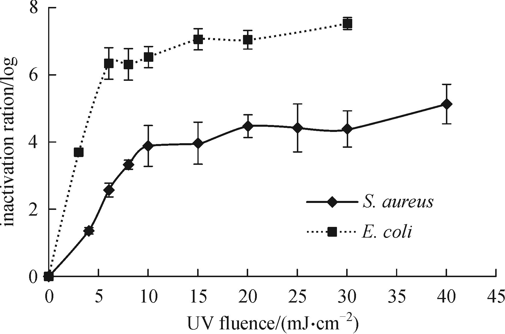
S. aureus showed more resistance to UV irradiation than E. coli by FCM.
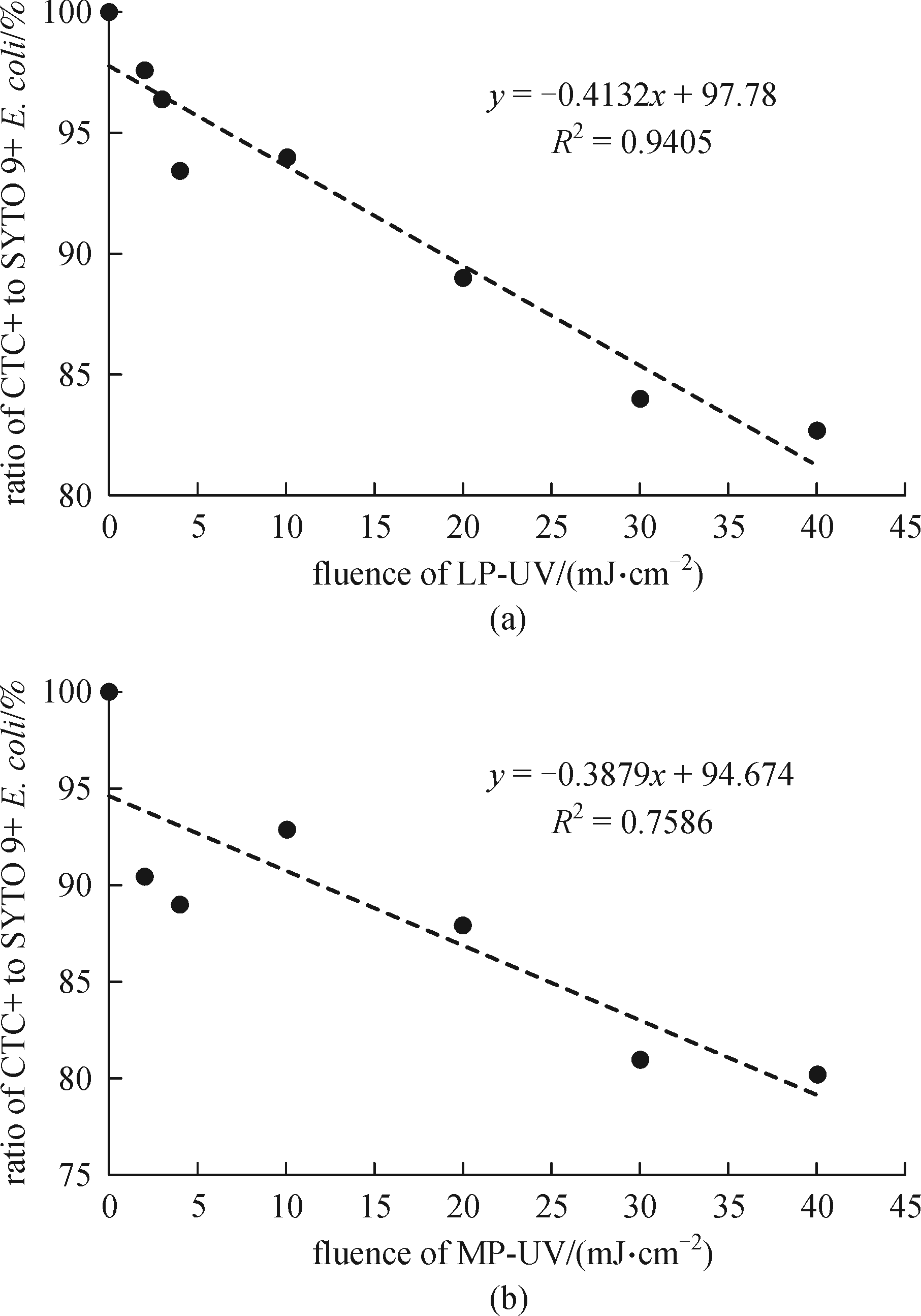
MP-UV was a stronger inhibitor of metabolic activity than LP-UV.
Flow cytometry (FCM) has been widely used in multi-parametric assessment of cells in various research fields, especially in environmental sciences. This study detected the metabolic activity of Escherichia coli and Staphylococcus aureus by using an FCM method based on 5-cyano-2,3-ditolyltetrazolium chloride (CTC); the accuracy of this method was enhanced by adding SYTO 9 and 10% R2A broth. The disinfection effects of chlorine, chloramine, and UV were subsequently evaluated by FCM methods. Chlorine demonstrated stronger and faster destructive effects on cytomembrane than chloramine, and nucleic acids decomposed afterwards. The metabolic activity of the bacteria persisted after the cytomembranewas damaged as detected using CTC. Low-pressure (LP) UV or medium-pressure (MP) UV treatments exerted no significant effects on membrane permeability. The metabolic activity of the bacteria decreased with increasing UV dosage, and MP-UV was a stronger inhibitor of metabolic activity than LP-UV. Furthermore, the membrane of Gram-positive S. aureus was more resistant to chlorine/chloramine than that of Gram-negative E. coli. In addition, S. aureus showed higher resistance to UV irradiation than E. coli.
|
| Keywords
Flow cytometry
Escherichia coli
Staphylococcusaureus
UV
CTC
SYTO 9
|
|
|
| Fund: |
|
Corresponding Author(s):
Wenjun Liu
|
|
Issue Date: 14 November 2016
|
|
| 1 |
Bogosian G, Bourneuf E V. A matter of bacterial life and death. EMBO Reports, 2001, 2(9): 770–774
|
| 2 |
Barcina I I, Ehu B, Arana I. The viable but nonculturable phenotype: a crossroads in the life-cycle of non-differentiating bacteria? Reviews in Environmental Science and Biotechnology, 2009, 8(3): 245–255
|
| 3 |
Robertson B R, Button D K. Characterizing auqatic bacteria according to population, cell-size, and apparent DNA content by flow-cytometry. Cytometry, 1989, 10(1): 70–76
|
| 4 |
Veal D A, Deere D, Ferrari B, Piper J, Attfield P V. Fluorescence staining and flow cytometry for monitoring microbial cells. Journal of Immunological Methods, 2000, 243(1–2): 191–210
|
| 5 |
Hammes F, Berney M, Wang Y, Vital M, Koester O, Egli T. Flow-cytometric total bacterial cell counts as a descriptive microbiological parameter for drinking water treatment processes. Water Research, 2008, 42(1–2): 269–277
|
| 6 |
Prest E I, El-Chakhtoura J, Hammes F, Saikaly P E, van Loosdrecht M, Vrouwenvelder J S. Combining flow cytometry and 16S rRNA gene pyrosequencing: a promising approach for drinking water monitoring and characterization. Water Research, 2014, 63(63): 179–189
|
| 7 |
Vives-Rego J, Lebaron P, Nebe-von Caron G. Current and future applications of flow cytometry in aquatic microbiology. FEMS Microbiology Reviews, 2000, 24(4): 429–448
|
| 8 |
Zipper H, Brunner H, Bernhagen J, Vitzthum F. Investigations on DNA intercalation and surface binding by SYBR Green I, its structure determination and methodological implications. Nucleic Acids Research, 2004, 32(12): 5227–5232
|
| 9 |
Smith J J, McFeters G A. Mechanisms of INT (2-(4-iodophenyl)-3-(4-nitrophenyl)-5-phenyl tetrazolium chloride), and CTC (5-cyano-2,3-ditolyl tetrazolium chloride) reduction in Escherichia coli K-12. Journal of Microbiological Methods, 1997, 29(3): 161–175
|
| 10 |
Dufour P, Colon M. The tetrazolium reduction method for assessing the viability of individual bacterial cells in aquatic environments: improvements, performance and applications. Hydrobiologia, 1992, 232(3): 211–218
|
| 11 |
Roslev P, King G M. Application of a tetrazolium salt with a water-soluble formazan as an indicator of viability in respiring bacteria. Applied and Environmental Microbiology, 1993, 59(9): 2891–2896
|
| 12 |
King L K, Parker B C. A simple, rapid method for enumerating total viable and metabolically active bacteria in groundwater. Applied and Environmental Microbiology, 1988, 54(6): 1630–1631
|
| 13 |
Hatzinger P B, Palmer P, Smith R L, Penarrieta C T, Yoshinari T. Applicability of tetrazolium salts for the measurement of respiratory activity and viability of groundwater bacteria. Journal of Microbiological Methods, 2003, 52(1): 47–58
|
| 14 |
Bartosch S, Mansch R, Knotzsch K, Bock E. CTC staining and counting of actively respiring bacteria in natural stone using confocal laser scanning microscopy. Journal of Microbiological Methods, 2003, 52(1): 75–84
|
| 15 |
Sabaeifard P, Abdi-Ali A, Soudi M R, Dinarvand R. Optimization of tetrazolium salt assay for Pseudomonas aeruginosa biofilm using microtiter plate method. Journal of Microbiological Methods, 2014, 105: 134–140
|
| 16 |
Schaule G, Flemming H C, Ridgway H F. Use of 5-cyano-2,3-ditolyl tetrazolium chloride for quantifying planktonic and sessile respring bacteria in drinking-water. Applied and Environmental Microbiology, 1993, 59(11): 3850–3857
|
| 17 |
Rodriguez G G, Phipps D, Ishiguro K, Ridgway H F. Use of a fluorescent redox probe for direct visualization of actively respiring bacteria. Applied and Environmental Microbiology, 1992, 58(6): 1801–1808
|
| 18 |
Maki J S, Remsen C C. Comparison of two direct-count methods for determining metabolizing bacteria in freshwater. Applied and Environmental Microbiology, 1981, 41(5): 1132–1138
|
| 19 |
Thom S M, Horobin R W, Seidler E, Barer M R. Factors affecting the selection and use of tetrazolium salts as cytochemical indicators of microbial viability and activity. Journal of Applied Bacteriology, 1993, 74(4): 433–443
|
| 20 |
Servais P, Agogue H, Courties C, Joux F, Lebaron P. Are the actively respiring cells (CTC+) those responsible for bacterial production in aquatic environments? FEMS Microbiology Ecology, 2001, 35(2): 171–179
|
| 21 |
Ullrich S, Karrasch B, Hoppe H G, Jeskulke K, Mehrens M. Toxic effects on bacterial metabolism of the redox dye 5-cyano-2,3-ditolyl tetrazolium chloride. Applied and Environmental Microbiology, 1996, 62(12): 4587–4593
|
| 22 |
Lin Y W, Yang T. Rapid detection of viable bacteria by integrated CTC (5-Cyano-2,3-ditoyl tetrazolium chloride) dying and flow cytometry assay (CTC-FCM). Acta Scientiae Circumstantiae, 2013, 33(9): 2511–2515
|
| 23 |
Rezaeinejad S, Ivanov V. Assessment of correlation between physiological states of Escherichia coli cells and their susceptibility to chlorine using flow cytometry. Water Science and Technology: Water Supply, 2013, 13(4): 1056–1062
|
| 24 |
Ramseier M K, von Gunten U, Freihofer P, Hammes F. Kinetics of membrane damage to high (HNA) and low (LNA) nucleic acid bacterial clusters in drinking water by ozone, chlorine, chlorine dioxide, monochloramine, ferrate(VI), and permanganate. Water Research, 2011, 45(3): 1490–1500
|
| 25 |
Olsen R O, Hoffmann F, Hess-Erga O K, Larsen A, Thuestad G, Hoell I A. Ultraviolet radiation as a ballast water treatment strategy: Inactivation of phytoplankton measured with flow cytometry. Marine Pollution Bulletin, 2015, 103(1–2): 270–275
|
| 26 |
Berney M, Hammes F, Bosshard F, Weilenmann H U, Egli T. Assessment and interpretation of bacterial viability by using the LIVE/DEAD BacLight kit in combination with flow cytometry. Applied and Environmental Microbiology, 2007, 73(10): 3283–3290
|
| 27 |
Caron G N, Stephens P, Badley R A. Assessment of bacterial viability status by flow cytometry and single cell sorting. Journal of Applied Microbiology, 1998, 84(6): 988–998
|
| 28 |
Hoefel D, Grooby W L, Monis P T, Andrews S, Saint C P. Enumeration of water-borne bacteria using viability assays and flow cytometry: a comparison to culture-based techniques. Journal of Microbiological Methods, 2003, 55(3): 585–597
|
|
Viewed |
|
|
|
Full text
|
|
|
|
|
Abstract
|
|
|
|
|
Cited |
|
|
|
|
| |
Shared |
|
|
|
|
| |
Discussed |
|
|
|
|