|
|
|
Using mRNA to investigate the effect of low-pressure ultraviolet disinfection on the viability of E. coli |
Chao Yang, Wenjun Sun( ), Xiuwei Ao ), Xiuwei Ao |
| School of Environment, Tsinghua University, Beijing 100084, China |
|
|
|
|
Abstract UV can induce damages on mRNA consistently among different genes. SOS response was more active after UV treatment. Programmed cell death was not found to be more active after UV treatment.
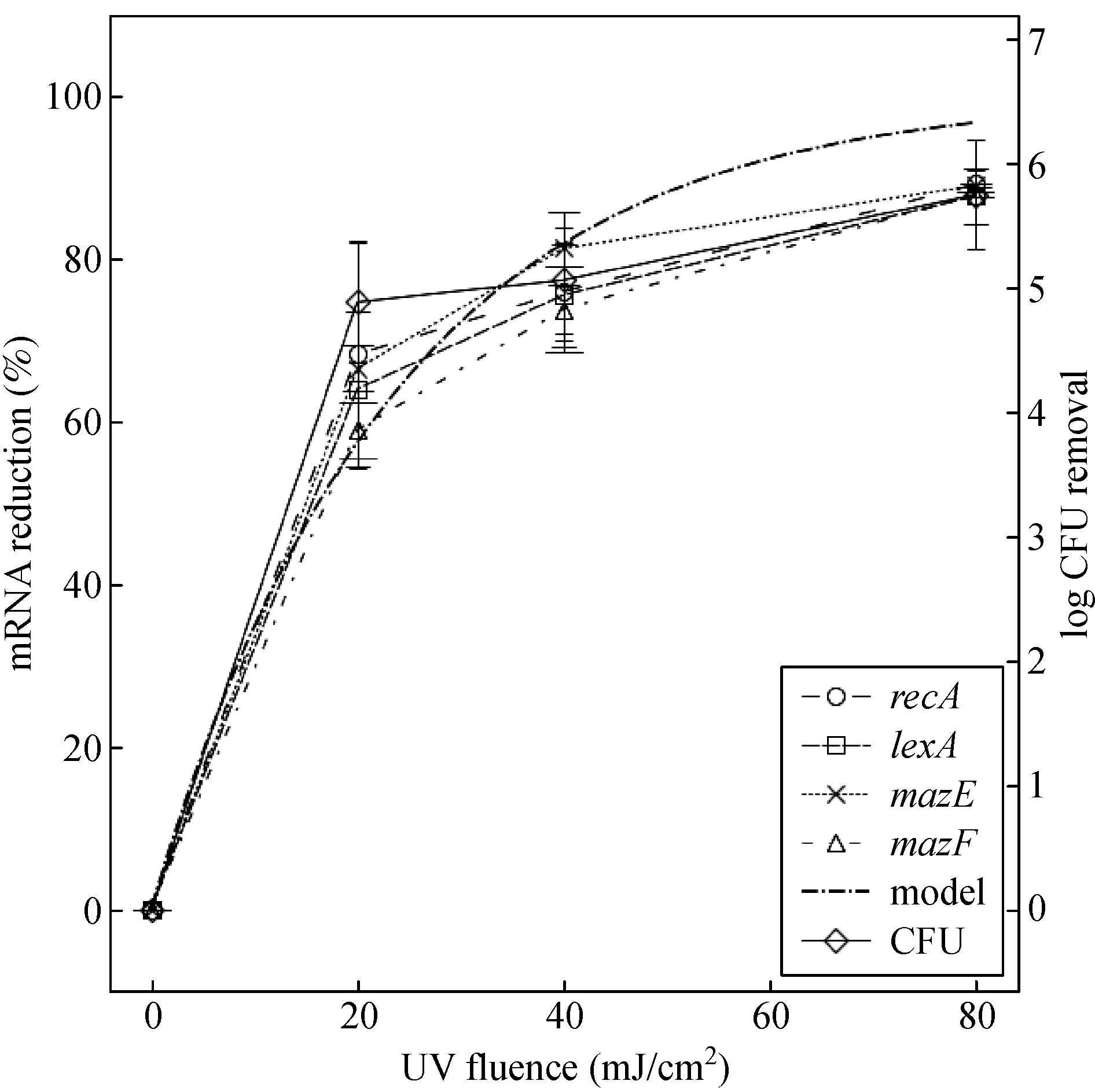
![]() The efficacy of ultraviolet (UV) disinfection has been analyzed and validated by numerous studies using culture-based methods, yet the discovery of the viable but nonculturable state necessitates the investigation of UV disinfection based on viability parameters. Paired regulators of the SOS response system, recA-lexA, and the programmed cell death system, mazEF, in Escherichia coli were chosen as the target genes, and the effect of UV irradiation on the mRNAs of the four genes was studied. This research showed that, after UV irradiation, the responses of the mRNAs were highly consistent, with reduction percentages of approximately 60% at 20 mJ/cm2, 70% at 40 mJ/cm2, and 90% at 80 mJ/cm2, and these reductions were believed to be the result of direct UV damage to nucleic acids. After 24 h of dark incubation, recA and lexA were both upregulated but to a lesser extent for repressor lexA; and mazE and mazF were both downregulated. This result implies that UV irradiation induces the dark repair system more actively, and the cells will proceed to death at a rate similar to that associated with natural decay.
|
| Keywords
UV disinfection
Viability
mRNA
SOS response
Programmed cell death
|
|
Corresponding Author(s):
Wenjun Sun
|
|
Issue Date: 21 March 2019
|
|
| 1 |
NAllocati, M Masulli, CDi Ilio, VDe Laurenzi (2015). Die for the community: An overview of programmed cell death in bacteria. Cell Death & Disease, 6(1): e1609
https://doi.org/10.1038/cddis.2014.570
pmid: 25611384
|
| 2 |
K WBayles (2014). Bacterial programmed cell death: making sense of a paradox. Nature Reviews. Microbiology, 12(1): 63–69
https://doi.org/10.1038/nrmicro3136
pmid: 24336185
|
| 3 |
MBelosevic, S A Craik, J L Stafford, N F Neumann, J Kruithof, D WSmith (2001). Studies on the resistance/reactivation of Giardia muris cysts and Cryptosporidium parvum oocysts exposed to medium-pressure ultraviolet radiation. FEMS Microbiology Letters, 204(1): 197–203
https://doi.org/10.1111/j.1574-6968.2001.tb10885.x
pmid: 11682201
|
| 4 |
E RBlatchley 3rd, NDumoutier, T NHalaby, YLevi, J M Laîné (2001). Bacterial responses to ultraviolet irradiation. Water Science and Technology, 43(10): 179–186
https://doi.org/10.2166/wst.2001.0614
pmid: 11436779
|
| 5 |
E RBlatchley, KOguma, RSommer (2017). Comment on ‘UV disinfection induces a VBNC state in Escherichia coli and Pseudomonas aeruginosa’. IUVA News, 18(3): 12–16
|
| 6 |
J RBolton, K G Linden (2003). Standardization of methods for fluence (UV dose) determination in bench-scale UV experiments. Journal of Environmental Engineering, 129(3): 209–215
https://doi.org/10.1061/(ASCE)0733-9372(2003)129:3(209)
|
| 7 |
S ACraik, G R Finch, J R Bolton, M Belosevic (2000). Inactivation of Giardia muris cysts using medium-pressure ultraviolet radiation in filtered drinking water. Water Research, 34(18): 4325–4332
https://doi.org/10.1016/S0043-1354(00)00207-4
|
| 8 |
AErental, Z Kalderon, ASaada, YSmith, HEngelberg-Kulka (2014). Apoptosis-like death, an extreme SOS response in Escherichia coli. mBio, 5(4): e01426–e14
https://doi.org/10.1128/mBio.01426-14
pmid: 25028428
|
| 9 |
JFang, H Liu, CShang, MZeng, M Ni, WLiu (2014). E. coli and bacteriophage MS2 disinfection by UV, ozone and the combined UV and ozone processes. Frontiers of Environmental Science & Engineering, 8(4): 547–552
https://doi.org/10.1007/s11783-013-0620-2
|
| 10 |
RGehr (2015). Comment on “UV disinfection induces a Vbnc state in Escherichia coli and Pseudomonas aeruginosa”. Environmental Science & Technology, 49(12): 7501
https://doi.org/10.1021/acs.est.5b00769
pmid: 26020586
|
| 11 |
MGuo, H Hu, J RBolton, M GEl-Din (2009). Comparison of low- and medium-pressure ultraviolet lamps: Photoreactivation of Escherichia coli and total coliforms in secondary effluents of municipal wastewater treatment plants. Water Research, 43(3): 815–821
https://doi.org/10.1016/j.watres.2008.11.028
pmid: 19081599
|
| 12 |
RHazan, B Sat, HEngelberg-Kulka (2004). Escherichia coli mazEF-mediated cell death is triggered by various stressful conditions. Journal of Bacteriology, 186(11): 3663–3669
https://doi.org/10.1128/JB.186.11.3663-3669.2004
pmid: 15150257
|
| 13 |
W AHijnen, E F Beerendonk, G J Medema (2006). Inactivation credit of UV radiation for viruses, bacteria and protozoan (oo)cysts in water: a review. Water Research, 40(1): 3–22
https://doi.org/10.1016/j.watres.2005.10.030
pmid: 16386286
|
| 14 |
CJungfer, T Schwartz, UObst (2007). UV-induced dark repair mechanisms in bacteria associated with drinking water. Water Research, 41(1): 188–196
https://doi.org/10.1016/j.watres.2006.09.001
pmid: 17055552
|
| 15 |
M JLehtola, I T Miettinen, T Vartiainen, PRantakokko, AHirvonen, P JMartikainen (2003). Impact of UV disinfection on microbially available phosphorus, organic carbon, and microbial growth in drinking water. Water Research, 37(5): 1064–1070
https://doi.org/10.1016/S0043-1354(02)00462-1
pmid: 12553981
|
| 16 |
K GLinden, N M Hull, R A Rodriguez (2015). Comment on “UV disinfection induces a VBNC state in Escherichia coli and Pseudomonas aeruginosa”. Environmental Science & Technology, 49(17): 10750–10751
https://doi.org/10.1021/acs.est.5b02534
pmid: 26270787
|
| 17 |
YLiu, Q Zhang, YHong (2017). Formation of disinfection byproducts from accumulated soluble products of oleaginous microalga after chlorination. Frontiers of Environmental Science & Engineering, 11(6): 1
|
| 18 |
S YLu, N Y Wang, C Wang (2018). Oxidation and biotoxicity assessment of microcystin-LR using different AOPs based on UV, O3 and H2O2. Frontiers of Environmental Science & Engineering, 12(3): 12
|
| 19 |
K EMurray, E I Manitou-Alvarez, E C Inniss, F G Healy, A A Bodour (2015). Assessment of oxidative and UV-C treatments for inactivating bacterial biofilms from groundwater wells. Frontiers of Environmental Science & Engineering, 9(1): 39–49
https://doi.org/10.1007/s11783-014-0699-0
|
| 20 |
XNie, W Liu, MChen, MLiu, L Ao (2016). Flow cytometric assessment of the effects of chlorine, chloramine, and UV on bacteria by using nucleic acid stains and 5-cyano-2, 3-ditolyltetrazolium chloride. Frontiers of Environmental Science & Engineering, 10(6): 12
https://doi.org/10.1007/s11783-016-0884-4
|
| 21 |
J D (Oliver 2000 ). The Public Health Significance of Viable but Nonculturable Bacteria. Boston: Springer, 277–300
|
| 22 |
J AParker, J L Darby (1995). Particle-associated coliform in secondary effluents: shielding from ultraviolet light disinfection. Water Environment Research, 67(7): 1065–1075
https://doi.org/10.2175/106143095X133310
|
| 23 |
B MPecson, M Ackermann, TKohn (2011). Framework for using quantitative PCR as a nonculture based method to estimate virus infectivity. Environmental Science & Technology, 45(6): 2257–2263
https://doi.org/10.1021/es103488e
pmid: 21322644
|
| 24 |
DPinto, M A Santos, L Chambel (2015). Thirty years of viable but nonculturable state research: unsolved molecular mechanisms. Critical Reviews in Microbiology, 41(1): 61–76
https://doi.org/10.3109/1040841X.2013.794127
pmid: 23848175
|
| 25 |
MPirnie, K G Linden, J P Malley, D Schmelling, O O WUsa (2006). Ultraviolet disinfection guidance manual for the final long term 2 enhanced surface water treatment rule: EPA 815-R-06–007. Washington, DC: EPA, 2–8
|
| 26 |
D AReckhow, K G Linden, J Kim, HShemer, GMakdissy (2010). Effect of UV treatment on DBP formation. Journal- American Water Works Association, 102(6): 100–113
https://doi.org/10.1002/j.1551-8833.2010.tb10134.x
|
| 27 |
R PSinha, D P Häder (2002). UV-induced DNA damage and repair: A review. Photochemical & Photobiological Sciences, 1(4): 225–236
https://doi.org/10.1039/b201230h
pmid: 12661961
|
| 28 |
FWang, W Y Li, Y Li, J PZhang, J PChen, WZhang, XWu (2018). Molecular analysis of bacterial community in the tap water with different water ages of a drinking water distribution system . Frontiers of Environmental Science & Engineering, 12(3): 6
|
| 29 |
N YWang, K Wang, CWang (2017). Comparison of different algicides on growth of Microcystis aeruginosa and microcystin release, as well as its removal pathway in riverways. Frontiers of Environmental Science & Engineering, 11(6): 3
|
| 30 |
H SXu, N Roberts, F LSingleton, R WAttwell, D JGrimes, R RColwell (1982). Survival and viability of nonculturable Escherichia coli and Vibrio cholerae in the estuarine and marine environment. Microbial Ecology, 8(4): 313–323
https://doi.org/10.1007/BF02010671
pmid: 24226049
|
| 31 |
LXu, C Zhang, PXu, X CWang (2018). Mechanisms of ultraviolet disinfection and chlorination of Escherichia coli: Culturability, membrane permeability, metabolism, and genetic damage. Journal of Environmental Sciences-China, 65: 356–366
pmid: 29548407
|
| 32 |
SZhang, C Ye, HLin, LLv, X Yu (2015). UV disinfection induces a VBNC state in Escherichia coli and Pseudomonas aeruginosa. Environmental Science & Technology, 49(3): 1721–1728
https://doi.org/10.1021/es505211e
pmid: 25584685
|
| 33 |
J LZimmer, R M Slawson (2002). Potential repair of Escherichia coli DNA following exposure to UV radiation from both medium- and low-pressure UV sources used in drinking water treatment. Applied and Environmental Microbiology, 68(7): 3293–3299
https://doi.org/10.1128/AEM.68.7.3293-3299.2002
pmid: 12089006
|
| 34 |
J LZimmer-Thomas, R MSlawson, P MHuck (2007). A comparison of DNA repair and survival of Escherichia coli O157:H7 following exposure to both low- and medium-pressure UV irradiation. Journal of Water and Health, 5(3): 407–415
https://doi.org/10.2166/wh.2007.036
pmid: 17878555
|
|
Viewed |
|
|
|
Full text
|
|
|
|
|
Abstract
|
|
|
|
|
Cited |
|
|
|
|
| |
Shared |
|
|
|
|
| |
Discussed |
|
|
|
|