|
|
|
p-Cresyl sulfate promotes the formation of atherosclerotic lesions and induces plaque instability by targeting vascular smooth muscle cells |
Hui Han1,2,Yanjia Chen1,2,Zhengbin Zhu1,Xiuxiu Su1,Jingwei Ni1,Run Du1,Ruiyan Zhang1,2,*( ),Wei Jin1,*() ),Wei Jin1,*() |
1. Department of Cardiology, Ruijin Hospital Affiliated to Shanghai Jiao Tong University School of Medicine, Shanghai 200025, China
2. Institute of Cardiovascular Diseases, Shanghai Jiao Tong University School of Medicine, Shanghai 200025, China |
|
|
|
|
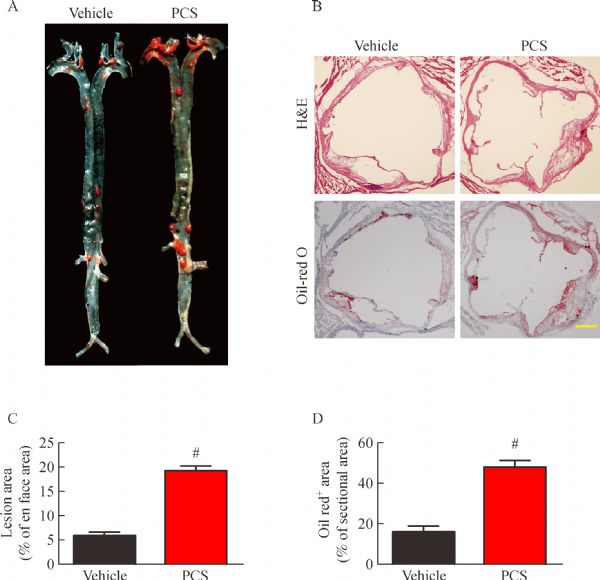
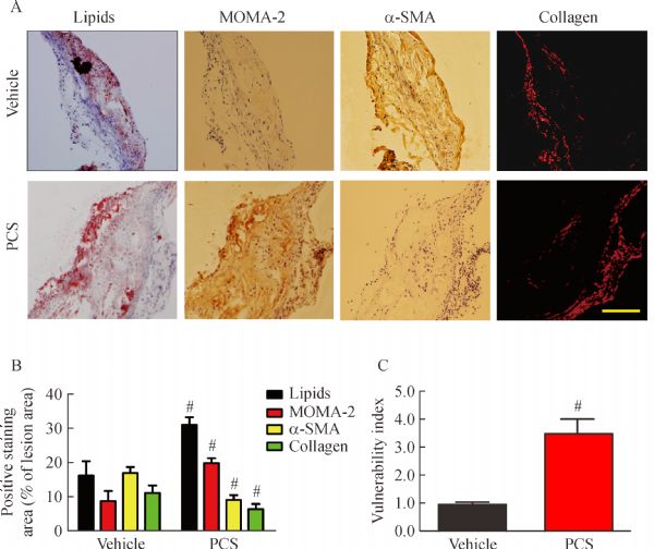
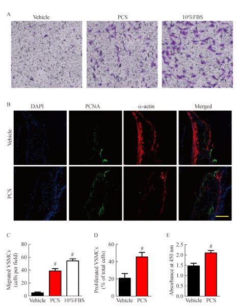
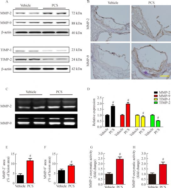
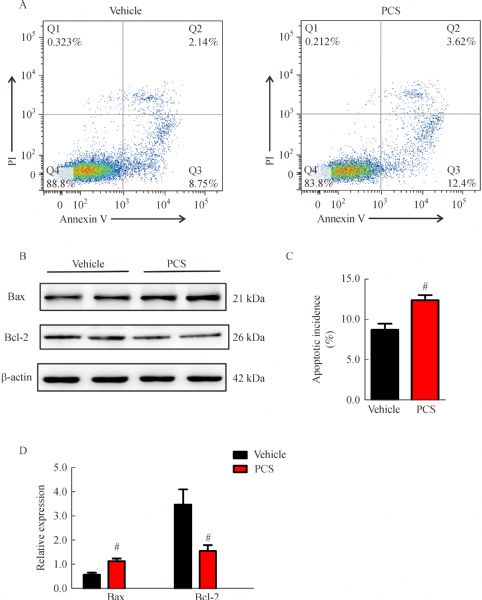
Abstract Coronary atherosclerosis is a major complication of chronic kidney disease. This condition contributes to the increased mortality in dialysis patients. p-Cresyl sulfate (PCS) is a prototype of protein-bound uremic toxins that cannot be efficiently removed through routine dialysis procedures. In the present study, ApoE−/− mice that underwent 5/6 nephrectomy were randomly divided into two groups, namely, vehicle-treated group (n = 20) and PCS-treated group (n = 20). Mice were sacrificed for en face and immunohistological analyses after 8 or 24 weeks of high-fat diet. Rat aortic vascular smooth muscle cells (VSMCs) were treated with phosphate buffer solution or 500 µmol/L PCS for in vitro evaluation. PCS-treated mice were observed to suffer increased atherosclerotic lesions after eight weeks of PCS administration. Moreover, 24 weeks of PCS administration also markedly increased the vulnerability index of aortic plaques. PCS was also observed to facilitate the migration and proliferation of VSMCs during the progression of the disease. Moreover, PCS disturbed the balance between matrix metalloproteinases and tissue inhibitor of metalloproteinases within the plaques. Thus, PCS played a vital role in promoting atherogenesis and disturbing the stability of formed plaques probably by targeting VSMCs.
|
| Keywords
p-cresyl sulfate
atherosclerosis
plaque stability
vascular smooth muscle cell
|
|
Corresponding Author(s):
Ruiyan Zhang,Wei Jin
|
|
Just Accepted Date: 19 July 2016
Online First Date: 12 August 2016
Issue Date: 30 August 2016
|
|
| 1 |
Collins AJ, Foley RN, Chavers B, Gilbertson D, Herzog C, Johansen K, Kasiske B, Kutner N, Liu J, St Peter W, Guo H, Gustafson S, Heubner B, Lamb K, Li S, Li S, Peng Y, Qiu Y, Roberts T, Skeans M, Snyder J, Solid C, Thompson B, Wang C, Weinhandl E, Zaun D, Arko C, Chen SC, Daniels F, Ebben J, Frazier E, Hanzlik C, Johnson R, Sheets D, Wang X, Forrest B, Constantini E, Everson S, Eggers P, Agodoa L. US Renal Data System 2011 Annual Data Report. Am J Kidney Dis 2012; 59(1 Suppl 1): A7, e1–420
https://doi.org/10.1053/j.ajkd.2011.11.015
|
| 2 |
Sarnak MJ, Levey AS, Schoolwerth AC, Coresh J, Culleton B, Hamm LL, McCullough PA, Kasiske BL, Kelepouris E, Klag MJ, Parfrey P, Pfeffer M, Raij L, Spinosa DJ, Wilson PW; American Heart Association Councils on Kidney in Cardiovascular Disease, High Blood Pressure Research, Clinical Cardiology, and Epidemiology and Prevention. Kidney disease as a risk factor for development of cardiovascular disease: a statement from the American Heart Association Councils on Kidney in Cardiovascular Disease, High Blood Pressure Research, Clinical Cardiology, and Epidemiology and Prevention. Circulation 2003; 108(17): 2154–2169
https://doi.org/10.1161/01.CIR.0000095676.90936.80
pmid: 14581387
|
| 3 |
Formanowicz D, Wanic-Kossowska M, Pawliczak E, Radom M, Formanowicz P. Usefulness of serum interleukin-18 in predicting cardiovascular mortality in patients with chronic kidney disease — systems and clinical approach. Sci Rep 2015; 5: 18332
https://doi.org/10.1038/srep18332
pmid: 26669254
|
| 4 |
Sirich TL, Meyer TW, Gondouin B, Brunet P, Niwa T. Protein-bound molecules: a large family with a bad character. Semin Nephrol 2014; 34(2): 106–117
https://doi.org/10.1016/j.semnephrol.2014.02.004
pmid: 24780467
|
| 5 |
Ito S, Yoshida M. Protein-bound uremic toxins: new culprits of cardiovascular events in chronic kidney disease patients. Toxins (Basel) 2014; 6(2): 665–678
https://doi.org/10.3390/toxins6020665
pmid: 24561478
|
| 6 |
Vanholder R, Schepers E, Pletinck A, Nagler EV, Glorieux G. The uremic toxicity of indoxyl sulfate and p-cresyl sulfate: a systematic review. J Am Soc Nephrol 2014; 25(9): 1897–1907
https://doi.org/10.1681/ASN.2013101062
pmid: 24812165
|
| 7 |
Itoh Y, Ezawa A, Kikuchi K, Tsuruta Y, Niwa T. Protein-bound uremic toxins in hemodialysis patients measured by liquid chromatography/tandem mass spectrometry and their effects on endothelial ROS production. Anal Bioanal Chem 2012; 403(7): 1841–1850
https://doi.org/10.1007/s00216-012-5929-3
pmid: 22447217
|
| 8 |
Miyamoto Y, Watanabe H, Noguchi T, Kotani S, Nakajima M, Kadowaki D, Otagiri M, Maruyama T. Organic anion transporters play an important role in the uptake of p-cresyl sulfate, a uremic toxin, in the kidney. Nephrol Dial Transplant 2011; 26(8): 2498–2502
https://doi.org/10.1093/ndt/gfq785
pmid: 21303967
|
| 9 |
Wu IW, Hsu KH, Hsu HJ, Lee CC, Sun CY, Tsai CJ, Wu MS. Serum free p-cresyl sulfate levels predict cardiovascular and all-cause mortality in elderly hemodialysis patients — a prospective cohort study. Nephrol Dial Transplant 2012; 27(3): 1169–1175
https://doi.org/10.1093/ndt/gfr453
pmid: 21891772
|
| 10 |
Liabeuf S, Barreto DV, Barreto FC, Meert N, Glorieux G, Schepers E, Temmar M, Choukroun G, Vanholder R, Massy ZA; European Uraemic Toxin Work Group (EUTox). Free p-cresylsulphate is a predictor of mortality in patients at different stages of chronic kidney disease. Nephrol Dial Transplant 2010; 25(4): 1183–1191
https://doi.org/10.1093/ndt/gfp592
pmid: 19914995
|
| 11 |
Curcio A, Torella D, Indolfi C. Mechanisms of smooth muscle cell proliferation and endothelial regeneration after vascular injury and stenting: approach to therapy. Circ J 2011; 75(6): 1287–1296
|
| 12 |
Newby AC, Libby P, van der Wal AC. Plaque instability — the real challenge for atherosclerosis research in the next decade? Cardiovasc Res 1999; 41(2): 321–322
pmid: 10341831
|
| 13 |
Muteliefu G, Enomoto A, Jiang P, Takahashi M, Niwa T. Indoxyl sulphate induces oxidative stress and the expression of osteoblast-specific proteins in vascular smooth muscle cells. Nephrol Dial Transplant 2009; 24(7): 2051–2058
https://doi.org/10.1093/ndt/gfn757
pmid: 19164326
|
| 14 |
Ito S, Osaka M, Higuchi Y, Nishijima F, Ishii H, Yoshida M. Indoxyl sulfate induces leukocyte-endothelial interactions through up-regulation of E-selectin. J Biol Chem 2010; 285(50): 38869–38875
https://doi.org/10.1074/jbc.M110.166686
pmid: 20937831
|
| 15 |
Feigenbaum J, Neuberg CA. Simplified method for the preparation of aromatic sulfuric acid esters. J Am Chem Soc 1941; 63: 3529–3530
|
| 16 |
Shimizu RT, Blank RS, Jervis R, Lawrenz-Smith SC, Owens GK. The smooth muscle α-actin gene promoter is differentially regulated in smooth muscle versus non-smooth muscle cells. J Biol Chem 1995; 270(13): 7631–7643
https://doi.org/10.1074/jbc.270.13.7631
pmid: 7706311
|
| 17 |
Ni J, Zhang W, Zhu Z, Zhu J, Du R, Jing Y, Lu L, Zhang R. In vivo kinetics of the uremic toxin p-cresyl sulfate in mice with variable renal function. Ther Apher Dial 2014; 18(6): 637–642
https://doi.org/10.1111/1744-9987.12185
pmid: 25256665
|
| 18 |
Han H, Zhu J, Zhu Z, Ni J, Du R, Dai Y, Chen Y, Wu Z, Lu L, Zhang R. p-Cresyl sulfate aggravates cardiac dysfunction associated with chronic kidney disease by enhancing apoptosis of cardiomyocytes. J Am Heart Assoc 2015; 4(6): e001852
https://doi.org/10.1161/JAHA.115.001852
pmid: 26066032
|
| 19 |
Cho KY, Miyoshi H, Kuroda S, Yasuda H, Kamiyama K, Nakagawara J, Takigami M, Kondo T, Atsumi T. The phenotype of infiltrating macrophages influences arteriosclerotic plaque vulnerability in the carotid artery. J Stroke Cerebrovasc Dis 2013; 22(7): 910–918
|
| 20 |
Ross R. Atherosclerosis — an inflammatory disease. N Engl J Med 1999; 340(2): 115–126
https://doi.org/10.1056/NEJM199901143400207
pmid: 9887164
|
| 21 |
Johnson JL. Emerging regulators of vascular smooth muscle cell function in the development and progression of atherosclerosis. Cardiovasc Res 2014; 103(4): 452–460
https://doi.org/10.1093/cvr/cvu171
pmid: 25053639
|
| 22 |
Gittenberger-de Groot AC, DeRuiter MC, Bergwerff M, Poelmann RE. Smooth muscle cell origin and its relation to heterogeneity in development and disease. Arterioscler Thromb Vasc Biol 1999; 19(7): 1589–1594
https://doi.org/10.1161/01.ATV.19.7.1589
pmid: 10397674
|
| 23 |
Rudijanto A. The role of vascular smooth muscle cells on the pathogenesis of atherosclerosis. Acta Med Indones 2007; 39(2): 86–93
pmid: 17933075
|
| 24 |
Visse R, Nagase H. Matrix metalloproteinases and tissue inhibitors of metalloproteinases: structure, function, and biochemistry. Circ Res 2003; 92(8): 827–839
https://doi.org/10.1161/01.RES.0000070112.80711.3D
pmid: 12730128
|
| 25 |
Newby AC. Matrix metalloproteinases regulate migration, proliferation, and death of vascular smooth muscle cells by degrading matrix and non-matrix substrates. Cardiovasc Res 2006; 69(3): 614–624
https://doi.org/10.1016/j.cardiores.2005.08.002
pmid: 16266693
|
| 26 |
Langer HF, Haubner R, Pichler BJ, Gawaz M. Radionuclide imaging: a molecular key to the atherosclerotic plaque. J Am Coll Cardiol 2008; 52(1): 1–12
https://doi.org/10.1016/j.jacc.2008.03.036
pmid: 18582628
|
| 27 |
Shiomi M, Ito T, Hirouchi Y, Enomoto M. Fibromuscular cap composition is important for the stability of established atherosclerotic plaques in mature WHHL rabbits treated with statins. Atherosclerosis 2001; 157(1): 75–84
https://doi.org/10.1016/S0021-9150(00)00708-5
pmid: 11427206
|
| 28 |
Yang JM, Dong M, Meng X, Zhao YX, Yang XY, Liu XL, Hao PP, Li JJ, Wang XP, Zhang K, Gao F, Zhao XQ, Zhang MX, Zhang Y, Zhang C. Angiotensin-(1-7) dose-dependently inhibits atherosclerotic lesion formation and enhances plaque stability by targeting vascular cells. Arterioscler Thromb Vasc Biol 2013; 33(8): 1978–1985
https://doi.org/10.1161/ATVBAHA.113.301320
pmid: 23723368
|
| 29 |
Schwarz U, Buzello M, Ritz E, Stein G, Raabe G, Wiest G, Mall G, Amann K. Morphology of coronary atherosclerotic lesions in patients with end-stage renal failure. Nephrol Dial Transplant 2000; 15(2): 218–223
https://doi.org/10.1093/ndt/15.2.218
pmid: 10648668
|
| 30 |
Rössig L, Dimmeler S, Zeiher AM. Apoptosis in the vascular wall and atherosclerosis. Basic Res Cardiol 2001; 96(1): 11–22
https://doi.org/10.1007/s003950170073
pmid: 11215528
|
|
Viewed |
|
|
|
Full text
|
|
|
|
|
Abstract
|
|
|
|
|
Cited |
|
|
|
|
| |
Shared |
|
|
|
|
| |
Discussed |
|
|
|
|