|
|
|
Inhibition of the nuclear export of p65 and IQCG in leukemogenesis by NUP98-IQCG |
Mengmeng Pan1,Qiyao Zhang1,2,Ping Liu1,Jinyan Huang1,Yueying Wang1( ),Saijuan Chen1,2() ),Saijuan Chen1,2() |
1. State Key Laboratory of Medical Genomics, Shanghai Institute of Hematology, Rui Jin Hospital Affiliated to Shanghai Jiao Tong University (SJTU) School of Medicine, Shanghai 200025, China
2. Institute of Health Sciences, Shanghai Institutes for Biological Sciences and Graduate School, Chinese Academy of Sciences and SJTU School of Medicine, Shanghai 200025, China |
|
|
|
|
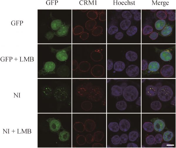
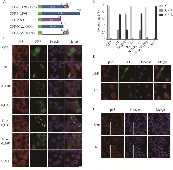
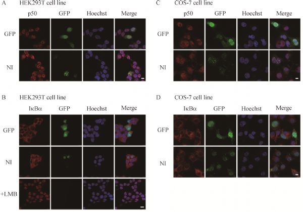
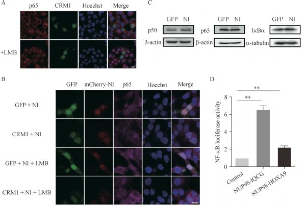
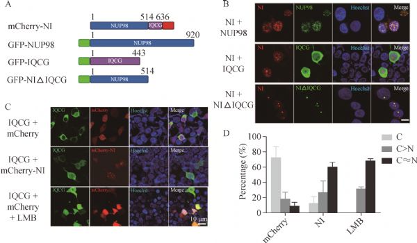
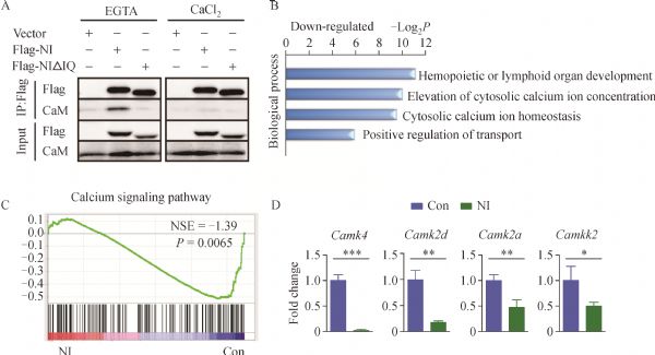
Abstract NUP98 fuses with approximately 34 different partner genes via translocation in hematological malignancies. Transgenic or retrovirus-mediated bone marrow transplanted mouse models reveal the leukemogenesis of some NUP98-related fusion genes. We previously reported the fusion protein NUP98-IQ motif containing G (IQCG) in a myeloid/T lymphoid bi-phenoleukemia patient with t(3;11) and confirmed its leukemogenic ability. Herein, we demonstrated the association of NUP98-IQCG with CRM1, and found that NUP98-IQCG expression inhibits the CRM1-mediated nuclear export of p65 and enhances the transcriptional activity of nuclear factor-κB. Moreover, IQCG could be entrapped in the nucleus by NUP98-IQCG, and the fusion protein interacts with calmodulin via the IQ motif in a calcium-independent manner. Therefore, the inhibition of nuclear exports of p65 and IQCG might contribute to the leukemogenesis of NUP98-IQCG.
|
| Keywords
NUP98-IQCG
nuclear export
NF-κB
CRM1
|
|
Corresponding Author(s):
Yueying Wang,Saijuan Chen
|
|
Just Accepted Date: 07 November 2016
Online First Date: 21 November 2016
Issue Date: 01 December 2016
|
|
| 1 |
Nakamura T, Largaespada DA, Lee MP, Johnson LA, Ohyashiki K, Toyama K, Chen SJ, Willman CL, Chen IM, Feinberg AP, Jenkins NA, Copeland NG, Shaughnessy JD Jr. Fusion of the nucleoporin gene NUP98 to HOXA9 by the chromosome translocation t(7;11)(p15;p15) in human myeloid leukaemia. Nat Genet 1996; 12(2): 154–158
https://doi.org/10.1038/ng0296-154
pmid: 8563753
|
| 2 |
Borrow J, Shearman AM, Stanton VP Jr, Becher R, Collins T, Williams AJ, Dubé I, Katz F, Kwong YL, Morris C, Ohyashiki K, Toyama K, Rowley J, Housman DE. The t(7;11)(p15;p15) translocation in acute myeloid leukaemia fuses the genes for nucleoporin NUP98 and class I homeoprotein HOXA9. Nat Genet 1996; 12(2): 159–167
https://doi.org/10.1038/ng0296-159
pmid: 8563754
|
| 3 |
Franks TM, Hetzer MW. The role of Nup98 in transcription regulation in healthy and diseased cells. Trends Cell Biol 2013; 23(3): 112–117
https://doi.org/10.1016/j.tcb.2012.10.013
pmid: 23246429
|
| 4 |
Takeda A, Sarma NJ, Abdul-Nabi AM, Yaseen NR. Inhibition of CRM1-mediated nuclear export of transcription factors by leukemogenic NUP98 fusion proteins. J Biol Chem 2010; 285(21): 16248–16257
https://doi.org/10.1074/jbc.M109.048785
pmid: 20233715
|
| 5 |
Fontoura BM, Blobel G, Yaseen NR. The nucleoporin Nup98 is a site for GDP/GTP exchange on ran and termination of karyopherin beta 2-mediated nuclear import. J Biol Chem 2000; 275(40): 31289–31296
https://doi.org/10.1074/jbc.M004651200
pmid: 10875935
|
| 6 |
Radu A, Moore MS, Blobel G. The peptide repeat domain of nucleoporin Nup98 functions as a docking site in transport across the nuclear pore complex. Cell 1995; 81(2): 215–222
https://doi.org/10.1016/0092-8674(95)90331-3
pmid: 7736573
|
| 7 |
Blevins MB, Smith AM, Phillips EM, Powers MA. Complex formation among the RNA export proteins Nup98, Rae1/Gle2, and TAP. J Biol Chem 2003; 278(23): 20979–20988
https://doi.org/10.1074/jbc.M302061200
pmid: 12637516
|
| 8 |
Taketani T, Taki T, Shibuya N, Kikuchi A, Hanada R, Hayashi Y. Novel NUP98-HOXC11 fusion gene resulted from a chromosomal break within exon 1 of HOXC11 in acute myeloid leukemia with t(11;12)(p15;q13). Cancer Res 2002; 62(16): 4571–4574
pmid: 12183408
|
| 9 |
Raza-Egilmez SZ, Jani-Sait SN, Grossi M, Higgins MJ, Shows TB, Aplan PD. NUP98-HOXD13 gene fusion in therapy-related acute myelogenous leukemia. Cancer Res 1998; 58(19): 4269–4273
pmid: 9766650
|
| 10 |
Jankovic D, Gorello P, Liu T, Ehret S, La Starza R, Desjobert C, Baty F, Brutsche M, Jayaraman PS, Santoro A, Mecucci C, Schwaller J. Leukemogenic mechanisms and targets of a NUP98/HHEX fusion in acute myeloid leukemia. Blood 2008; 111(12): 5672–5682
https://doi.org/10.1182/blood-2007-09-108175
pmid: 18388181
|
| 11 |
Nakamura T, Yamazaki Y, Hatano Y, Miura I. NUP98 is fused to PMX1 homeobox gene in human acute myelogenous leukemia with chromosome translocation t(1;11)(q23;p15). Blood 1999; 94(2): 741–747
pmid: 10397741
|
| 12 |
Gough SM, Slape CI, Aplan PD. NUP98 gene fusions and hematopoietic malignancies: common themes and new biologic insights. Blood 2011; 118(24): 6247–6257
https://doi.org/10.1182/blood-2011-07-328880
pmid: 21948299
|
| 13 |
Yassin ER, Abdul-Nabi AM, Takeda A, Yaseen NR. Effects of the NUP98-DDX10 oncogene on primary human CD34+ cells: role of a conserved helicase motif. Leukemia 2010; 24(5): 1001–1011
https://doi.org/10.1038/leu.2010.42
pmid: 20339440
|
| 14 |
Wang GG, Cai L, Pasillas MP, Kamps MP. NUP98-NSD1 links H3K36 methylation to Hox-A gene activation and leukaemogenesis. Nat Cell Biol 2007; 9(7): 804–812
https://doi.org/10.1038/ncb1608
pmid: 17589499
|
| 15 |
Pan Q, Zhu YJ, Gu BW, Cai X, Bai XT, Yun HY, Zhu J, Chen B, Weng L, Chen Z, Xue YQ, Chen SJ. A new fusion gene NUP98-IQCG identified in an acute T-lymphoid/myeloid leukemia with a t(3;11)(q29q13;p15)del(3)(q29) translocation. Oncogene 2008; 27(24): 3414–3423
https://doi.org/10.1038/sj.onc.1210999
pmid: 18084320
|
| 16 |
Hussey DJ, Dobrovic A. Recurrent coiled-coil motifs in NUP98 fusion partners provide a clue to leukemogenesis. Blood 2002; 99(3): 1097–1098
https://doi.org/10.1182/blood.V99.3.1097
pmid: 11822362
|
| 17 |
Pan MM, Zhang QY, Wang YY, Liu P, Ren RB, Huang JY, Chen LT, Xi XD, Chen Z, Chen SJ. Human NUP98-IQCG fusion protein induces acute myelomonocytic leukemia in mice by dysregulating the Hox/Pbx3 pathway. Leukemia 2016; 30(7): 1590–1593 doi:10.1038/leu.2015.347
pmid: 26675333
|
| 18 |
Baldwin AS Jr. The NF-kB and IkB proteins: new discoveries and insights. Annu Rev Immunol 1996; 14(1): 649–681
https://doi.org/10.1146/annurev.immunol.14.1.649
pmid: 8717528
|
| 19 |
Wang Y, Mo X, Piper MG, Wang H, Parinandi NL, Guttridge D, Marsh CB. M-CSF induces monocyte survival by activating NF-kB p65 phosphorylation at Ser276 via protein kinase C. PLoS ONE 2011; 6(12): e28081
https://doi.org/10.1371/journal.pone.0028081
pmid: 22216091
|
| 20 |
Oka M, Asally M, Yasuda Y, Ogawa Y, Tachibana T, Yoneda Y. The mobile FG nucleoporin Nup98 is a cofactor for Crm1-dependent protein export. Mol Biol Cell 2010; 21(11): 1885–1896
https://doi.org/10.1091/mbc.E09-12-1041
pmid: 20375145
|
| 21 |
Li RK, Tan JL, Chen LT, Feng JS, Liang WX, Guo XJ, Liu P, Chen Z, Sha JH, Wang YF, Chen SJ. Iqcg is essential for sperm flagellum formation in mice. PLoS ONE 2014; 9(5): e98053
https://doi.org/10.1371/journal.pone.0098053
pmid: 24849454
|
| 22 |
Chen LT, Liang WX, Chen S, Li RK, Tan JL, Xu PF, Luo LF, Wang L, Yu SH, Meng G, Li KK, Liu TX, Chen Z, Chen SJ. Functional and molecular features of the calmodulin-interacting protein IQCG required for haematopoiesis in zebrafish. Nat Commun 2014; 5(5): 3811
pmid: 24787902
|
| 23 |
Kitsos CM, Sankar U, Illario M, Colomer-Font JM, Duncan AW, Ribar TJ, Reya T, Means AR. Calmodulin-dependent protein kinase IV regulates hematopoietic stem cell maintenance. J Biol Chem 2005; 280(39): 33101–33108
https://doi.org/10.1074/jbc.M505208200
pmid: 16020540
|
| 24 |
Monaco S, Rusciano MR, Maione AS, Soprano M, Gomathinayagam R, Todd LR, Campiglia P, Salzano S, Pastore L, Leggiero E, Wilkerson DC, Rocco M, Selleri C, Iaccarino G, Sankar U, Illario M. A novel crosstalk between calcium/calmodulin kinases II and IV regulates cell proliferation in myeloid leukemia cells. Cell Signal 2015; 27(2): 204–214
https://doi.org/10.1016/j.cellsig.2014.11.007
pmid: 25446257
|
| 25 |
Kim D, Pertea G, Trapnell C, Pimentel H, Kelley R, Salzberg SL. TopHat2: accurate alignment of transcriptomes in the presence of insertions, deletions and gene fusions. Genome Biol 2013; 14(4): R36
https://doi.org/10.1186/gb-2013-14-4-r36
pmid: 23618408
|
| 26 |
Anders S, Pyl PT, Huber W. HTSeq—a Python framework to work with high-throughput sequencing data. Bioinformatics 2015; 31(2): 166–169
https://doi.org/10.1093/bioinformatics/btu638
pmid: 25260700
|
| 27 |
Robinson MD, McCarthy DJ, Smyth GK. edgeR: a Bioconductor package for differential expression analysis of digital gene expression data. Bioinformatics 2010; 26(1): 139–140
https://doi.org/10.1093/bioinformatics/btp616
pmid: 19910308
|
| 28 |
Huang DW, Sherman BT, Lempicki RA. Systematic and integrative analysis of large gene lists using DAVID bioinformatics resources. Nat Protoc 2009; 4(1): 44–57
https://doi.org/10.1038/nprot.2008.211
pmid: 19131956
|
| 29 |
Subramanian A, Tamayo P, Mootha VK, Mukherjee S, Ebert BL, Gillette MA, Paulovich A, Pomeroy SL, Golub TR, Lander ES, Mesirov JP. Gene set enrichment analysis: a knowledge-based approach for interpreting genome-wide expression profiles. Proc Natl Acad Sci USA 2005; 102(43): 15545–15550
https://doi.org/10.1073/pnas.0506580102
pmid: 16199517
|
| 30 |
Huang TT, Kudo N, Yoshida M, Miyamoto S. A nuclear export signal in the N-terminal regulatory domain of IkBα controls cytoplasmic localization of inactive NF-kB/IkBα complexes. Proc Natl Acad Sci USA 2000; 97(3): 1014–1019
https://doi.org/10.1073/pnas.97.3.1014
pmid: 10655476
|
| 31 |
Minucci S, Maccarana M, Cioce M, De Luca P, Gelmetti V, Segalla S, Di Croce L, Giavara S, Matteucci C, Gobbi A, Bianchini A, Colombo E, Schiavoni I, Badaracco G, Hu X, Lazar MA, Landsberger N, Nervi C, Pelicci PG. Oligomerization of RAR and AML1 transcription factors as a novel mechanism of oncogenic activation. Mol Cell 2000; 5(5): 811–820
https://doi.org/10.1016/S1097-2765(00)80321-4
pmid: 10882117
|
| 32 |
Tran QK, Black DJ, Persechini A. Intracellular coupling via limiting calmodulin. J Biol Chem 2003; 278(27): 24247–24250
https://doi.org/10.1074/jbc.C300165200
pmid: 12738782
|
| 33 |
Colomer J, Means AR. Physiological roles of the Ca2+/CaM-dependent protein kinase cascade in health and disease. Subcell Biochem 2007; 45: 169–214
https://doi.org/10.1007/978-1-4020-6191-2_7
pmid: 18193638
|
| 34 |
Stewart TA, Yapa KT, Monteith GR. Altered calcium signaling in cancer cells. Biochim Biophys Acta 2015; 1848(10 Pt B): 2502–2511
https://doi.org/10.1016/j.bbamem.2014.08.016
pmid: 25150047
|
|
Viewed |
|
|
|
Full text
|
|
|
|
|
Abstract
|
|
|
|
|
Cited |
|
|
|
|
| |
Shared |
|
|
|
|
| |
Discussed |
|
|
|
|