|
|
|
Sub-cytotoxic concentrations of ionic silver promote the proliferation of human keratinocytes by inducing the production of reactive oxygen species |
Xiaodong Duan1,2, Daizhi Peng1,3( ), Yilan Zhang1, Yalan Huang1, Xiao Liu1, Ruifu Li1, Xin Zhou1, Jing Liu1 ), Yilan Zhang1, Yalan Huang1, Xiao Liu1, Ruifu Li1, Xin Zhou1, Jing Liu1 |
1. Institute of Burn Research, Southwest Hospital, Third Military Medical University, Chongqing 400038, China
2. Burn and Plastic Surgery Department, 209 Hospital of PLA, Mudanjiang 157011, China
3. Tissue Engineering Research Unit, State Key Laboratory of Trauma, Burns and Combined Injury, Third Military Medical University, Chongqing 400038, China |
|
|
|
|
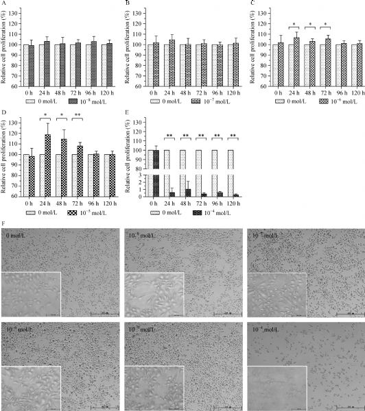
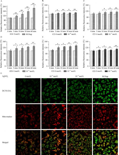
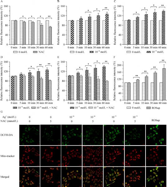
Abstract Silver-containing preparations are widely used in the management of skin wounds, but the effects of silver ions on skin wound healing remain poorly understood. This study investigated the effects of silver ions (Ag+) on the proliferation of human skin keratinocytes (HaCaT) and the production of intracellular reactive oxygen species (ROS). After treating HaCaT cells with Ag+ and/or the active oxygen scavenger N-acetyl cysteine (NAC), cell proliferation and intracellular ROS generation were assessed using CCK-8 reagent and DCFH-DA fluorescent probe, respectively. In addition, 5-bromo-2-deoxyUridine (BrdU) incorporation assays, cell cycle flow cytometry, and proliferating cell nuclear antigen (PCNA) immunocytochemistry were conducted to further evaluate the effects of sub-cytotoxic Ag+ concentrations on HaCaT cells. The proliferation of HaCaT cells was promoted in the presence of 10−6 and 10−5 mol/L Ag+ at 24, 48, and 72 h. Intracellular ROS generation also significantly increased for 5–60 min after exposure to Ag+. The number of BrdU-positive cells and the presence of PCNA in HaCaT cells increased 48 h after the addition of 10−6 and 10−5 mol/L Ag+, with 10−5 mol/L Ag+ markedly increasing the cell proliferation index. These effects of sub-cytotoxic Ag+ concentrations were repressed by 5 mmol/L NAC. Our results suggest that sub-cytotoxic Ag+ concentrations promote the proliferation of human keratinocytes and might be associated with a moderate increase in intracellular ROS levels. This study provides important experimental evidence for developing novel silver-based wound agents or dressings with few or no cytotoxicity.
|
| Keywords
ionic silver
human keratinocyte
cell proliferation
reactive oxygen species
active oxygen scavenger
NAC
|
|
Corresponding Author(s):
Daizhi Peng
|
|
Just Accepted Date: 04 August 2017
Online First Date: 03 November 2017
Issue Date: 04 May 2018
|
|
| 1 |
Moyer CA, Brentano L, Gravens DL, Margraf HW Jr, Monafo WW Jr. Treatment of large human burns with 0.5 per cent silver nitrate solution. Arch Surg 1965; 90(6): 812–867
https://doi.org/10.1001/archsurg.1965.01320120014002
pmid: 14333527
|
| 2 |
Fox CL Jr. Silver sulfadiazine—a new topical therapy for Pseudomonas in burns. Therapy of Pseudomonas infection in burns. Arch Surg 1968; 96(2): 184–188
https://doi.org/10.1001/archsurg.1968.01330200022004
pmid: 5638080
|
| 3 |
Chopra I. The increasing use of silver-based products as antimicrobial agents: a useful development or a cause for concern? J Antimicrob Chemother 2007; 59(4): 587–590
https://doi.org/10.1093/jac/dkm006
pmid: 17307768
|
| 4 |
Swathy JR, Sankar MU, Chaudhary A, Aigal S, Anshup T, Pradeep. Antimicrobial silver: an unprecedented anion effect. Sci Rep 2014; 4: 7161
https://doi.org/10.1038/srep07161
pmid: 25418185
|
| 5 |
Mijnendonckx K, Leys N, Mahillon J, Silver S, Van Houdt R. Antimicrobial silver: uses, toxicity and potential for resistance. Biometals 2013; 26(4): 609–621
https://doi.org/10.1007/s10534-013-9645-z
pmid: 23771576
|
| 6 |
Lansdown A, Williams A. Bacterial resistance to silver-based antibiotics. Nurs Times 2007; 103(9): 48–49
pmid: 17375724
|
| 7 |
Burd A, Kwok CH, Hung SC, Chan HS, Gu H, Lam WK, Huang L. A comparative study of the cytotoxicity of silver-based dressings in monolayer cell, tissue explant, and animal models. Wound Repair Regen 2007; 15(1): 94–104
https://doi.org/10.1111/j.1524-475X.2006.00190.x
pmid: 17244325
|
| 8 |
Berger TJ, Spadaro JA, Chapin SE, Becker RO. Electrically generated silver ions: quantitative effects on bacterial and mammalian cells. Antimicrob Agents Chemother 1976; 9(2): 357–358
https://doi.org/10.1128/AAC.9.2.357
pmid: 944551
|
| 9 |
Arora S, Jain J, Rajwade JM, Paknikar KM. Interactions of silver nanoparticles with primary mouse fibroblasts and liver cells. Toxicol Appl Pharmacol 2009; 236(3): 310–318
https://doi.org/10.1016/j.taap.2009.02.020
pmid: 19269301
|
| 10 |
Zanette C, Pelin M, Crosera M, Adami G, Bovenzi M, Larese FF, Florio C. Silver nanoparticles exert a long-lasting antiproliferative effect on human keratinocyte HaCaT cell line. Toxicol In Vitro 2011; 25(5): 1053–1060
https://doi.org/10.1016/j.tiv.2011.04.005
pmid: 21501681
|
| 11 |
Poon VKM, Burd A. In vitro cytotoxity of silver: implication for clinical wound care. Burns 2004; 30(2): 140–147
https://doi.org/10.1016/j.burns.2003.09.030
pmid: 15019121
|
| 12 |
Morones-Ramirez JR, Winkler JA, Spina CS, Collins JJ. Silver enhances antibiotic activity against gram-negative bacteria. Sci Transl Med 2013; 5(190): 190ra81
https://doi.org/10.1126/scitranslmed.3006276
pmid: 23785037
|
| 13 |
Thomas GW, Rael LT, Bar-Or R, Shimonkevitz R, Mains CW, Slone DS, Craun ML, Bar-Or D. Mechanisms of delayed wound healing by commonly used antiseptics. J Trauma 2009, 66:82–90, discussion 90–91
https://doi.org/10.1097/TA.0b013e31818b146d
|
| 14 |
Michaels JA, Campbell B, King B, Palfreyman SJ, Shackley P, Stevenson M. Randomized controlled trial and cost-effectiveness analysis of silver-donating antimicrobial dressings for venous leg ulcers (VULCAN trial). Br J Surg 2009; 96(10): 1147–1156
https://doi.org/10.1002/bjs.6786
pmid: 19787753
|
| 15 |
White R, Cutting K, Ousey K, Butcher M, Gray D, Flanagan M, Donnelly J, McIntosh C, Kingsley A, Fletcher J, Chadwick P, Gethin G, Beldon P. Randomized controlled trial and cost-effectiveness analysis of silver-donating antimicrobial dressings for venous leg ulcers (VULCAN trial) (Br J Surg 2009; 96: 1147-1156). Br J Surg 2010; 97(3): 459–460
https://doi.org/10.1002/bjs.7017
pmid: 20140943
|
| 16 |
Vermeulen H, van Hattem JM, Storm-Versloot MN, Ubbink DT. Topical silver for treating infected wounds. Cochrane Database Syst Rev 2007; (1): CD005486
pmid: 17253557
|
| 17 |
Storm-Versloot MN, Vos CG, Ubbink DT, Vermeulen H. Topical silver for preventing wound infection. Cochrane Database Syst Rev 2010; (3): CD006478
pmid: 20238345
|
| 18 |
Aziz Z, Abu SF, Chong NJ. A systematic review of silver-containing dressings and topical silver agents (used with dressings) for burn wounds. Burns 2012; 38(3): 307–318
https://doi.org/10.1016/j.burns.2011.09.020
pmid: 22030441
|
| 19 |
Gordon O, Vig Slenters T, Brunetto PS, Villaruz AE, Sturdevant DE, Otto M, Landmann R, Fromm KM. Silver coordination polymers for prevention of implant infection: thiol interaction, impact on respiratory chain enzymes, and hydroxyl radical induction. Antimicrob Agents Chemother 2010; 54(10): 4208– 4218
https://doi.org/10.1128/AAC.01830-09
pmid: 20660682
|
| 20 |
Foldbjerg R, Olesen P, Hougaard M, Dang DA, Hoffmann HJ, Autrup H. PVP-coated silver nanoparticles and silver ions induce reactive oxygen species, apoptosis and necrosis in THP-1 monocytes. Toxicol Lett 2009; 190(2): 156–162
https://doi.org/10.1016/j.toxlet.2009.07.009
pmid: 19607894
|
| 21 |
Miyayama T, Arai Y, Suzuki N, Hirano S. Mitochondrial electron transport is inhibited by disappearance of metallothionein in human bronchial epithelial cells following exposure to silver nitrate. Toxicology 2013; 305: 20–29
https://doi.org/10.1016/j.tox.2013.01.004
pmid: 23333424
|
| 22 |
Allen RG, Tresini M. Oxidative stress and gene regulation. Free Radic Biol Med 2000; 28(3): 463–499
https://doi.org/10.1016/S0891-5849(99)00242-7
pmid: 10699758
|
| 23 |
Yamamoto A, Honma R, Tanaka A, Sumita M. Generic tendency of metal salt cytotoxicity for six cell lines. J Biomed Mater Res 1999; 47(3): 396–403
https://doi.org/10.1002/(SICI)1097-4636(19991205)47:3<396::AID-JBM15>3.0.CO;2-R
pmid: 10487892
|
| 24 |
Martin KR, Barrett JC. Reactive oxygen species as double-edged swords in cellular processes: low-dose cell signaling versus high-dose toxicity. Hum Exp Toxicol 2002; 21(2): 71–75
https://doi.org/10.1191/0960327102ht213oa
pmid: 12102499
|
| 25 |
Sena LA, Chandel NS. Physiological roles of mitochondrial reactive oxygen species. Mol Cell 2012; 48(2): 158–167
https://doi.org/10.1016/j.molcel.2012.09.025
pmid: 23102266
|
| 26 |
Sauer H, Wartenberg M, Hescheler J. Reactive oxygen species as intracellular messengers during cell growth and differentiation. Cell Physiol Biochem 2001; 11(4): 173–186
https://doi.org/10.1159/000047804
pmid: 11509825
|
| 27 |
Samuni Y, Goldstein S, Dean OM, Berk M. The chemistry and biological activities of N-acetylcysteine. Biochim Biophys Acta. 2013;1830(8): 4117–4129
https://doi.org/10.1016/j.bbagen.2013.04.016
|
| 28 |
Bateman DN, Dear JW, Thanacoody HK, Thomas SH, Eddleston M, Sandilands EA, Coyle J, Cooper JG, Rodriguez A, Butcher I, Lewis SC, Vliegenthart AD, Veiraiah A, Webb DJ, Gray A. Reduction of adverse effects from intravenous acetylcysteine treatment for paracetamol poisoning: a randomised controlled trial. Lancet 2014; 383(9918): 697–704
https://doi.org/10.1016/S0140-6736(13)62062-0
pmid: 24290406
|
| 29 |
Tominaga H, Ishiyama M, Ohseto F, Sasamoto K, Hamamoto T, Suzuki K, Watanabe M. A water-soluble tetrazolium salt useful for colorimetric cell viability assay. Anal Commun 1999; 36(2): 47–50
https://doi.org/10.1039/a809656b
|
| 30 |
Bosq J, Bourhis J. Bromodeoxyuridine (BrdU). Analysis of cellular proliferation. Ann Pathol 1997; 17(3): 171–178 (in French)
pmid: 9296576
|
| 31 |
Chen AC, Arany PR, Huang YY, Tomkinson EM, Sharma SK, Kharkwal GB, Saleem T, Mooney D, Yull FE, Blackwell TS, Hamblin MR. Low-level laser therapy activates NF-κB via generation of reactive oxygen species in mouse embryonic fibroblasts. PLoS One 2011; 6(7): e22453
https://doi.org/10.1371/journal.pone.0022453
pmid: 21814580
|
| 32 |
Burdon RH. Superoxide and hydrogen peroxide in relation to mammalian cell proliferation. Free Radic Biol Med 1995; 18(4): 775–794
https://doi.org/10.1016/0891-5849(94)00198-S
pmid: 7750801
|
| 33 |
Ruiz-Ginés JA, López-Ongil S, González-Rubio M, González-Santiago L, Rodrµguez-Puyol M, Rodrµguez-Puyol D. Reactive oxygen species induce proliferation of bovine aortic endothelial cells. J Cardiovasc Pharmacol 2000; 35(1): 109–113
https://doi.org/10.1097/00005344-200001000-00014
pmid: 10630740
|
| 34 |
Egelkrout EM, Mariconti L, Settlage SB, Cella R, Robertson D, Hanley-Bowdoin L. Two E2F elements regulate the proliferating cell nuclear antigen promoter differently during leaf development. Plant Cell 2002; 14(12): 3225–3236
https://doi.org/10.1105/tpc.006403
pmid: 12468739
|
| 35 |
Koundrioukoff S, Jónsson ZO, Hasan S, de Jong RN, van der Vliet PC, Hottiger MO, Hübscher U. A direct interaction between proliferating cell nuclear antigen (PCNA) and Cdk2 targets PCNA-interacting proteins for phosphorylation. J Biol Chem 2000; 275(30): 22882–22887
https://doi.org/10.1074/jbc.M001850200
pmid: 10930425
|
|
Viewed |
|
|
|
Full text
|
|
|
|
|
Abstract
|
|
|
|
|
Cited |
|
|
|
|
| |
Shared |
|
|
|
|
| |
Discussed |
|
|
|
|