|
|
|
Application of BIB polishing technology in cross-section preparation of porous, layered and powder materials: A review |
Rongrong JIANG1, Ming LI1,2, Yirong YAO1, Jianmin GUAN1, Huanming LU1( ) ) |
1. Test Center, Ningbo Institute of Materials Technology and Engineering, Chinese Academy of Sciences, Ningbo 315201, China
2. University of Chinese Academy of Sciences, Beijing 100049, China |
|
|
|
|
Abstract For the accuracy of experimental results, preparing a high quality polished surface and cross-section of the materials for further analysis using electron backscattered diffraction (EBSD), electron probe microanalysis (EPMA), and scanning probe microscopy (SPM) is extremely important. Broad ion beam (BIB) polishing, a method based on the principle of ion bombardment, has irreplaceable advantages. It makes up for the drawbacks and limitations of traditional polishing methods such as mechanical polishing, electrochemical polishing, and chemical polishing. The ions will not leave the bombardment area during polishing, which makes the BIB method suitable for porous materials. The energy of the ion beam can be adjusted according to the sample to reduce the deformation and strain of the polishing area, especially for fragile, soft, and hard materials. The conditions that need to be controlled during BIB polishing are simple. This paper demonstrated the unique advantages of BIB polishing technology in porous, layered and powder materials characterization through some typical application examples, and guided more researchers to understand and utilize BIB polishing technology in the development of new applications.
|
| Keywords
broad ion beam
polishing
layered material
powder
cross-section
porous material
|
|
Corresponding Author(s):
Huanming LU
|
|
Online First Date: 06 May 2019
Issue Date: 19 June 2019
|
|
| 1 |
Y L Chen, S M Zhu, S J Lee, et al.. The technology combined electrochemical mechanical polishing. Journal of Materials Processing Technology, 2003, 140(1‒3): 203–205
https://doi.org/10.1016/S0924-0136(03)00826-4
|
| 2 |
S J Lee, Y M Lee, M F Du. The polishing mechanism of electrochemical mechanical polishing technology. Journal of Materials Processing Technology, 2003, 140(1‒3): 280–286
https://doi.org/10.1016/S0924-0136(03)00720-9
|
| 3 |
Y Ein-Eli, D Starosvetsky. Review on copper chemical–mechanical polishing (CMP) and post-CMP cleaning in ultra large system integrated (ULSI) — An electrochemical perspective. Electrochimica Acta, 2007, 52(5): 1825–1838
https://doi.org/10.1016/j.electacta.2006.07.039
|
| 4 |
F O Barbosa, J A Gomes, M C de Araújo. Influence of electrochemical polishing on the mechanical properties of K3 nickel‒titanium rotary instruments. Journal of Endodontics, 2008, 34(12): 1533–1536
https://doi.org/10.1016/j.joen.2008.08.023
pmid: 19026889
|
| 5 |
X Chu, L Bai, T Chen. Investigation on the electrochemical‒mechanical polishing of NiP substrate of hard disk. Rare Metal Materials and Engineering, 2011, 40(11): 1906–1909
https://doi.org/10.1016/S1875-5372(12)60012-5
|
| 6 |
L R Skubal, D R Walters. Chemical polishing of aluminum coupons in support of vacuum chambers. Vacuum, 2013, 96: 1–6
https://doi.org/10.1016/j.vacuum.2013.03.002
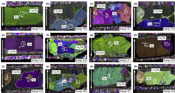
|
| 7 |
K Shan, P Zhou, J Cai, et al.. Electrogenerated chemical polishing of copper. Precision Engineering, 2015, 39: 161–166
https://doi.org/10.1016/j.precisioneng.2014.08.004
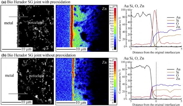
|
| 8 |
P Janoš, J Ederer, V Pilařová, et al.. Chemical mechanical glass polishing with cerium oxide: Effect of selected physico-chemical characteristics on polishing efficiency. Wear, 2016, 362–363: 114–120
https://doi.org/10.1016/j.wear.2016.05.020
|
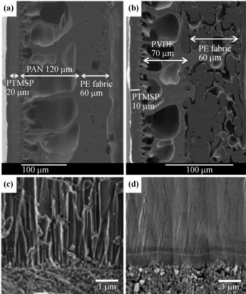
| 9 |
N Tatsumi, K Harano, T Ito, et al.. Polishing mechanism and surface damage analysis of type IIa single crystal diamond processed by mechanical and chemical polishing methods. Diamond and Related Materials, 2016, 63: 80–85
https://doi.org/10.1016/j.diamond.2015.11.021
|
| 10 |
H Deng, R Huang, K Liu, et al.. Abrasive-free polishing of tungsten alloy using electrochemical etching. Electrochemistry Communications, 2017, 82: 80–84
https://doi.org/10.1016/j.elecom.2017.07.030
|
| 11 |
L Zhang, B Zhang, B Pan, et al.. Germanium electrochemical study and its CMP application. Applied Surface Science, 2017, 422: 247–256
https://doi.org/10.1016/j.apsusc.2017.05.220
|
| 12 |
N Erdman, R Campbell, S Asahina. Precise SEM cross-section polishing via argon beam milling. Microscopy Today, 2006, 14(3): 22–25
https://doi.org/10.1017/S155192950005762X
|
| 13 |
Y Takahashi, M Tanaka, K Higashida, et al.. High-voltage electron-microscopic observation of cyclic slip behavior around a fatigue crack tip in an iron alloy. Scripta Materialia, 2009, 60(8): 717–720
https://doi.org/10.1016/j.scriptamat.2009.01.002
|
| 14 |
R G Loucks, R M Reed, S C Ruppel, et al.. Morphology, genesis, and distribution of nanometer-scale pores in siliceous mudstones of the Mississippian Barnett Shale. Journal of Sedimentary Research, 2009, 79(12): 848–861
https://doi.org/10.2110/jsr.2009.092
|
| 15 |
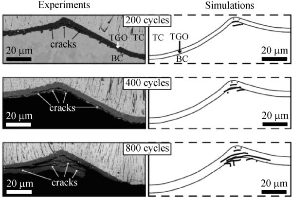
H Takahashi, A Sato, M Takakura, et al.. A new method of surface preparation for high spatial resolution EPMA/SEM with an argon ion beam. Mikrochimica Acta, 2006, 155(1‒2): 295–300
https://doi.org/10.1007/s00604-006-0559-0
|
| 16 |
A Yasuhara. Development of ion slicer (thin-film specimen preparation equipment). JEOL News, 2005, 40(1): 46–49
|
| 17 |
P Sigmund. Sputtering by ion bombardment: Theoretical concept. In: R Behrisch, ed. Sputtering by Particle Bombardment I. Berlin/Heidelberg, Germany: Springer, 1981 doi:10.1007/3540105212_7
|
| 18 |
K Gamo. Ion beam assisted etching and deposition. Journal of Vacuum Science & Technology B, 1990, 8: 1927 doi:10.1116/1.584876
|
| 19 |
A Barna. Topographic kinetics and practice of low angle ion beam thinning. Proceedings of the Materials Research Society, 1991, 254: 3–22
https://doi.org/10.1557/PROC-254-3
|
| 20 |
A Barna, B Pécz. Simple method for the preparation of InP based samples for TEM investigation. Journal of Electron Microscopy Technique, 1991, 18(3): 325–328
https://doi.org/10.1002/jemt.1060180315
pmid: 1880605
|
| 21 |
A G Cullis, N G Chew. Ion milling of compound semiconductors for transmission electron microscopy. Proceedings of the Materials Research Society, 1987, 115: 3–14
https://doi.org/10.1557/PROC-115-3
|
| 22 |
W Hauffe. Production of microstructures by ion beam sputtering. In: S B Heidelberg, ed. Sputtering by Particle Bombardment III. Berlin/Heidelberg, Germany: Springer, 1991, 305–338 doi:10.1007/3540534288_20
|
| 23 |
T Ishitani, T Yaguchi. Cross-sectional sample preparation by focused ion beam: a review of ion-sample interaction. Microscopy Research and Technique, 1996, 35(4): 320–333
https://doi.org/10.1002/(SICI)1097-0029(19961101)35:4<320::AID-JEMT3>3.0.CO;2-Q
pmid: 8987026
|
| 24 |
G Desbois, J L Urai, P A Kukla, et al.. High-resolution 3D fabric and porosity model in a tight gas sandstone reservoir: A new approach to investigate microstructures from mm- to nm-scale combining argon beam cross-sectioning and SEM imaging. Journal of Petroleum Science and Engineering, 2011, 78(2): 243–257
https://doi.org/10.1016/j.petrol.2011.06.004
|
| 25 |
S Hemes, G Desbois, J L Urai, et al.. Multi-scale characterization of porosity in Boom Clay (HADES-level, Mol, Belgium) using a combination of X-ray μ-CT, 2D BIB-SEM and FIB-SEM tomography. Microporous and Mesoporous Materials, 2015, 208: 1–20
https://doi.org/10.1016/j.micromeso.2015.01.022
|
| 26 |
M E Houben, G Desbois, J L Urai. A comparative study of representative 2D microstructures in Shaly and Sandy facies of Opalinus Clay (Mont Terri, Switzerland) inferred form BIB-SEM and MIP methods. Marine and Petroleum Geology, 2014, 49: 143–161
https://doi.org/10.1016/j.marpetgeo.2013.10.009
|
| 27 |
G Desbois, J L Urai, S Hemes, et al.. Nanometer-scale pore fluid distribution and drying damage in preserved clay cores from Belgian clay formations inferred by BIB-cryo-SEM. Engineering Geology, 2014, 179: 117–131
https://doi.org/10.1016/j.enggeo.2014.07.004
|
| 28 |
M E Houben, G Desbois, J L Urai. Pore morphology and distribution in the Shalyfacies of Opalinus Clay (Mont Terri, Switzerland): Insights from representative 2D BIB-SEM investigations on mm to nm scale. Applied Clay Science, 2013, 71: 82–97
https://doi.org/10.1016/j.clay.2012.11.006
|
| 29 |
S Giffin, R Littke, J Klaver, et al.. Application of BIB-SEM technology to characterize macropore morphology in coal. International Journal of Coal Geology, 2013, 114: 85–95
https://doi.org/10.1016/j.coal.2013.02.009
|
| 30 |
J Klaver, G Desbois, R Littke, et al.. BIB-SEM pore characterization of mature and post mature Posidonia Shale samples from the Hils area, Germany. International Journal of Coal Geology, 2016, 158: 78–89
https://doi.org/10.1016/j.coal.2016.03.003
|
| 31 |
G Desbois, J L Urai, S Hemes, et al.. Multi-scale analysis of porosity in diagenetically altered reservoir sandstone from the Permian Rotliegend (Germany). Journal of Petroleum Science and Engineering, 2016, 140: 128–148
https://doi.org/10.1016/j.petrol.2016.01.019
|
| 32 |
J Klaver, G Desbois, R Littke, et al.. BIB-SEM characterization of pore space morphology and distribution in postmature to overmature samples from the Haynesville and Bossier Shales. Marine and Petroleum Geology, 2015, 59: 451–466
https://doi.org/10.1016/j.marpetgeo.2014.09.020
|
| 33 |
J Klaver, G Desbois, J L Urai, et al.. BIB-SEM study of the pore space morphology in early mature Posidonia Shale from the Hils area, Germany. International Journal of Coal Geology, 2012, 103: 12–25
https://doi.org/10.1016/j.coal.2012.06.012
|
| 34 |
G Desbois, J L Urai, J H P de Bresser. Fluid distribution in grain boundaries of natural fine-grained rock salt deformed at low differential stress (Qom Kuh salt fountain, central Iran): Implications for rheology and transport properties. Journal of Structural Geology, 2012, 43: 128–143
https://doi.org/10.1016/j.jsg.2012.07.002
|
| 35 |
G Desbois, J L Urai, P A Kukla. Morphology of the pore space in claystones — evidence from BIB/FIB ion beam sectioning and cryo-SEM observations. eEarth, 2009, 4(1): 15–22 doi:10.5194/ee-4-15-2009
|
| 36 |
B J Olanipekun, K Azmy. Genesis and morphology of intracrystalline nanopores and mineral micro inclusions hosted in burial dolomite crystals: Application of broad ion beam-scanning electron microscope (BIB-SEM). Marine and Petroleum Geology, 2016, 74: 1–11
https://doi.org/10.1016/j.marpetgeo.2016.03.029
|
| 37 |
G R Chalmers, R M Bustin, I M Power. Characterization of gas shale pore systems by porosimetry, pycnometry, surface area, and field emission scanning electron microscopy/transmission electron microscopy image analyses: Examples from the Barnett, Woodford, Haynesville, Marcellus, and Doig units. AAPG Bulletin, 2012, 96(6): 1099–1119
https://doi.org/10.1306/10171111052
|
| 38 |
G Chalmers, R M Bustin, I Powers. A pore by any other name would be as small the importance of meso- and microporosity in shale gas capacity. In: Proceedings of the AAPG Annual Convention and Exhibition, Denver, CO, USA, 7–10 June 2009
|
| 39 |
G Desbois, J L Urai, F Pérez-Willard, et al.. Argon broad ion beam tomography in a cryogenic scanning electron microscope: a novel tool for the investigation of representative microstructures in sedimentary rocks containing pore fluid. Journal of Microscopy, 2013, 249(3): 215–235
https://doi.org/10.1111/jmi.12011
pmid: 23323728
|
| 40 |
S Hemes, G Desbois, J L Urai, et al.. Variations in the morphology of porosity in the boom clay formation: Insights from 2D high resolution BIB-SEM imaging and mercury injection porosimetry. Netherlands Journal of Geosciences, 2014, 92: 275–300 doi:10.1017/S0016774600000214
|
| 41 |
H Dong, M J Blunt. Pore-network extraction from micro-computerized-tomography images. Physical Review E, 2009, 80(3): 036307
https://doi.org/10.1103/PhysRevE.80.036307
pmid: 19905212
|
| 42 |
M E Davis. New vistas in zeolite and molecular sieve catalysis. Accounts of Chemical Research, 1993, 26(3): 111–115
https://doi.org/10.1021/ar00027a006
|
| 43 |
T Kikuchi, J Kawashima, S Natsui, et al.. Fabrication of porous tungsten oxide via anodizing in an ammonium nitrate/ethylene glycol/water mixture for visible light-driven photocatalyst. Applied Surface Science, 2017, 422: 130–137
https://doi.org/10.1016/j.apsusc.2017.05.256
|
| 44 |
S M Stevens, A R Loiola, P Cubillas, et al.. Hierarchical porous materials: Internal structure revealed by argon ion-beam cross-section polishing, HRSEM and AFM. Solid State Sciences, 2011, 13(4): 745–749
https://doi.org/10.1016/j.solidstatesciences.2010.04.027
|
| 45 |
A Grobe, J Schmatz, R Littke, et al.. Enhanced surface flatness of vitrinite particles by broad ion beam polishing and implications for reflectance measurements. International Journal of Coal Geology, 2017, 180: 113–121
https://doi.org/10.1016/j.coal.2017.05.006
|
| 46 |
K De Sitter, C Dotremont, I Genné, et al.. The use of nanoparticles as alternative pore former for the production of more sustainable polyethersulfone ultrafiltration membranes. Journal of Membrane Science, 2014, 471: 168–178
https://doi.org/10.1016/j.memsci.2014.06.061
|
| 47 |
T Honma, H Kamata, J Tatami. Microstructure of seal formed between a Nb wire and an Al2O3 capillary melted by YAG laser radiation. Ceramics International, 2013, 39(5): 4861–4875
https://doi.org/10.1016/j.ceramint.2012.11.079
|
| 48 |
T Sen, G J T Tiddy, J L Casci, et al.. Meso-cellular silica foams, macro-cellular silica foams and mesoporous solids: A study of emulsion-mediated synthesis. Microporous and Mesoporous Materials, 2005, 78(2‒3): 255–263
https://doi.org/10.1016/j.micromeso.2004.09.022
|
| 49 |
C Liu, M Xiang, Z Fu, et al.. Microstructural refinement in spark plasma sintering 3Y-TZP nanoceramics. Journal of the European Ceramic Society, 2016, 36(10): 2565–2571
https://doi.org/10.1016/j.jeurceramsoc.2016.03.019
|
| 50 |
K Asama, T Matsuda, T Ogura, et al.. Low-temperature metal-to-alumina direct bonding process utilizing redox reaction between silver oxide and organic agent. Materials Science and Engineering A, 2017, 702: 398–405
https://doi.org/10.1016/j.msea.2017.07.034
|
| 51 |
M S Kim, H Nishikawa. Effects of bonding temperature on microstructure, fracture behavior and joint strength of Ag nanoporous bonding for high temperature die attach. Materials Science and Engineering A, 2015, 645: 264–272
https://doi.org/10.1016/j.msea.2015.08.015
|
| 52 |
M Pekarčíková, M Skarba, P Konopka, et al.. Investigation of defects in functional layer of high temperature superconducting tapes. Physica C, 2014, 497: 24–29 doi:10.1016/j.physc.2013.10.010
|
| 53 |
S Romankov, Y Hayasaka, I V Shchetinin, et al.. Joining and microstructural development of Ni–Al–Ti sheets under ball collisions. Acta Materialia, 2012, 60(5): 2196–2208
https://doi.org/10.1016/j.actamat.2011.12.015
|
| 54 |
K Lee, K S Kim, Y Tsukada, et al.. Influence of crystallographic orientation of Sn–Ag–Cu on electromigration in flip‒chip joint. Microelectronics and Reliability, 2011, 51(12): 2290–2297
https://doi.org/10.1016/j.microrel.2011.05.003
|
| 55 |
J Liao, N Yamamoto, H Liu, et al.. Microstructure at friction stir lap joint interface of pure titanium and steel. Materials Letters, 2010, 64(21): 2317–2320
https://doi.org/10.1016/j.matlet.2010.07.049
|
| 56 |
S Romankov, Y C Park, I V Shchetinin. Deformation-induced plastic flow and mechanical intermixing of intentionally introduced impurities into a Ni sheet under ball collisions. Journal of Alloys and Compounds, 2017, 694: 1121–1132
https://doi.org/10.1016/j.jallcom.2016.10.137
|
| 57 |
S Romankov, I V Shchetinin, Y C Park. Aluminizing a Ni sheet through severe plastic deformation induced by ball collisions. Applied Surface Science, 2015, 343: 94–105
https://doi.org/10.1016/j.apsusc.2015.03.047
|
| 58 |
M Asakura, Y Kominami, T Hayashi, et al.. The effect of zinc levels in a gold-based alloy on porcelain-metal bonding. Dental Materials, 2012, 28(5): e35–e41
https://doi.org/10.1016/j.dental.2012.02.003
pmid: 22418286
|
| 59 |
C Y Yu, W Y Chen, J G Duh. Improving the impact toughness of Sn–Ag–Cu/Cu–Zn Pb-free solder joints under high speed shear testing. Journal of Alloys and Compounds, 2014, 586: 633–638
https://doi.org/10.1016/j.jallcom.2013.10.113
|
| 60 |
S A Paknejad, G Dumas, G West, et al.. Microstructure evolution during 300 °C storage of sintered Ag nanoparticles on Ag and Au substrates. Journal of Alloys and Compounds, 2014, 617: 994–1001
https://doi.org/10.1016/j.jallcom.2014.08.062
|
| 61 |
S M Hwang, Y G Lim, J G Kim, et al.. A case study on fibrous porous SnO2 anode for robust, high-capacity lithium-ion batteries. Nano Energy, 2014, 10: 53–62
https://doi.org/10.1016/j.nanoen.2014.08.020
|
| 62 |
T Yoon, S Park, J Mun, et al.. Failure mechanisms of LiNi0.5Mn1.5O4 electrode at elevated temperature. Journal of Power Sources, 2012, 215: 312–316
https://doi.org/10.1016/j.jpowsour.2012.04.103
|
| 63 |
S Claes, P Vandezande, S Mullens, et al.. High flux composite PTMSP‒silica nanohybrid membranes for the pervaporation of ethanol/water mixtures. Journal of Membrane Science, 2010, 351(1‒2): 160–167
https://doi.org/10.1016/j.memsci.2010.01.043
|
| 64 |
M Ravelingien, S Mullens, J Luyten, et al.. Thermal decomposition of bioactive sodium titanate surfaces. Applied Surface Science, 2009, 255(23): 9539–9542
https://doi.org/10.1016/j.apsusc.2009.07.091
|
| 65 |
H Voß, A Diefenbacher, G Schuch, et al.. Butene isomers separation on titania supported MFI membranes at conditions relevant for practice. Journal of Membrane Science, 2009, 329(1‒2): 11–17
https://doi.org/10.1016/j.memsci.2008.11.039
|
| 66 |
S Romankov, Y Hayasaka, G Kalikova, et al.. TEM study of TiN coatings fabricated by mechanical milling using vibration technique. Surface and Coatings Technology, 2009, 203(13): 1879–1884
https://doi.org/10.1016/j.surfcoat.2009.01.011
|
| 67 |
S V Komarov, S H Son, N Hayashi, et al.. Development of a novel method for mechanical plating using ultrasonic vibrations. Surface and Coatings Technology, 2007, 201(16‒17): 6999–7006
https://doi.org/10.1016/j.surfcoat.2007.01.002
|
| 68 |
Y S Al Jabbari, T Koutsoukis, S Al Hadlaq, et al.. Surface and cross-sectional characterization of titanium-nitride coated nickel–titanium endodontic files. Journal of Dental Sciences, 2016, 11(1): 48–53
https://doi.org/10.1016/j.jds.2015.07.004
|
| 69 |
S Romankov, S V Komarov, E Vdovichenko, et al.. Fabrication of TiN coatings using mechanical milling techniques. International Journal of Refractory Metals & Hard Materials, 2009, 27(2): 492–497
https://doi.org/10.1016/j.ijrmhm.2008.10.005
|
| 70 |
G M Song, T Vystavel, N van der Pers, et al.. Relation between microstructure and adhesion of hot dip galvanized zinc coatings on dual phase steel. Acta Materialia, 2012, 60(6‒7): 2973–2981
https://doi.org/10.1016/j.actamat.2012.02.003
|
| 71 |
A Lafort, H Kebaili, S Goumri-Said, et al.. Optical properties of thermochromic VO2 thin films on stainless steel: Experimental and theoretical studies. Thin Solid Films, 2011, 519(10): 3283–3287
https://doi.org/10.1016/j.tsf.2010.12.122
|
| 72 |
M H Hong, D H Lee, K M Kim, et al.. Study on bioactivity and bonding strength between Ti alloy substrate and TiO2 film by micro-arc oxidation. Thin Solid Films, 2011, 519(20): 7065–7070
https://doi.org/10.1016/j.tsf.2011.01.223
|
| 73 |
S Arai, T Sato, M Endo. Fabrication of various electroless Ni–P alloy/multiwalled carbon nanotube composite films on an acrylonitrile butadiene styrene resin. Surface and Coatings Technology, 2011, 205(10): 3175–3181
https://doi.org/10.1016/j.surfcoat.2010.11.030
|
| 74 |
C M Parish, C S Snow, D R Kammler, et al.. Processing effects on microstructure in Er and ErD2 thin-films. Journal of Nuclear Materials, 2010, 403(1‒3): 191–197
https://doi.org/10.1016/j.jnucmat.2010.06.026
|
| 75 |
Z Jiao, A Ueno, Y Suzuki, et al.. Study on the influences of reduction temperature on nickel‒yttria-stabilized zirconia solid oxide fuel cell anode using nickel oxide-film electrode. Journal of Power Sources, 2016, 328: 377–384
https://doi.org/10.1016/j.jpowsour.2016.08.043
|
| 76 |
S Romankov, Y Hayasaka, E Kasai, et al.. Fabrication of nanostructured Mo coatings on Al and Ti substrates by ball impact cladding. Surface and Coatings Technology, 2010, 205(7): 2313–2321
https://doi.org/10.1016/j.surfcoat.2010.09.014
|
| 77 |
S Romankov, S D Kaloshkin, Y Hayasaka, et al.. Structural evolution of the Ti–Al coatings produced by mechanical alloying technique. Journal of Alloys and Compounds, 2009, 483(1‒2): 386–388
https://doi.org/10.1016/j.jallcom.2008.07.199
|
| 78 |
S Romankov, S D Kaloshkin, Y Hayasaka, et al.. Effect of process parameters on the formation of Ti–Al coatings fabricated by mechanical milling. Journal of Alloys and Compounds, 2009, 484(1‒2): 665–673
https://doi.org/10.1016/j.jallcom.2009.05.016
|
| 79 |
S Romankov, Y Hayasaka, N Hayashi, et al.. Ball impact cladding of metals with dissimilar metallic foils. Surface and Coatings Technology, 2009, 204(1‒2): 125–130
https://doi.org/10.1016/j.surfcoat.2009.06.047
|
| 80 |
H Falk-Windisch, J Claquesin, J E Svensson, et al.. The effect of metallic Co-coating thickness on ferritic stainless steels intended for use as interconnect material in intermediate temperature solid oxide fuel cells. Oxidation of Metals, 2018, 89(1‒2): 233–250
https://doi.org/10.1007/s11085-017-9782-9
|
| 81 |
H Falk-Windisch, J Claquesin, M Sattari, et al.. Co- and Ce/Co-coated ferritic stainless steel as interconnect material for intermediate temperature solid oxide fuel cells. Journal of Power Sources, 2017, 343: 1–10
https://doi.org/10.1016/j.jpowsour.2017.01.045
|
| 82 |
T Liu, C Wang, H Shen, et al.. The effects of Cr and Al concentrations on the oxidation behavior of oxide dispersion strengthened ferritic alloys. Corrosion Science, 2013, 76: 310–316
https://doi.org/10.1016/j.corsci.2013.07.004
|
| 83 |
C Y Yu, J G Duh. Stabilization of hexagonal Cu6(Sn,Zn)5 by minor Zn doping of Sn-based solder joints. Scripta Materialia, 2011, 65(9): 783–786
https://doi.org/10.1016/j.scriptamat.2011.07.029
|
| 84 |
X Cheng, K W Putz, C D Wood, et al.. Characterization of local elastic modulus in confined polymer films via AFM indentation. Macromolecular Rapid Communications, 2015, 36(4): 391–397
https://doi.org/10.1002/marc.201400487
pmid: 25537230
|
| 85 |
A Thomas, J Andersson, D Grüner, et al.. Direct observation of bone coherence with dental implants. Journal of the European Ceramic Society, 2012, 32(11): 2607–2612
https://doi.org/10.1016/j.jeurceramsoc.2012.02.042
|
| 86 |
J Fahlteich, C Steiner, N Schiller, et al.. Roll-to-roll thin film coating on fluoropolymer webs — Status, challenges and applications. Surface and Coatings Technology, 2017, 314: 160–168
https://doi.org/10.1016/j.surfcoat.2016.11.106
|
| 87 |
M Shimazu, K Yamaji, H Kishimoto, et al.. Stability of Sc2O3 and CeO2 co-doped ZrO2 electrolyte during the operation of solid oxide fuel cells: Part III. Detailed mechanism of the decomposition. Solid State Ionics, 2012, 224: 6–14
https://doi.org/10.1016/j.ssi.2012.06.025
|
| 88 |
T S Hille, T J Nijdam, A S J Suiker, et al.. Damage growth triggered by interface irregularities in thermal barrier coatings. Acta Materialia, 2009, 57(9): 2624–2630
https://doi.org/10.1016/j.actamat.2009.01.022
|
| 89 |
Y N Jo, Y Kim, J S Kim, et al.. Si–graphite composites as anode materials for lithium secondary batteries. Journal of Power Sources, 2010, 195(18): 6031–6036
https://doi.org/10.1016/j.jpowsour.2010.03.008
|
| 90 |
J H Kim, S Yonezawa, M Takashima. Preparation and characterization of carbon composite plates using Ni‒PTFE composite nano-plating. Applied Surface Science, 2013, 279: 329–333
https://doi.org/10.1016/j.apsusc.2013.04.093
|
| 91 |
J H Kim, S Yonezawa, M Takashima. Preparation and characterization of C/Ni–PTFE electrode using Ni–PTFE composite plating for alkaline fuel cells. International Journal of Hydrogen Energy, 2011, 36(2): 1720–1729
https://doi.org/10.1016/j.ijhydene.2010.10.078
|
| 92 |
S G Woo, J H Kim, H R Kim, et al.. Failure mechanism analysis of LiNi0.88Co0.09Mn0.03O2 cathodes in Li-ion full cells. Journal of Electroanalytical Chemistry, 2017, 799: 315–320
https://doi.org/10.1016/j.jelechem.2017.06.034
|
| 93 |
H R Kim, S G Woo, J H Kim, et al.. Capacity fading behavior of Ni-rich layered cathode materials in Li-ion full cells. Journal of Electroanalytical Chemistry, 2016, 782: 168–173
https://doi.org/10.1016/j.jelechem.2016.10.032
|
| 94 |
Y Xiong, J Hu, Z Shen. Dynamic pore coalescence in nanoceramic consolidated by two-step sintering procedure. Journal of the European Ceramic Society, 2013, 33(11): 2087–2092
https://doi.org/10.1016/j.jeurceramsoc.2013.03.015
|
| 95 |
T Oertel, F Hutter, R Tänzer, et al.. Primary particle size and agglomerate size effects of amorphous silica in ultra-high performance concrete. Cement and Concrete Composites, 2013, 37: 61–67
https://doi.org/10.1016/j.cemconcomp.2012.12.005
|
| 96 |
S Arai, Y Suzuki, J Nakagawa, et al.. Fabrication of metal coated carbon nanotubes by electroless deposition for improved wettability with molten aluminum. Surface and Coatings Technology, 2012, 212: 207–213
https://doi.org/10.1016/j.surfcoat.2012.09.051
|
| 97 |
Y S Lin, J G Duh. Facile synthesis of mesoporous lithium titanate spheres for high rate lithium-ion batteries. Journal of Power Sources, 2011, 196(24): 10698–10703
https://doi.org/10.1016/j.jpowsour.2011.09.007
|
| 98 |
K Cho, R Ryoo, S Asahina, et al.. Mesopore generation by organosilane surfactant during LTA zeolite crystallization, investigated by high-resolution SEM and Monte Carlo simulation. Solid State Sciences, 2011, 13(4): 750–756
https://doi.org/10.1016/j.solidstatesciences.2010.04.022
|
| 99 |
J H Kim, S Yonezawa, M Takashima. Preparation and characterization of Ni–PTFE plate as an electrode for alkaline fuel cell: Effects of conducting materials on the performance of electrode. International Journal of Hydrogen Energy, 2010, 35(16): 8707–8714
https://doi.org/10.1016/j.ijhydene.2010.05.110
|
|
Viewed |
|
|
|
Full text
|
|
|
|
|
Abstract
|
|
|
|
|
Cited |
|
|
|
|
| |
Shared |
|
|
|
|
| |
Discussed |
|
|
|
|