|
|
|
Structural shifts in the intestinal microbiota of rats treated with cyclosporine A after orthotropic liver transplantation |
Junjun Jia1,2,3, Xinyao Tian1,2,3, Jianwen Jiang1,2,3, Zhigang Ren1,2,3, Haifeng Lu2,4, Ning He1,2,3, Haiyang Xie1,2,3, Lin Zhou1,2,3, Shusen Zheng1,2,3( ) ) |
1. Department of Hepatobiliary and Pancreatic Surgery, the First Affiliated Hospital, Zhejiang University School of Medicine, Hangzhou 310003, China
2. Key Laboratory of Combined Multi-Organ Transplantation, Ministry of Public Health
3. Collaborative Innovation Center for Diagnosis and Treatment of Infectious Diseases, the First Affiliated Hospital, Zhejiang University School of Medicine, Hangzhou 310003, China
4. State Key Laboratory for Diagnosis and Treatment of Infectious Diseases, the First Affiliated Hospital, Zhejiang University School of Medicine, Hangzhou 310003, China |
|
|
|
|
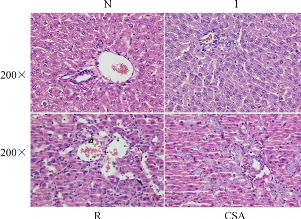
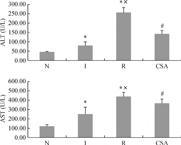
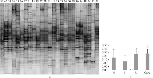
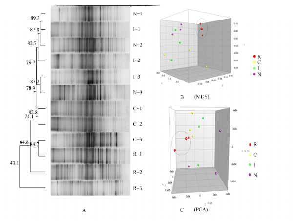
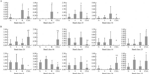
Abstract Understanding the effect of immunosuppressive agents on intestinal microbiota is important to reduce the mortality and morbidity from orthotopic liver transplantation (OLT). We investigated the relationship between the commonly used immunosuppressive agent cyclosporine A (CSA) and the intestinal microbial variation in an OLT model. The rat samples were divided as follows: (1) N group (normal control); (2) I group (isograft LT, Brown Norway [BN] rat to BN); (3) R group (allograft LT, Lewis to BN rat); and (4) CSA group (R group treated with CSA). The intestinal microbiota was assayed by denaturing gradient gel electrophoresis profiles and by using real-time polymerase chain reaction. The liver histopathology and the alanine/aspartate aminotransferase ratio after LT were both ameliorated by CSA. In the CSA group, the numbers of rDNA gene copies of Clostridium cluster I, Clostridium cluster XIV, and Enterobacteriaceae decreased, whereas those of Faecalibacterium prausnitzii increased compared with the R group. Cluster analysis indicated that the samples from the N, I, and CSA groups were clustered, whereas the other clusters contained the samples from the R group. Hence, CSA ameliorates hepatic graft injury and partially restores gut microbiota following LT, and these may benefit hepatic graft rejection.
|
| Keywords
microbial community
liver transplantation
immunosuppressive agents
cyclosporine A
|
|
Corresponding Author(s):
Shusen Zheng
|
|
Just Accepted Date: 04 March 2019
Online First Date: 24 April 2019
Issue Date: 02 August 2019
|
|
| 1 |
EY Cheng, MJ Everly. Trends of Immunosuppression and Outcomes Following Liver Transplantation: An Analysis of the United Network for Organ Sharing Registry. In: Everly MJ, Terasaki PI. Clinical Transplants 2014. LA: UCLA Immunogenetics Center, 2015: 13–26 (Chapter 2)
pmid: 26281123
|
| 2 |
L Barkholt, BG Ericzon, J Tollemar, AS Malmborg, A Ehrnst, H Wilczek, J Andersson. Infections in human liver recipients: different patterns early and late after transplantation. Transpl Int 1993; 6(2): 77–84
https://doi.org/10.1111/j.1432-2277.1993.tb00755.x
pmid: 8447929
|
| 3 |
K Tanaka, S Uemoto, H Egawa, Y Takada, K Ozawa, S Teramukai, M Kasahara, K Ogawa, M Ono, H Sato, K Takai, M Fukushima, K Inaba. Cytotoxic T-cell-mediated defense against infections in human liver transplant recipients. Liver Transpl 2007; 13(2): 287–293
https://doi.org/10.1002/lt.21065
pmid: 17256783
|
| 4 |
LV Hooper, T Midtvedt, JI Gordon. How host-microbial interactions shape the nutrient environment of the mammalian intestine. Annu Rev Nutr 2002; 22(1): 283–307
https://doi.org/10.1146/annurev.nutr.22.011602.092259
pmid: 12055347
|
| 5 |
F Bäckhed, RE Ley, JL Sonnenburg, DA Peterson, JI Gordon. Host-bacterial mutualism in the human intestine. Science 2005; 307(5717): 1915–1920
https://doi.org/10.1126/science.1104816
pmid: 15790844
|
| 6 |
F Guarner, JR Malagelada. Gut flora in health and disease. Lancet 2003; 361(9356): 512–519
https://doi.org/10.1016/S0140-6736(03)12489-0
pmid: 12583961
|
| 7 |
Y Chen, F Yang, H Lu, B Wang, Y Chen, D Lei, Y Wang, B Zhu, L Li. Characterization of fecal microbial communities in patients with liver cirrhosis. Hepatology 2011; 54(2): 562–572
https://doi.org/10.1002/hep.24423
pmid: 21574172
|
| 8 |
JS Bajaj, PB Hylemon, JM Ridlon, DM Heuman, K Daita, MB White, P Monteith, NA Noble, M Sikaroodi, PM Gillevet. Colonic mucosal microbiome differs from stool microbiome in cirrhosis and hepatic encephalopathy and is linked to cognition and inflammation. Am J Physiol Gastrointest Liver Physiol 2012; 303(6): G675–G685
https://doi.org/10.1152/ajpgi.00152.2012
pmid: 22821944
|
| 9 |
K Atarashi, K Honda. Microbiota in autoimmunity and tolerance. Curr Opin Immunol 2011; 23(6): 761–768
https://doi.org/10.1016/j.coi.2011.11.002
pmid: 22115876
|
| 10 |
Y Xie, Z Luo, Z Li, M Deng, H Liu, B Zhu, B Ruan, L Li. Structural shifts of fecal microbial communities in rats with acute rejection after liver transplantation. Microb Ecol 2012; 64(2): 546–554
https://doi.org/10.1007/s00248-012-0030-1
pmid: 22430504
|
| 11 |
VM Dong, KL Womer, MH Sayegh. Transplantation tolerance: the concept and its applicability. Pediatr Transplant 1999; 3(3): 181–192
https://doi.org/10.1034/j.1399-3046.1999.00042.x
pmid: 10487277
|
| 12 |
AL Taylor, CJ Watson, JA Bradley. Immunosuppressive agents in solid organ transplantation: mechanisms of action and therapeutic efficacy. Crit Rev Oncol Hematol 2005; 56(1): 23–46
https://doi.org/10.1016/j.critrevonc.2005.03.012
pmid: 16039869
|
| 13 |
M Malinowski, P Martus, JF Lock, P Neuhaus, M Stockmann. Systemic influence of immunosuppressive drugs on small and large bowel transport and barrier function. Transpl Int 2011; 24(2): 184–193
https://doi.org/10.1111/j.1432-2277.2010.01167.x
pmid: 21208295
|
| 14 |
MJ Ferris, G Muyzer, DM Ward. Denaturing gradient gel electrophoresis profiles of 16S rRNA-defined populations inhabiting a hot spring microbial mat community. Appl Environ Microbiol 1996; 62(2): 340–346
pmid: 8593039
|
| 15 |
EG Zoetendal, CT Collier, S Koike, RI Mackie, HR Gaskins. Molecular ecological analysis of the gastrointestinal microbiota: a review. J Nutr 2004; 134(2): 465–472
https://doi.org/10.1093/jn/134.2.465
pmid: 14747690
|
| 16 |
V Mai, JG Morris Jr. Colonic bacterial flora: changing understandings in the molecular age. J Nutr 2004; 134(2): 459–464
https://doi.org/10.1093/jn/134.2.459
pmid: 14747689
|
| 17 |
X Tian, Z Yang, F Luo , S Zheng. Gut microbial balance and liver transplantation: alteration, management, and prediction. Front Med 2018; 12 (2): 123–129
https://doi.org/10.1007/s11684-017-0563-2
|
| 18 |
G Zaza, A Dalla Gassa, G Felis, S Granata, S Torriani, A Lupo. Impact of maintenance immunosuppressive therapy on the fecal microbiome of renal transplant recipients: comparison between an everolimus- and a standard tacrolimus-based regimen. PLoS One 2017; 12(5): e0178228
https://doi.org/10.1371/journal.pone.0178228
pmid: 28542523
|
| 19 |
Z Ren, G Cui, H Lu, X Chen, J Jiang, H Liu, Y He, S Ding, Z Hu, W Wang, S Zheng. Liver ischemic preconditioning (IPC) improves intestinal microbiota following liver transplantation in rats through 16s rDNA-based analysis of microbial structure shift. PLoS One 2013; 8(10): e75950
https://doi.org/10.1371/journal.pone.0075950
pmid: 24098410
|
| 20 |
H Lu, Z Wu, W Xu, J Yang, Y Chen, L Li. Intestinal microbiota was assessed in cirrhotic patients with hepatitis B virus infection. Intestinal microbiota of HBV cirrhotic patients. Microb Ecol 2011; 61(3): 693–703
https://doi.org/10.1007/s00248-010-9801-8
pmid: 21286703
|
| 21 |
PB Eckburg, EM Bik, CN Bernstein, E Purdom, L Dethlefsen, M Sargent, SR Gill, KE Nelson, DA Relman. Diversity of the human intestinal microbial flora. Science 2005; 308(5728): 1635–1638
https://doi.org/10.1126/science.1110591
pmid: 15831718
|
| 22 |
PD Cani, NM Delzenne. The role of the gut microbiota in energy metabolism and metabolic disease. Curr Pharm Des 2009; 15(13): 1546–1558
https://doi.org/10.2174/138161209788168164
pmid: 19442172
|
| 23 |
SR Gill, M Pop, RT Deboy, PB Eckburg, PJ Turnbaugh, BS Samuel, JI Gordon, DA Relman, CM Fraser-Liggett, KE Nelson. Metagenomic analysis of the human distal gut microbiome. Science 2006; 312(5778): 1355–1359
https://doi.org/10.1126/science.1124234
pmid: 16741115
|
| 24 |
RE Ley, DA Peterson, JI Gordon. Ecological and evolutionary forces shaping microbial diversity in the human intestine. Cell 2006; 124(4): 837–848
https://doi.org/10.1016/j.cell.2006.02.017
pmid: 16497592
|
| 25 |
HC Xing, LJ Li, KJ Xu, T Shen, YB Chen, JF Sheng, Y Chen, SZ Fu, CL Chen, JG Wang, D Yan, FW Dai, SS Zheng. Protective role of supplement with foreign Bifidobacterium and Lactobacillus in experimental hepatic ischemia-reperfusion injury. J Gastroenterol Hepatol 2006; 21(4): 647–656
https://doi.org/10.1111/j.1440-1746.2006.04306.x
pmid: 16677148
|
| 26 |
B Chassaing, L Etienne-Mesmin, AT Gewirtz. Microbiota-liver axis in hepatic disease. Hepatology 2014; 59(1): 328–339
https://doi.org/10.1002/hep.26494
pmid: 23703735
|
| 27 |
Z Ren, A Li, J Jiang, L Zhou, Z Yu, H Lu, H Xie, X Chen, L Shao, R Zhang, S Xu, H Zhang, G Cui, X Chen, R Sun, H Wen, JP Lerut, Q Kan, L Li, S Zheng. Gut microbiome analysis as a tool towards targeted non-invasive biomarkers for early hepatocellular carcinoma. Gut 2018 Jul 25. [Epub ahead of print] doi: 10.1136/gutjnl-2017-315084
https://doi.org/10.1136/gutjnl-2017-315084
pmid: 30045880
|
| 28 |
H Lu, J He, Z Wu, W Xu, H Zhang, P Ye, J Yang, S Zhen, L Li. Assessment of microbiome variation during the perioperative period in liver transplant patients: a retrospective analysis. Microb Ecol 2013; 65(3): 781–791
https://doi.org/10.1007/s00248-013-0211-6
pmid: 23504024
|
| 29 |
A Müller, H Jungen, S Iwersen-Bergmann, M Sterneck, H Andresen-Streichert. Analysis of cyclosporin A in hair samples from liver transplanted patients. Ther Drug Monit 2013; 35(4): 450–458
https://doi.org/10.1097/FTD.0b013e31828abb1d
pmid: 23783168
|
| 30 |
IB Jeffery, PW O’Toole, L Öhman, MJ Claesson, J Deane, EM Quigley, M Simrén. An irritable bowel syndrome subtype defined by species-specific alterations in faecal microbiota. Gut 2012; 61(7): 997–1006
https://doi.org/10.1136/gutjnl-2011-301501
pmid: 22180058
|
| 31 |
CM Surawicz, LJ Brandt, DG Binion, AN Ananthakrishnan, SR Curry, PH Gilligan, LV McFarland, M Mellow, BS Zuckerbraun. Guidelines for diagnosis, treatment, and prevention of Clostridium difficile infections. Am J Gastroenterol 2013; 108(4): 478–498, quiz 499
https://doi.org/10.1038/ajg.2013.4
pmid: 23439232
|
| 32 |
LP Smits, KE Bouter, WM de Vos, TJ Borody, M Nieuwdorp. Therapeutic potential of fecal microbiota transplantation. Gastroenterology 2013; 145(5): 946–953
https://doi.org/10.1053/j.gastro.2013.08.058
pmid: 24018052
|
| 33 |
E van Nood, A Vrieze, M Nieuwdorp, S Fuentes, EG Zoetendal, WM de Vos, CE Visser, EJ Kuijper, JF Bartelsman, JG Tijssen, P Speelman, MG Dijkgraaf, JJ Keller. Duodenal infusion of donor feces for recurrent Clostridium difficile. N Engl J Med 2013; 368(5): 407–415
https://doi.org/10.1056/NEJMoa1205037
pmid: 23323867
|
|
Viewed |
|
|
|
Full text
|
|
|
|
|
Abstract
|
|
|
|
|
Cited |
|
|
|
|
| |
Shared |
|
|
|
|
| |
Discussed |
|
|
|
|