|
|
|
Comparison of the clinical features and therapeutics of COVID-19 in cardio-cerebrovascular disease (CCVD) and non-CCVD patients |
Yu Wang1, Lan Li1, Yuanjiang Pan2, Yu He3, Zuhua Chen5, Yunhao Xun5, Yuhan Xu1, Yilei Guo1, Jiehong Yang4( ), Jianchun Guo5(), Haitong Wan1() ), Jianchun Guo5(), Haitong Wan1() |
1. Institute of Cardio-cerebrovascular Disease, Zhejiang Chinese Medical University, Hangzhou 310053, China
2. Department of Chemistry, Zhejiang University, Hangzhou 310027, China
3. College of Pharmaceutical Science, Zhejiang Chinese Medical University, Hangzhou 310053, China
4. School of Basic Medical Sciences and Public Health, Zhejiang Chinese Medical University, Hangzhou 310053, China
5. Integrated TCM & Western Medicine Department, Xixi Hospital of Hangzhou, Hangzhou 310023, China |
|
|
|
|
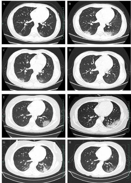
Abstract Cardio-cerebrovascular disease (CCVD) is a major comorbidity of coronavirus disease 2019 (COVID-19). However, the clinical characteristics and outcomes remain unclear. In this study, 102 cases of COVID-19 from January 22, 2020 to March 26, 2020 in Xixi Hospital of Hangzhou were included. Twenty cases had pre-existing CCVD. Results showed that compared with non-CCVD patients, those with CCVD are more likely to develop severe disease (15% versus 1%), and the proportion of pneumonia severity index grade IV was significantly higher (25% versus 3.6%). Computed tomography images demonstrated that the proportion of multiple lobe lesion involvement was significantly higher in the CCVD group than in the non-CCVD group (90% versus 63.4%). Compared with non-CCVD group, the levels of C-reactive protein, fibrinogen, D-dimer, and serum amyloid-A were higher, whereas the total protein and arterial partial PaO2 were lower in the CCVD group. Although no statistical difference was observed in the outcomes between groups, CCVD patients received more intensive comprehensive treatment to improve COVID-19 symptoms compared with non-CCVD patients. Integrated Chinese and Western medicine treatments have certain advantages in controlling the severe conversion rate and mortality of COVID-19. In addition, given that COVID-19 patients are usually related to coagulation disorders and thrombosis risk, the application of Chinese medicine in promoting blood circulation and removing stasis should be strengthened.
|
| Keywords
COVID-19
cardio-cerebrovascular disease
traditional Chinese medicine
clinical features
clinical therapeutics
|
|
Corresponding Author(s):
Jiehong Yang,Jianchun Guo,Haitong Wan
|
|
Just Accepted Date: 13 January 2021
Online First Date: 27 April 2021
Issue Date: 23 September 2021
|
|
| 1 |
X Wei, X Li, J Cui. Evolutionary perspectives on novel coronaviruses identified in pneumonia cases in China. Natl Sci Rev 2020; 7(2): 239–242
https://doi.org/10.1093/nsr/nwaa009
pmid: 32288962
|
| 2 |
Z Wu, JM McGoogan. Characteristics of and important lessons from the coronavirus disease 2019 (COVID-19) outbreak in China: summary of a report of 72 314 cases from the Chinese Center for Disease Control and Prevention. JAMA 2020; 323(13): 1239
https://doi.org/10.1001/jama.2020.2648
|
| 3 |
N Zhu, D Zhang, W Wang, X Li, B Yang, J Song, X Zhao, B Huang, W Shi, R Lu, P Niu, F Zhan, X Ma, D Wang, W Xu, G Wu, GF Gao, W; Tan China Novel Coronavirus Investigating and Research Team. A novel coronavirus from patients with pneumonia in china, 2019. N Engl J Med 2020; 382(8): 727–733
https://doi.org/10.1056/NEJMoa2001017
pmid: 31978945
|
| 4 |
WHO Director-General/Speeches. WHO Director-General’s remarks at the media briefing on 2019-nCoV on 11 February 2020. 2020.
|
| 5 |
Eurosurveillance Editorial Team. Note from the editors: World Health Organization declares novel coronavirus (2019-nCoV) sixth public health emergency of international concern. Euro Surveill 2020; 25(5): pii=200131e
https://doi.org/10.2807/1560-7917.ES.2020.25.5.200131e
|
| 6 |
World Health Organization. Coronavirus disease (COVID-19) situation report-161. 2020.
|
| 7 |
F Wang, S Zheng, C Zheng, X Sun. Attaching clinical significance to COVID-19-associated diarrhea. Life Sci 2020; 260: 118312
https://doi.org/10.1016/j.lfs.2020.118312
pmid: 32846165
|
| 8 |
JY Li, Z You, Q Wang, ZJ Zhou, Y Qiu, R Luo, XY Ge. The epidemic of 2019-novel-coronavirus (2019-nCoV) pneumonia and insights for emerging infectious diseases in the future. Microbes Infect 2020; 22(2): 80–85
https://doi.org/10.1016/j.micinf.2020.02.002
pmid: 32087334
|
| 9 |
F Zhou, T Yu, R Du, G Fan, Y Liu, Z Liu, J Xiang, Y Wang, B Song, X Gu, L Guan, Y Wei, H Li, X Wu, J Xu, S Tu, Y Zhang, H Chen, B Cao. Clinical course and risk factors for mortality of adult inpatients with COVID-19 in Wuhan, China: a retrospective cohort study. Lancet 2020; 395(10229): 1054–1062
https://doi.org/10.1016/S0140-6736(20)30566-3
pmid: 32171076
|
| 10 |
J Chen, T Qi, L Liu, Y Ling, Z Qian, T Li, F Li, Q Xu, Y Zhang, S Xu, Z Song, Y Zeng, Y Shen, Y Shi, T Zhu, H Lu. Clinical progression of patients with COVID-19 in Shanghai, China. J Infect 2020; 80(5): e1–e6
https://doi.org/10.1016/j.jinf.2020.03.004
pmid: 32171869
|
| 11 |
National Health Commission of the People’s Republic of China. Diagnosis and Treatment Protocol for COVID-19 (Trial Version 7). 2020.
|
| 12 |
World Health Organization. Clinical management of severe acute respiratory infection when novel coronavirus (2019-nCoV) infection is suspected: interim guidance, 28 January 2020. 2020.
|
| 13 |
K Liu, Y Chen, R Lin, K Han. Clinical features of COVID-19 in elderly patients: a comparison with young and middle-aged patients. J Infect 2020; 80(6): e14–e18
https://doi.org/10.1016/j.jinf.2020.03.005
pmid: 32171866
|
| 14 |
World Health Organization. Laboratory testing for coronavirus disease 2019 in suspected human cases: interim guidance, 2 March 2020. 2020.
|
| 15 |
World Health Organization. Coronavirus disease 2019 (COVID-19): situation report, 63. 2020.
|
| 16 |
DG Ahn, HJ Shin, MH Kim, S Lee, HS Kim, J Myoung, BT Kim, SJ Kim. Current status of epidemiology, diagnosis, therapeutics, and vaccines for novel coronavirus disease 2019 (COVID-19). J Microbiol Biotechnol 2020; 30(3): 313–324
https://doi.org/10.4014/jmb.2003.03011
pmid: 32238757
|
| 17 |
Y Li, Y Hu, J Yu, T Ma. Retrospective analysis of laboratory testing in 54 patients with severe- or critical-type 2019 novel coronavirus pneumonia. Lab Invest 2020; 100(6): 794–800
https://doi.org/10.1038/s41374-020-0431-6
pmid: 32341519
|
| 18 |
L Zhu, ZG She, X Cheng, JJ Qin, XJ Zhang, J Cai, F Lei, H Wang, J Xie, W Wang, H Li, P Zhang, X Song, X Chen, M Xiang, C Zhang, L Bai, D Xiang, MM Chen, Y Liu, Y Yan, M Liu, W Mao, J Zou, L Liu, G Chen, P Luo, B Xiao, C Zhang, Z Zhang, Z Lu, J Wang, H Lu, X Xia, D Wang, X Liao, G Peng, P Ye, J Yang, Y Yuan, X Huang, J Guo, BH Zhang, H Li. Association of blood glucose control and outcomes in patients with COVID-19 and pre-existing type 2 diabetes. Cell Metab 2020; 31(6): 1068–1077.e3
https://doi.org/10.1016/j.cmet.2020.04.021
pmid: 32369736
|
| 19 |
MA Lake. What we know so far: COVID-19 current clinical knowledge and research. Clin Med (Lond) 2020; 20(2): 124–127
https://doi.org/10.7861/clinmed.2019-coron
pmid: 32139372
|
| 20 |
WJ Guan, ZY Ni, Y Hu, WH Liang, CQ Ou, JX He, L Liu, H Shan, CL Lei, DSC Hui, B Du, LJ Li, G Zeng, KY Yuen, RC Chen, CL Tang, T Wang, PY Chen, J Xiang, SY Li, JL Wang, ZJ Liang, YX Peng, L Wei, Y Liu, YH Hu, P Peng, JM Wang, JY Liu, Z Chen, G Li, ZJ Zheng, SQ Qiu, J Luo, CJ Ye, SY Zhu, NS; China Medical Treatment Expert Group for Covid-19 Zhong. Clinical characteristics of coronavirus disease 2019 in China. N Engl J Med 2020; 382(18): 1708–1720
https://doi.org/10.1056/NEJMoa2002032
pmid: 32109013
|
| 21 |
D Wang, B Hu, C Hu, F Zhu, X Liu, J Zhang, B Wang, H Xiang, Z Cheng, Y Xiong, Y Zhao, Y Li, X Wang, Z Peng. Clinical characteristics of 138 hospitalized patients with 2019 novel coronavirus-infected pneumonia in Wuhan, China. JAMA 2020; 323(11): 1061–1069
https://doi.org/10.1001/jama.2020.1585
pmid: 32031570
|
| 22 |
M Dolhnikoff, AN Duarte-Neto, RA de Almeida Monteiro, LFF da Silva, EP de Oliveira, PHN Saldiva, T Mauad, EM Negri. Pathological evidence of pulmonary thrombotic phenomena in severe COVID-19. J Thromb Haemost 2020; 18(6): 1517–1519
https://doi.org/10.1111/jth.14844
pmid: 32294295
|
| 23 |
W Thomas, J Varley, A Johnston, E Symington, M Robinson, K Sheares, A Lavinio, M Besser. Thrombotic complications of patients admitted to intensive care with COVID-19 at a teaching hospital in the United Kingdom. Thromb Res 2020; 191: 76–77
https://doi.org/10.1016/j.thromres.2020.04.028
pmid: 32402996
|
| 24 |
FA Klok, MJHA Kruip, NJM van der Meer, MS Arbous, D Gommers, KM Kant, FHJ Kaptein, J van Paassen, MAM Stals, MV Huisman, H Endeman. Confirmation of the high cumulative incidence of thrombotic complications in critically ill ICU patients with COVID-19: an updated analysis. Thromb Res 2020; 191: 148–150
https://doi.org/10.1016/j.thromres.2020.04.041
pmid: 32381264
|
| 25 |
J Helms, C Tacquard, F Severac, I Leonard-Lorant, M Ohana, X Delabranche, H Merdji, R Clere-Jehl, M Schenck, F Fagot Gandet, S Fafi-Kremer, V Castelain, F Schneider, L Grunebaum, E Anglés-Cano, L Sattler, PM Mertes, F; Meziani CRICS TRIGGERSEP Group (Clinical Research in Intensive Care and Sepsis Trial Group for Global Evaluation and Research in Sepsis). High risk of thrombosis in patients with severe SARS-CoV-2 infection: a multicenter prospective cohort study. Intensive Care Med 2020; 46(6): 1089–1098
https://doi.org/10.1007/s00134-020-06062-x
pmid: 32367170
|
| 26 |
K Wang, D Zhang, J Wu, S Liu, X Zhang, B Zhang. A comparative study of Danhong injection and Salvia miltiorrhiza injection in the treatment of cerebral infarction: a systematic review and meta-analysis. Medicine (Baltimore) 2017; 96(22): e7079
https://doi.org/10.1097/MD.0000000000007079
pmid: 28562578
|
| 27 |
X Feng, Y Li, Y Wang, L Li, PJ Little, SW Xu, S Liu. Danhong injection in cardiovascular and cerebrovascular diseases: pharmacological actions, molecular mechanisms, and therapeutic potential. Pharmacol Res 2019; 139: 62–75
https://doi.org/10.1016/j.phrs.2018.11.006
pmid: 30408571
|
| 28 |
M Lyu, CL Yan, HX Liu, TY Wang, XH Shi, JP Liu, J Orgah, GW Fan, JH Han, XY Wang, Y Zhu. Network pharmacology exploration reveals endothelial inflammation as a common mechanism for stroke and coronary artery disease treatment of Danhong injection. Sci Rep 2017; 7(1): 15427
https://doi.org/10.1038/s41598-017-14692-3
pmid: 29133791
|
| 29 |
S Zhao, Y Tang, H Cai, W Liu, L Zhang, D Chen, B Chen. Treatment of Danhong Injection combined with Naoxintong Capsule in acute coronary syndrome patients undergoing PCI operation: study for a randomized controlled and double-blind trial. Evid Based Complement Alternat Med 2018; 2018: 8485472
https://doi.org/10.1155/2018/8485472
pmid: 29707035
|
| 30 |
JB Zou, XF Zhang, J Wang, F Wang, JX Cheng, FY Yang, X Song, Y Wang, YL Liang, YJ Shi. The therapeutic efficacy of Danhong injection combined with percutaneous coronary intervention in acute coronary syndrome: a systematic review and meta-analysis. Front Pharmacol 2018; 9: 550
https://doi.org/10.3389/fphar.2018.00550
pmid: 29915535
|
|
Viewed |
|
|
|
Full text
|
|
|
|
|
Abstract
|
|
|
|
|
Cited |
|
|
|
|
| |
Shared |
|
|
|
|
| |
Discussed |
|
|
|
|