|
|
|
Platelet membrane-based and tumor-associated platelet- targeted drug delivery systems for cancer therapy |
Yinlong Zhang1,2, Guangna Liu1,2, Jingyan Wei1( ), Guangjun Nie2,3() ), Guangjun Nie2,3() |
1. College of Pharmaceutical Science, Jilin University, Changchun 130021, China
2. CAS Key Laboratory for Biomedical Effects of Nanomaterials and Nanosafety, CAS Center for Excellence in Nanoscience, National Center for Nanoscience and Technology, Beijing 100190, China
3. University of Chinese Academy of Sciences, Beijing 100049, China |
|
|
|
|
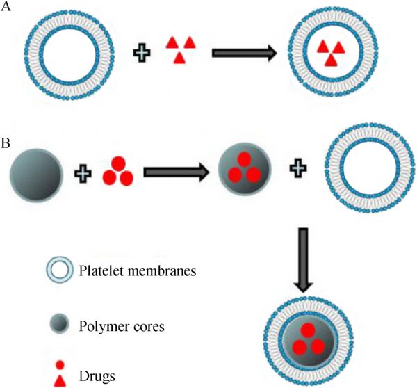
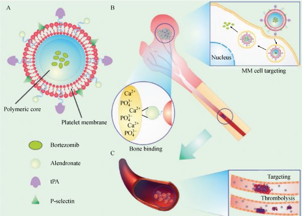
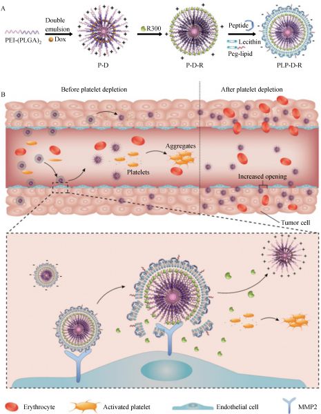
Abstract Platelets have long been known to play critical roles in hemostasis by clumping and clotting blood vessel injuries. Recent experimental evidence strongly indicates that platelets can also interact with tumor cells by direct binding or secreting cytokines. For example, platelets have been shown to protect circulating cancer cells in blood circulation and to promote tumor metastasis. In-depth understanding of the role of platelets in cancer progression and metastasis provides promising approaches for platelet biomimetic drug delivery systems and functional platelet-targeting strategies for effective cancer treatment. This review highlights recent progresses in platelet membrane-based drug delivery and unique strategies that target tumor-associated platelets for cancer therapy. The paper also discusses future development opportunities and challenges encountered for clinical translation.
|
| Keywords
platelet-mimicking delivery systems
tumor-associated platelets
cancer therapy
EPR effect
|
|
Corresponding Author(s):
Jingyan Wei,Guangjun Nie
|
|
Just Accepted Date: 03 January 2018
Online First Date: 09 April 2018
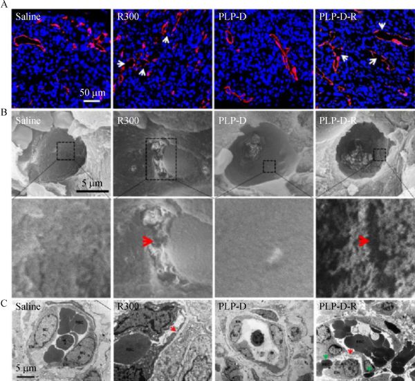
Issue Date: 03 December 2018
|
|
| 1 |
Gurtner GC, Werner S, Barrandon Y, Longaker MT. Wound repair and regeneration. Nature 2008; 453(7193): 314–321
https://doi.org/10.1038/nature07039
pmid: 18480812
|
| 2 |
Furie B, Furie BC. Thrombus formation in vivo. J Clin Invest 2005; 115(12): 3355–3362
https://doi.org/10.1172/JCI26987
pmid: 16322780
|
| 3 |
Ho-Tin-Noé B, Goerge T, Cifuni SM, Duerschmied D, Wagner DD. Platelet granule secretion continuously prevents intratumor hemorrhage. Cancer Res 2008; 68(16): 6851–6858
https://doi.org/10.1158/0008-5472.CAN-08-0718
pmid: 18701510
|
| 4 |
Gay LJ, Felding-Habermann B. Contribution of platelets to tumour metastasis. Nat Rev Cancer 2011; 11(2): 123–134
https://doi.org/10.1038/nrc3004
pmid: 21258396
|
| 5 |
Quail DF, Joyce JA. Microenvironmental regulation of tumor progression and metastasis. Nat Med 2013; 19(11): 1423–1437
https://doi.org/10.1038/nm.3394
pmid: 24202395
|
| 6 |
Mammadovabach E, Zigrino P, Brucker C, Bourdon C, Freund M, Arcangelis AD, Abrams SI, Orend G, Gachet C, Mangin PH. Platelet integrin a6b1 controls lung metastasis through direct binding to cancer cell–derived ADAM9. JCI Insight 2016; 1(14): e88245
https://doi.org/10.1172/jci.insight.88245
pmid: 27699237
|
| 7 |
Yu LX, Yan L, Yang W, Wu FQ, Ling Y, Chen SZ, Tang L, Tan YX, Cao D, Wu MC, Yan HX, Wang HY. Platelets promote tumour metastasis via interaction between TLR4 and tumour cell-released high-mobility group box1 protein. Nat Commun 2014; 5: 5256
https://doi.org/10.1038/ncomms6256
pmid: 25348021
|
| 8 |
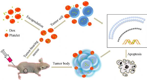
Xu P, Zuo H, Chen B, Wang R, Ahmed A, Hu Y, Ouyang J. Doxorubicin-loaded platelets as a smart drug delivery system: an improved therapy for lymphoma. Sci Rep 2017; 7: 42632
https://doi.org/10.1038/srep42632
pmid: 28198453
|
| 9 |
Nurden AT, Nurden P, Sanchez M, Andia I, Anitua E. Platelets and wound healing. Front Biosci 2008; 13: 3532–3548
pmid: 18508453
|
| 10 |
Nieswandt B, Hafner M, Echtenacher B, Männel DN. Lysis of tumor cells by natural killer cells in mice is impeded by platelets. Cancer Res 1999; 59(6): 1295–1300
pmid: 10096562
|
| 11 |
Wang C, Sun W, Ye Y, Hu Q, Bomba HN, Gu Z. In situ activation of platelets with checkpoint inhibitors for post-surgical cancer immunotherapy. Nat Biomed Eng 2017; 1: 0011
|
| 12 |
Daly ME. Determinants of platelet count in humans. Haematologica 2011; 96(1): 10–13
https://doi.org/10.3324/haematol.2010.035287
pmid: 21193429
|
| 13 |
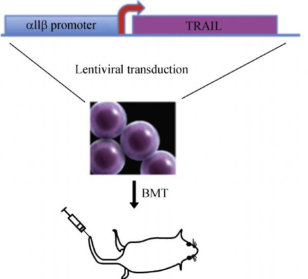
Li J, Sharkey CC, Wun B, Liesveld JL, King MR. Genetic engineering of platelets to neutralize circulating tumor cells. J Control Release 2016; 228: 38–47
https://doi.org/10.1016/j.jconrel.2016.02.036
pmid: 26921521
|
| 14 |
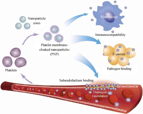
Hu CM, Fang RH, Wang KC, Luk BT, Thamphiwatana S, Dehaini D, Nguyen P, Angsantikul P, Wen CH, Kroll AV, Carpenter C, Ramesh M, Qu V, Patel SH, Zhu J, Shi W, Hofman FM, Chen TC, Gao W, Zhang K, Chien S, Zhang L. Nanoparticle biointerfacing by platelet membrane cloaking. Nature 2015; 526(7571): 118–121
https://doi.org/10.1038/nature15373
pmid: 26374997
|
| 15 |
Dehaini D, Wei X, Fang RH, Masson S, Angsantikul P, Luk BT, Zhang Y, Ying M, Jiang Y, Kroll AV, Gao W, Zhang L. Erythrocyte-platelet hybrid membrane coating for enhanced nanoparticle functionalization. Adv Mater 2017 29(16):1606209
https://doi.org/10.1002/adma.201606209
pmid: 28199033
|
| 16 |
Hu Q, Sun W, Qian C, Wang C, Bomba HN, Gu Z. Anticancer platelet-mimicking nanovehicles. Adv Mater 2015; 27(44): 7043–7050
https://doi.org/10.1002/adma.201503323
pmid: 26416431
|
| 17 |
Hu Q, Qian C, Sun W, Wang J, Chen Z, Bomba HN, Xin H, Shen Q, Gu Z. Engineered nanoplatelets for enhanced treatment of multiple myeloma and thrombus. Adv Mater 2016; 28(43): 9573–9580
https://doi.org/10.1002/adma.201603463
pmid: 27626769
|
| 18 |
Hu Q, Sun W, Qian C, Bomba HN, Xin H, Gu Z. Relay drug delivery for amplifying targeting signal and enhancing anticancer efficacy. Adv Mater 2017; 29(13): 1605803
https://doi.org/10.1002/adma.201605803
pmid: 28160337
|
| 19 |
Li J, Ai Y, Wang L, Bu P, Sharkey CC, Wu Q, Wun B, Roy S, Shen X, King MR. Targeted drug delivery to circulating tumor cells via platelet membrane-functionalized particles. Biomaterials 2016; 76: 52–65
https://doi.org/10.1016/j.biomaterials.2015.10.046
pmid: 26519648
|
| 20 |
Cho MS, Bottsford-Miller J, Vasquez HG, Stone R, Zand B, Kroll MH, Sood AK, Afshar-Kharghan V. Platelets increase the proliferation of ovarian cancer cells. Blood 2012; 120(24): 4869–4872
https://doi.org/10.1182/blood-2012-06-438598
pmid: 22966171
|
| 21 |
Cooke NM, Spillane CD, Sheils O, O’Leary J, Kenny D. Aspirin and P2Y12 inhibition attenuate platelet-induced ovarian cancer cell invasion. BMC Cancer 2015; 15(1): 627
https://doi.org/10.1186/s12885-015-1634-x
pmid: 26353776
|
| 22 |
Klement GL, Yip TT, Cassiola F, Kikuchi L, Cervi D, Podust V, Italiano JE, Wheatley E, Abou-Slaybi A, Bender E, Almog N, Kieran MW, Folkman J. Platelets actively sequester angiogenesis regulators. Blood 2009; 113(12): 2835–2842
https://doi.org/10.1182/blood-2008-06-159541
pmid: 19036702
|
| 23 |
Rachidi S, Metelli A, Riesenberg B, Wu BX, Nelson MH, Wallace C, Paulos CM, Rubinstein MP, Garrett-Mayer E, Hennig M, Bearden DW, Yang Y, Liu B, Li Z. Platelets subvert T cell immunity against cancer via GARP-TGFb axis. Sci Immunol 2017; 2(11): 7911
https://doi.org/10.1126/sciimmunol.aai7911
pmid: 28763790
|
| 24 |
Zhang Y, Wei J, Liu S, Wang J, Han X, Qin H, Lang J, Cheng K, Li Y, Qi Y, Anderson GJ, Sukumar S, Li S, Nie G. Inhibition of platelet function using liposomal nanoparticles blocks tumor metastasis. Theranostics 2017; 7(5): 1062–1071
https://doi.org/10.7150/thno.17908
pmid: 28435448
|
| 25 |
Ho-Tin-Noé B, Goerge T, Wagner DD. Platelets: guardians of tumor vasculature. Cancer Res 2009; 69(14): 5623–5626
https://doi.org/10.1158/0008-5472.CAN-09-1370
pmid: 19584268
|
| 26 |
Kisucka J, Butterfield CE, Duda DG, Eichenberger SC, Saffaripour S, Ware J, Ruggeri ZM, Jain RK, Folkman J, Wagner DD. Platelets and platelet adhesion support angiogenesis while preventing excessive hemorrhage. Proc Natl Acad Sci USA 2006; 103(4): 855–860
https://doi.org/10.1073/pnas.0510412103
pmid: 16418262
|
| 27 |
Li S, Zhang Y, Wang J, Zhao Y, Ji T, Zhao X, Ding Y, Zhao X, Zhao R, Li F, Yang X, Liu S, Liu Z, Lai J, Whittaker AK, Anderson GJ, Wei J, Nie G. Nanoparticle-enabled local depletion of tumor-associated platelets disrupts tumor vascular barriers and augments tumor drug accumulation. Nat Biomed Eng 2017;1:667–679
https://doi.org/DOI: 10.1038/s41551-017-0115-8
|
| 28 |
Ishikawa S, Miyashita T, Inokuchi M, Hayashi H, Oyama K, Tajima H, Takamura H, Ninomiya I, Ahmed AK, Harman JW, Fushida S, Ohta T. Platelets surrounding primary tumor cells are related to chemoresistance. Oncol Rep 2016; 36(2): 787–794
https://doi.org/10.3892/or.2016.4898
pmid: 27349611
|
|
Viewed |
|
|
|
Full text
|
|
|
|
|
Abstract
|
|
|
|
|
Cited |
|
|
|
|
| |
Shared |
|
|
|
|
| |
Discussed |
|
|
|
|