


Blood pressure monitoring has come a long way from the initial observations made by Reverend Hales in the 18th century. There are none that deny the importance of monitoring perioperative blood pressure; however, the limited ability of the current prevalent technology (oscillometric blood pressure monitoring) to offer continuous blood pressure measurements leaves room for improvement. Invasive monitoring is able to detect beat-to-beat blood pressure measurement, but the risks inherent to the procedure make it unsuitable for routine use except when this risk is outweighed by the benefits. This review focuses on the discoveries which have led up to the current blood pressure monitoring technologies, and especially the creation of those offering non-invasive but continuous blood pressure monitoring capabilities, including their methods of measurement and limitations.


The standard monitoring of blood pressure in the perioperative setting is a recognized necessity especially because of acute fluctuations in hemodynamic status occurring from anesthesia, surgical stimulation, hemorrhage, pain, etc. The current use of automated digital sphygmomanometers is fairly reliable, safe and convenient for standard use intraoperatively and is recommended by the ASA to cycle every 5 min [ 1]. However, for any patient with multiple comorbidities undergoing a high-risk surgery, it is frequent practice to place invasive monitoring devices for improved time sensitivity. Accordingly, in a survey conducted among North American and European anesthesiologists, the vast majority (>90%) used a thin catheter inserted into an easily accessible artery (invasive arterial line) for high risk surgeries [ 2]. While a clear definition of the term needs to be further delineated [ 3], periods of hypotension have been associated with poor outcomes in several studies [ 4, 5, 6, 7] which makes it important to detect blood pressure changes as quickly as possible. Due to the inherent risks associated with invasive lines, arterial catheters should be placed only when clinically indicated. Thus, there has been and will continue to be an opportunity for the development of technologies which can offer beat-to-beat blood pressure measurements without the associated risks of invasive monitoring. This review will focus on current technologies that can potentially bridge this gap.
The first description of blood pressure is often attributed to a country parson, Reverend Stephen Hales, in the 18th century (Fig. 1). By cannulating the femoral artery of a horse and attaching a glass tube, he observed the pulsatile rise and fall of blood indicating the presence of blood pressure and pulse pressure.
 | Fig.1 The first description of blood pressure is often attributed to a country parson, Revered Stephen Hales, in the 18th century. (This image is in the public domain and has been downloaded from the US National Library of Medicine website, http://www.nlm.nih.gov/hmd/ihm/, accessed on April 5, 2012.) |
It was not until the 1820s that Poiseulle, a physician-physicist (Fig. 2), created the mercury manometer. This then enabled Carl Ludwig (Fig. 3) to develop the kymograph in 1847. The kymograph consisted of a cannula, mercury manometer and a float attached to a pen which was arranged to write on a revolving drum, thus creating the first recording of an arterial wave form.
 | Fig.2 Mercury manometer developed by Poiseuille. (This image is in the public domain and has been downloaded from the US National Library of Medicine website, http://www.nlm.nih.gov/hmd/ihm/, accessed on April 5, 2012.) |
 | Fig.3 Kymograph developed by Ludwig in 1847. (This image is in the public domain and has been downloaded from the US National Library of Medicine website, http://www.nlm.nih.gov/hmd/ihm/, accessed on April 5, 2012.) |
By 1855, Vierordt created a non-invasive technique of measuring blood pressure with the sphygmograph (Fig. 4). His approach consisted of a combination of weights and levers utilizing a probe placed on the skin superficial to the radial artery. The force required to inhibit blood flow was continuously measured and recorded.
 | Fig.4 In 1855, Vierordt created a non-invasive technique of measuring blood pressure with the sphygmograph. (This image is in the public domain and has been downloaded from the US National Library of Medicine website, http://www.nlm.nih.gov/hmd/ihm/, accessed on April 5, 2012.) |
Throughout the remainder of the 19th century, several investigators advanced methods of non-invasive blood pressure measurements, which consisted of slow and gradual improvements until finally, in 1895, Scipione Riva-Rocci created the non-invasive device that is still seen today: the inflatable cuff armband. In 1905, a Russian surgeon named Korotkoff originally discovered the method in use for over a century—the auscultatory method—which required an inflatable blood pressure cuff and stethoscope (Fig. 5).
 | Fig.5 In 1905, Korotkoff, a Russian surgeon, discovered the method in use for over a century — the auscultatory method — which required the inflatable blood pressure cuff and stethoscope. (This image is in the public domain and has been downloaded from Wikimedia website, http://commons.wikimedia.org/wiki/Main_Page, accessed on April 5, 2012.) |
However, current automated blood pressure measuring equipment in operating rooms use the oscillometric principle first noted by Marey in 1885 prior to the advent of the auscultatory method (Fig. 6).
 | Fig.6 Sphygomanometer. (This image is in the public domain and has been downloaded from Wikimedia website, http://commons.wikimedia.org/wiki/Main_Page, accessed on April 5, 2012.) |
Blood pressure is traditionally reported with three values: systolic, diastolic and mean pressure. These are easily defined in physiologic terms as the (1) systolic pressure, pressure exerted on the wall of blood vessels by blood at the end of systolic contraction of ventricles, (2) diastolic pressure, pressure exerted on the blood vessel walls at the end of relaxation or diastole and (3) mean pressure, the pressure determining the average rate of blood flow through systemic vessels. However, due to the development of several methods of blood pressure determination, these definitions may be associated with different values. For example, the arterial line, which is known as to be the “gold standard” of blood pressure measurement, has relatively straightforward definitions of these values. A pulsatile waveform is displayed and the maximum pressure is systolic, the minimum pressure diastolic and the mean pressure is the area under the waveform-time curve divided by the time interval for one or more beats (calculated by the available software). If using a manual blood pressure cuff, the use of Kortokoff sounds (a system with five phases to determine systolic and diastolic pressure) introduces problems with inter-observer error. Although phase I sounds are said to correlate with systolic BP and phase V sounds correlate with diastolic blood pressure, auscultation is such a subjective measure that it is difficult to accurately reproduce measurements between different observers. Additionally, the use of phase IV and phase V sounds to determine diastolic pressure has been debated, as occasionally the sounds never quite disappear to determine the start of phase V.
Finally, in the oscillometric technique, oscillations begin prior to systolic pressure and continue after diastolic pressure, and only mean arterial pressure corresponds to maximum oscillation amplitude [ 8]. Software algorithms to determine systolic and diastolic pressure values are patented and not shared among manufacturers which may be the basis for wide variability in values between different devices.
Nonetheless, although the determination of blood pressure may be fraught with issues, the standard unit of measure for blood pressure in the United States remains millimeters of mercury (mmHg) because of the original measurements done by mercury manometers. In other parts of the world, the kilopascal (kPa), equal to 7.5 mmHg, is used. In the United States, monitors will display readings to within 1 mmHg which has no additional clinical value since the actual precision of these devices using the oscillometric technique is close to 5 to 10 mmHg [ 9].
The “gold standard” for blood pressure measurements is the invasive arterial line which typically consists of a cannula inserted into a peripheral artery. In clinical studies, it is considered the clinical standard of comparison for all other blood pressure measurement devices. This is true despite the fact that central aortic pressure likely reflects a more accurate representation of blood pressure with a variable degree of amplification of the pulse pressure wave from the aortic root to peripheral arteries [ 10]. Even so, measurements from the peripheral arteries continue to be the most frequently used since measurement of aortic root pressure has safety considerations precluding it from clinical use. A list of the most commonly used techniques for blood pressure monitoring is provided (Table 1) and will each be more thoroughly explained in the following sections.
| Tab.1 Different techniques for blood pressure monitoring |
Riva-Rocci’s blood pressure cuff was a simple device consisting of a rubber bag, surrounded by an inelastic material, which could be inflated by air with an attached rubber bulb along with a manometer to measure pressure. Korotkoff clearly outlined the different stages of “sounds” which could be heard when taking measurements by deflating an air-inflatable cuff. Surprisingly, this method has changed very little over the past 100 years since its advent and it continues to be a simple and inexpensive method of obtaining blood pressure. The cuff is placed on the proximal arm, inflated to a pressure estimated to be above systolic pressure and deflated slowly while a stethoscope is placed over the brachial artery to appreciate Korotkoff sounds which consists of five distinct phases. The first clear “tapping” sounds heard are phase I which corresponds with systolic blood pressure. In phase II, sounds become softer and longer while phase III is characterized by sounds that are crisper and louder. Phase II and III have no clinical significance. Phase IV is generally described as muffled sounds which disappear when phase V is reached. The beginning of phase V is currently accepted as the diastolic pressure despite the fact that this transition may be difficult to determine. Mean blood pressure is calculated by the following Eq. [ 9]:
Original blood pressure devices used mercury sphygmomanometers but due to concerns of mercury exposure, whether real or imagined, aneroid and hybrid devices have become increasingly popular. Mercury sphygmomanometers, as the name suggests, use columns of mercury to display cuff pressure while aneroid devices use a system of metal bellows and a circular scale to display pressures. Hybrid devices do not utilize mercury or a circular scale but instead use an electronic pressure gauge and display values digitally. There has been significant concern over the fact that aneroid and hybrid devices with more moving parts have a potential to introduce inaccuracy into obtained values. These devices require more maintenance and should be calibrated to a mercury sphygmomanometer every 6 months [ 11]. If used appropriately, both devices have been shown to be comparable to a mercury sphygmomanometer with the added benefit of being easier to use.
In Riva-Rocci’s original experiments, the cuff was only 5 cm wide which would have resulted in an error reporting higher than actual values. The ideal width of a cuff should be 46% of the arm circumference [ 12]; inappropriately sized cuffs will result in inaccurate reporting, either too high or low, of blood pressure values. Cuffs must also be appropriately applied with the proper tension. Even with cuff size and placement optimized, Riva-Rocci-Kortokoff measurements are known to underestimate systolic blood pressure and overestimate diastolic blood pressure when compared to invasive arterial lines [ 13]. Additionally, although a simple skill to learn, the time consumed obtaining blood pressure values when frequent measurements were necessary made the development of an automated technique desirable. A short-lived attempt in the 1970s was made to automate the Kortokoff technique with a small microphone or Doppler transducer built into the cuff and placed over the brachial artery. Many issues related to motion and noise artifacts [ 14] impeded the use of these devices and eventually led to their disappearance. Presently, automated blood pressure cuffs are ubiquitous in operating rooms across the world but are based on a different principle—the oscillometric technique.
As previously mentioned, the oscillometric technique predates the auscultatory method described by Korotkoff when a French physiologist, Marey, noted the presence of oscillations in pressure associated with the pulse. The oscillometric method uses this pressure variation to estimate systolic, diastolic and mean pressures. The cuff is inflated to a point above systolic blood pressure and slowly deflated while the presence of oscillations is detected by a pressure sensor. Oscillations begin approximately at systolic blood pressure and continue below diastolic pressure with maximal oscillations occurring at mean arterial pressure when the arterial wall is considered maximally unloaded. Since the oscillations do not correspond with systolic or diastolic pressure, the oscillometric technique can only indirectly estimate these values. This technique was commonly used in children and obese patients where Korotkoff sounds are difficult to hear.
Automated devices based on the oscillometric device are present in operating rooms worldwide consisting of a cuff with a sensor that measures oscillations electronically. Computations of systolic and diastolic pressure have been estimated to be when oscillations reach 0.5 and 0.66 of the maximum amplitude respectively [ 8]. Unfortunately, each manufacturer designates these as proprietary values and they are seldom made public. These devices also have the advantage of taking repeated measurements at predetermined time intervals.
Automation offers several advantages over manual techniques. Most importantly, clinician subjectivity is eliminated with the possibility of attaining repeatable results while saving clinicians the time it would have taken to obtain results manually. However, the choice of cuff size and application continues to be a problem, as this is still left to human judgment. Inappropriate cuff size as well as loose cuffs can have a significant impact on obtaining erroneous results, as described by Bur et al.[ 15]. Additionally, values obtained by different machines have been shown to differ based on the algorithms used by the manufacturers [ 16, 17]. Finally, at its best, automated oscillometric devices can only offer a blood pressure reading every 30 s [ 18] which is less than ideal in situations where rapid blood pressure changes need to be closely monitored. It should also be noted that “stat” cycling mode has been loosely associated with compression injuries in the arm [ 19, 20]. While rare, these injuries can be best avoided by only using the maximum cycling frequency during acute clinical situations where it is necessary and justified.
Invasive arterial blood pressure monitoring is considered the gold standard for measurement of blood pressure. Arterial catheterization will often be performed for high-risk surgical patients undergoing procedures with expected large fluid shifts, necessary frequent blood sampling and/or the possibility of acute blood pressure changes necessitating beat-to-beat blood pressure monitoring. Invasive blood pressure monitoring is typically performed through the cannulization of a peripheral artery with a catheter. The most common site is the radial artery due to ease of access, ease of actual cannulation and low rate of complications [ 21]. The catheter is connected by a long, thin and liquid-filled tubing to a transducer, consisting of a diaphragm which records the transmitted blood pressure through the fluid column. The transducers are single use disposable units encased in a clear plastic case placed at heart level which translates the pressure changes into electrical signals displayed on a monitor. Although this method of direct measurement offers reliable and accurate measurements, there are disadvantages associated with invasive monitoring, namely the risk incurred by the patients. While complications are infrequent, arterial catheterization has been associated with bleeding, hematoma, arterial thrombosis, infection, accidental injection of intravenous drugs, nerve damage and even distal limb ischemia [ 22] (Table 2).
| Tab.2 Radial artery catheterization complications* |
Blood pressure values are highly dependent on the site that is being measured [ 23]. Typically, radial artery pressures do correlate well with central aortic pressures. Systolic values tend to be higher and diastolic lower in peripheral arteries due to distal pulse amplification but mean values are comparable. Pressure waveforms recorded simultaneously from different sites will also display different morphologies due to this pulse amplification. Several characteristics of wave tracings from increasingly peripheral arteries include a steeper arterial upstroke, a wider pulse pressure (higher systolic peak and lower diastolic pressure), and a delayed dicrotic notch when compared with central aortic pressure waveforms [ 24]. Additionally, pulse amplification may be influenced by a number of factors including arterial stiffness, heart rate, height, gender and drug therapies [ 25]. In the case of arterial stiffness, for example, a 2006 expert consensus determined brachial pulse pressure would not accurately represent aortic pulse pressure [ 26].
Invasive arterial line waveforms also contain a large host of additional information than just blood pressure alone. Lamia et al. reported the information which can be obtained from a tracing such as continuous pulse pressure values representing arterial stiffness, diastolic pressures representing systemic vascular tone and respiratory variation of blood pressure reflecting degrees of hypovolemia [ 27]. Pulse pressure variation (PPV), which can be calculated using a variety of devices and their respect algorithms, has been shown to be able to detect fluid responsiveness in patients and therefore useful for cardiac output optimization [ 28].
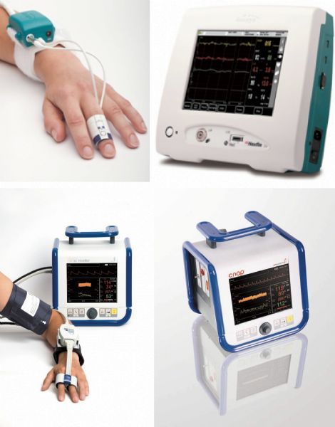
A non-invasive method of measuring beat-to-beat blood pressure, the technique described in 1973 by Peñáz was appealing as it did not incur the risk of arterial catheterization but could still offer the benefits of continuous blood pressure monitoring (Fig. 7) [ 29]. Peñáz used the volume-clamp method, which employs the idea of “vascular unloading.” The basic device consists of a small finger cuff containing a photoplethysmograph —a light source on one side of the cuff and infrared receiver on the opposite side—with the ability to estimate the blood volume of the finger via the infrared light absorbance. Thus, the signal obtained from the plethysmograph is used in a feedback loop allowing for adjustment of the cuff to keep blood volume constant and the vessels in a constant state of “vascular unloading.” Furthermore, the “Finapres” (an acronym for FINger Arterial PRESsure) first modified this technique in the 1990s by assuming the cuff pressure is equal to arterial pressure and then using a formula to reconstruct brachial artery pressures. This is then sent to a display system showing a waveform similar to the one obtained with invasive arterial monitoring. Another commonly used approach is calibrating the Peñás-technique values with a non-invasive cuff places on the upper arm. Currently, the two most widely tested devices employing these variations of the Peñás’ approach include the NexfinTM (Finapres) and CNAPTM (Upper arm calibration) (Fig. 7).
 | Fig.7 Demonstration of non-invasive and beat-to-beat arterial pressure monitoring systems (“Nexfin,” BMeye [top], and “CNAP,” CNSystem [bottom]). |
As with every device, there are some limitations associated with these technologies, namely any state causing low peripheral perfusion which reduces the ability of the feedback system to function appropriately. In fact, vascular disease, cold temperature, Raynaud’s disease as well as other factors have been reported to contribute to finger plethysmogram failure in about 1% of patients when looking specifically at the Finapres technology [ 30]. Systemic vasoactive drugs also have a role in inducing error in the device, which was shown by Imholz et al. in their report on the significant effect that intravenous phenylephrine had on the reliability of the arterial volume clamp method [ 30, 31]. Additionally, cooling the finger upon which the cuff was placed may cause a higher systolic and diastolic pressure bias [ 31]. Once again, similar to all technologies employing a cuff, proper application of the cuff (including appropriate size and placement) can affect outcome.
Another technique which provides non-invasive beat-to-beat measurement is arterial tonometry, which produces a waveform similar to Peñás technique but their principles of operation are very different. Tonometers are placed over a superficial artery with sufficient bony support and utilize the “pressure pulse” method. Basically, the tonometer sensor is placed over the radial artery to compress it until the vessel is flattened against the bone but not occluded (applanation). A pressure transducer on the skin surface then should be able to measure arterial blood pressure via contact pressure. The amount of pressure needed to flatten but not occlude the vessel is known as the “proper hold-down pressure” and this is calculated by an algorithm which includes the systolic, diastolic and pulse pressures over a range of hold down pressures [ 32] (Fig. 8).
 | Fig.8 Demonstration of an arterial tonometry device (T-Line, Tensys Medical). |
Theoretically, arterial tonometry offers some benefits over Peñás technique. Due to the larger size of the radial artery, the technology should be less sensitive to inaccuracies caused by vascular disease and vasoconstriction. In addition, measurements of radial arterial pressure may be more reflective of central arterial pressure than that of the finger [ 23].
One of the pitfalls of this technology is the difficulty in manually positioning a single sensor over the radial artery but the development of devices with an array of tonometers addresses this problem. Additionally, tonometry requires calibration via an initial blood pressure measurement by another technique to be considered completely accurate. These shortcomings have been underlined by previous studies [ 33]. It is worth mentioning that the only currently available commercial device utilizing this technology, the T-line from Tensys Medical, has had much more positive findings recently demonstrated [ 34, 35].
In the push to establish non-invasive continuous means of blood pressure monitoring, pulse transit time (PTT), or the photometric method, is one of the newest technologies receiving attention. This method is based upon the observations first described by Bramwell and Hill in the early 20th century of pulse wave velocity which describes the propagation of the systolic pressure wave through the aorta. To use this technique, there needs to be pulse transducers at two separate arterial sites. These transducers measure the time between the arterial pulses at the two sites. Pulse transit time has also been defined as the time interval between the ECG R-wave and the arrival of the pulse at a peripheral site. The arrival of the pulse at a peripheral site can be detected non-invasively with the use of the finger photoplethysmographic pulse oximetry, a standard intraoperative monitor. Propagation of the arterial pulse is dependent on arterial wall stiffness and vessel size. Pulse transit time has an inverse relationship to systolic arterial blood pressure [ 36, 37] but diastolic and mean pressures are not as easily extrapolated [ 38]. As a “cuffless” technique, it avoids the problems that are inherent in cuff based blood pressure monitoring mentioned previously but more work needs to be done before exact blood pressure measurements can be correlated to specific pulse transit times.
Currently, the most successful method of non-invasive continuous blood pressure monitoring has been embodied by the NexfinTM and CNAPTM, both of which employ the Peñás system. Over the past several decades, numerous studies have been published validating this technique for monitoring blood pressure when compared to invasive monitoring [ 39, 40, 41, 42, 43, 44, 45, 46, 47, 48, 49, 50] as well as non-invasive monitoring [ 51, 52, 53] in different patient populations. The NexfinTM monitor is able to track changes in autonomic function testing [ 54] and has even been shown to be feasible for use for patients undergoing cardiac bypass who have reduced arterial pulsatility [ 55]. Both of these devices have shown the potential to detect hypotensive events earlier than a standard non-invasive blood pressure monitor [ 56, 57], which typically takes blood pressure values every five minutes. However, as a great deal of evidence suggests that hypotension is linked to morbidity and mortality [ 4, 5, 6, 7], the possibility of detecting these events earlier may have a significant role in improving clinical care. In addition to blood pressure, the NexfinTM device also uses an algorithm to calculate continuous cardiac output with some conflicting results as to its potential usefulness [ 58, 59, 60] with recent studies showing promising results in regards to its ability to trend when compared to pulmonary artery catheter values [ 61], transthoracic echography [ 62], and esophageal Doppler [ 63]. Alternatively, Fischer et al. have concluded that while the strong potential for measuring blood pressure non-invasively is valid, such extrapolated cardiac index monitoring cannot replace transpulmonary thermodilution [ 64]. Overall, this device shows strong indications that it will be a significant competitor for the future market of non-invasive blood pressure monitoring and cardiac output estimation.
The CNAPTM device has also had a limited amount of clinical research detailed in the past few years. The main conclusions have been that it is reasonably accurate for general anesthesia [ 47], but still needs some minor improvements. It was also shown to be more accurate for MAP when compared to systolic arterial pressure and diastolic arterial pressure for patients undergoing vascular surgery [ 39]. It is worth mentioning that a recent study done on the CNAPTM showed that during caesarian sections, this monitor performed poorly in obtaining accurate and continuous blood pressure measurements [ 65].
Although the method of arterial tonometry came into practice earlier than that of Peñás, it has not gained a foothold in current market for non-invasive continuous blood pressure monitors. In the 1990s, several studies were conducted with devices employing this technique showing reasonable agreement with invasive arterial lines [ 32, 66, 67, 68, 69] but some had significant concerns as to its accuracy and ability to detect rapid and large transient changes in blood pressure [ 67, 68]. A more recent study in 2004 showed its lack of reliability in elderly patients [ 70]. This technology has also been commonly used to allow for the measurement of pulse wave velocity (PWV) in order to measure arterial stiffness [ 71]. A renewed interest in PWV has been observed in the past decade due to its potential for assisting in the diagnosing and treatment of hypertension and cardiovascular disease [ 72, 73]. More recently, there have been two manuscripts describing the positive potential for this specific technology (T-line©) to be used in the intensive care [ 74] and operating room [ 75] settings. As mentioned earlier, there have also been studies describing improved accuracy when compared to invasive arterial pressures [ 34, 35].
Finally, pulse transit time, which is an offshoot of pulse wave analysis, started being investigated in the early 1970s. There are no current devices which utilize this technology but studies have been ongoing in the past decade to develop this method into a device which can non-invasively and continuously measure blood pressure. Several recent studies have shown the potential of this technology with pulse transit time correlating to blood pressures [ 76, 77] but research into the development of an algorithm that can do this is ongoing [ 78].
As mentioned previously, invasive arterial monitoring offers the prospect of using PPV, and although it is early to tell, there is hope that some of the non-invasive technologies above may also offer this ability to predict fluid responsiveness in patients without contributing the risk [79, 80].
Several methods for non-invasive continuous blood pressure measurements have been studied in the latter half of the 20th century until the present decade. These devices have the potential to bridge the gap between traditional sphygmamometry, which is unable to monitor short-term dynamic variability of blood pressure, with invasive arterial lines, without their associated complication risk. Although some of the devices have demonstrated reliability and ease of use, the expense associated with alternative technologies when compared to intermittent BP monitoring by automated blood pressure cuffs continue to limit their widespread acceptance. The 21st century will surely see the further advancement and refinement of these technologies with the exciting prospect that routine care will include non-invasive continuous blood pressure monitoring.
| 1 |
|
| 2 |
|
| 3 |
|
| 4 |
|
| 5 |
|
| 6 |
|
| 7 |
|
| 8 |
|
| 9 |
|
| 10 |
|
| 11 |
|
| 12 |
|
| 13 |
|
| 14 |
|
| 15 |
|
| 16 |
|
| 17 |
|
| 18 |
|
| 19 |
|
| 20 |
|
| 21 |
|
| 22 |
|
| 23 |
|
| 24 |
|
| 25 |
|
| 26 |
|
| 27 |
|
| 28 |
|
| 29 |
|
| 30 |
|
| 31 |
|
| 32 |
|
| 33 |
|
| 34 |
|
| 35 |
|
| 36 |
|
| 37 |
|
| 38 |
|
| 39 |
|
| 40 |
|
| 41 |
|
| 42 |
|
| 43 |
|
| 44 |
|
| 45 |
|
| 46 |
|
| 47 |
|
| 48 |
|
| 49 |
|
| 50 |
|
| 51 |
|
| 52 |
|
| 53 |
|
| 54 |
|
| 55 |
|
| 56 |
|
| 57 |
|
| 58 |
|
| 59 |
|
| 60 |
|
| 61 |
|
| 62 |
|
| 63 |
|
| 64 |
|
| 65 |
|
| 66 |
|
| 67 |
|
| 68 |
|
| 69 |
|
| 70 |
|
| 71 |
|
| 72 |
|
| 73 |
|
| 74 |
|
| 75 |
|
| 76 |
|
| 77 |
|
| 78 |
|
| 79 |
|
| 80 |
|