|
|
|
Cell surface protein engineering for high-performance whole-cell catalysts |
Hajime Nakatani,Katsutoshi Hori( ) ) |
| Department of Biotechnology, Graduate School of Engineering, Nagoya University, Nagoya 464-8603, Japan |
|
|
|
|
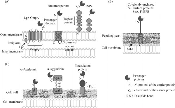
Abstract Cell surface protein engineering facilitated by accumulation of information on genome and protein structure involves heterologous production and modification of cell surface proteins using genetic engineering, and is important for the development of high-performance whole-cell catalysts. In this field, cell surface display is a major technology by exposing target proteins, such as enzymes, on the cell surface using a carrier protein. The target proteins are fused to the carrier proteins that transport and tether them to the cell surface, as well as to a secretion signal. This paper reviews cell surface display systems for prokaryotic and eukaryotic cells from the perspective of carrier proteins, which determine the number of displayed molecules, and the localization, size, and direction (N- or C-terminal anchoring) of the passengers. We also discuss advanced methods for displaying multiple enzymes and a new method for the immobilization of whole-cell catalysts using adhesive surface proteins.
|
| Keywords
cell surface engineering
surface display
whole-cell catalysts
bioprocess
|
|
Corresponding Author(s):
Katsutoshi Hori
|
|
Online First Date: 13 February 2017
Issue Date: 17 March 2017
|
|
| 1 |
Liljeqvist S, Samuelson P, Hansson M, Nguyen T N, Binz H, Stahl S. Surface display of the cholera toxin B subunit on Staphylococcus xylosus and Staphylococcus carnosus. Applied and Environmental Microbiology, 1997, 63(7): 2481–2488
|
| 2 |
Lee J S, Shin K S, Pan J G, Kim C J. Surface-displayed viral antigens on Salmonella carrier vaccine. Nature Biotechnology, 2000, 18(6): 645–648
https://doi.org/10.1038/76494
|
| 3 |
Martineau P, Charbit A, Leclerc C, Werts C, O’Callaghan D, Hofnung M. A genetic system to elicit and monitor antipeptide antibodies without peptide synthesis. Bio/Technology, 1991, 9(2): 170–172
https://doi.org/10.1038/nbt0291-170
|
| 4 |
Westerlund-Wikstrom B, Tanskanen J, Virkola R, Hacker J, Lindberg M, Skurnik M, Korhonen T K. Functional expression of adhesive peptides as fusions to Escherichia coli flagellin. Protein Engineering, 1997, 10(11): 1319–1326
https://doi.org/10.1093/protein/10.11.1319
|
| 5 |
Boder E T, Wittrup K D. Yeast surface display for screening combinatorial polypeptide libraries. Nature Biotechnology, 1997, 15(6): 553–557
https://doi.org/10.1038/nbt0697-553
|
| 6 |
Xu Z, Lee S Y. Display of polyhistidine peptides on the Escherichia coli cell surface by using outer membrane protein C as an anchoring motif. Applied and Environmental Microbiology, 1999, 65(11): 5142–5147
|
| 7 |
Sousa C, Kotrba P, Ruml T, Cebolla A, De Lorenzo V. Metalloadsorption by Escherichia coli cells displaying yeast and mammalian metallothioneins anchored to the outer membrane protein LamB. Journal of Bacteriology, 1998, 180(9): 2280–2284
|
| 8 |
Bae W, Mulchandani A, Chen W. Cell surface display of synthetic phytochelatins using ice nucleation protein for enhanced heavy metal bioaccumulation. Journal of Inorganic Biochemistry, 2002, 88(2): 223–227
https://doi.org/10.1016/S0162-0134(01)00392-0
|
| 9 |
Liu C, Yang B, Gan J, Zhang Y, Liang M, Shu X, Shu J. Heterogeneous reactions of suspended parathion, malathion, and fenthion particles with NO(3) radicals. Chemosphere, 2012, 87(5): 470–476
https://doi.org/10.1016/j.chemosphere.2011.12.031
|
| 10 |
Smith G P. Filamentous fusion phage: Novel expression vectors that display cloned antigens on the virion surface. Science, 1985, 228(4705): 1315–1317
https://doi.org/10.1126/science.4001944
|
| 11 |
Li M. Applications of display technology in protein analysis. Nature Biotechnology, 2000, 18(12): 1251–1256
https://doi.org/10.1038/82355
|
| 12 |
Freudl R, MacIntyre S, Degen M, Henning U. Cell surface exposure of the outer membrane protein OmpA of Escherichia coli K-12. Journal of Molecular Biology, 1986, 188(3): 491–494
https://doi.org/10.1016/0022-2836(86)90171-3
|
| 13 |
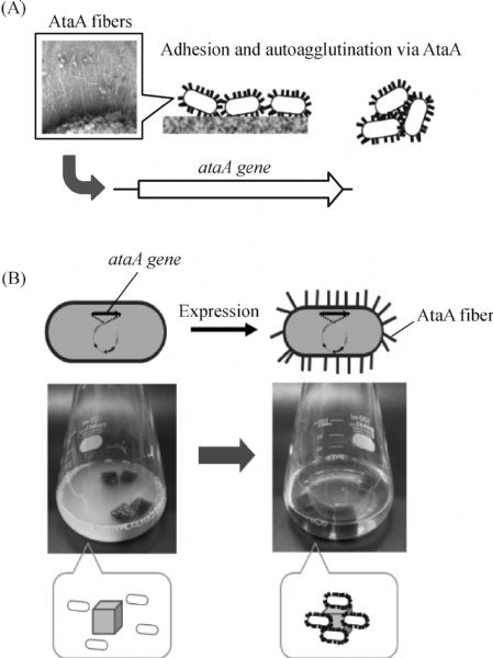
Ishikawa M, Shigemori K, Hori K. Application of the adhesive bacterionanofiber AtaA to a novel microbial immobilization method for the production of indigo as a model chemical. Biotechnology and Bioengineering, 2014, 111(1): 16–24
https://doi.org/10.1002/bit.25012
|
| 14 |
Hori K, Ohara Y, Ishikawa M, Nakatani H. Effectiveness of direct immobilization of bacterial cells onto material surfaces using the bacterionanofiber protein AtaA. Applied Microbiology and Biotechnology, 2015, 99(12): 5025–5032
https://doi.org/10.1007/s00253-015-6554-9
|
| 15 |
Xu X, Gao C, Zhang X, Che B, Ma C, Qiu J, Tao F, Xu P. Production of N-acetyl-D-neuraminic acid by use of an efficient spore surface display system. Applied and Environmental Microbiology, 2011, 77(10): 3197–3201
https://doi.org/10.1128/AEM.00151-11
|
| 16 |
Smith M R, Khera E, Wen F. Engineering novel and improved biocatalysts by cell surface display. Industrial & Engineering Chemistry Research, 2015, 54(16): 4021–4032
https://doi.org/10.1021/ie504071f
|
| 17 |
Beerli R R, Bauer M, Buser R B, Gwerder M, Muntwiler S, Maurer P, Saudan P, Bachmann M F. Isolation of human monoclonal antibodies by mammalian cell display. Proceedings of the National Academy of Sciences of the United States of America, 2008, 105(38): 14336–14341
https://doi.org/10.1073/pnas.0805942105
|
| 18 |
Ernst W, Grabherr R, Wegner D, Borth N, Grassauer A, Katinger H. Baculovirus surface display: Construction and screening of a eukaryotic epitope library. Nucleic Acids Research, 1998, 26(7): 1718–1723
https://doi.org/10.1093/nar/26.7.1718
|
| 19 |
Schneewind O, Missiakas D M. Protein secretion and surface display in Gram-positive bacteria. Philosophical Transactions of the Royal Society of London. Series B: Biological Sciences, 2012, 367(1592): 1123–1139
|
| 20 |
van Bloois E, Winter R T, Kolmar H, Fraaije M W. Decorating microbes: Surface display of proteins on Escherichia coli. Trends in Biotechnology, 2011, 29(2): 79–86
https://doi.org/10.1016/j.tibtech.2010.11.003
|
| 21 |
Levin A M, Weiss G A. Optimizing the affinity and specificity of proteins with molecular display. Molecular BioSystems, 2006, 2(1): 49–57
https://doi.org/10.1039/B511782H
|
| 22 |
Francisco J A, Earhart C F, Georgiou G. Transport and anchoring of beta-lactamase to the external surface of Escherichia coli. Proceedings of the National Academy of Sciences of the United States of America, 1992, 89(7): 2713–2717
https://doi.org/10.1073/pnas.89.7.2713
|
| 23 |
Francisco J A, Campbell R, Iverson B L, Georgiou G. Production and fluorescence-activated cell sorting of Escherichia coli expressing a functional antibody fragment on the external surface. Proceedings of the National Academy of Sciences of the United States of America, 1993, 90(22): 10444–10448
https://doi.org/10.1073/pnas.90.22.10444
|
| 24 |
Francisco J A, Stathopoulos C, Warren R A, Kilburn D G, Georgiou G. Specific adhesion and hydrolysis of cellulose by intact Escherichia coli expressing surface anchored cellulase or cellulose binding domains. Bio/Technology, 1993, 11(4): 491–495
https://doi.org/10.1038/nbt0493-491
|
| 25 |
Wei W, Liu X, Sun P, Wang X, Zhu H, Hong M, Mao Z W, Zhao J. Simple whole-cell biodetection and bioremediation of heavy metals based on an engineered lead-specific operon. Environmental Science & Technology, 2014, 48(6): 3363–3371
https://doi.org/10.1021/es4046567
|
| 26 |
Yang C, Zhao Q, Liu Z, Li Q, Qiao C, Mulchandani A, Chen W. Cell surface display of functional macromolecule fusions on Escherichia coli for development of an autofluorescent whole-cell biocatalyst. Environmental Science & Technology, 2008, 42(16): 6105–6110
https://doi.org/10.1021/es800441t
|
| 27 |
Qu W, Xue Y, Ding Q. Display of fungi xylanase on Escherichia coli cell surface and use of the enzyme in xylan biodegradation. Current Microbiology, 2015, 70(6): 779–785
https://doi.org/10.1007/s00284-015-0781-2
|
| 28 |
Richins R D, Kaneva I, Mulchandani A, Chen W. Biodegradation of organophosphorus pesticides by surface-expressed organophosphorus hydrolase. Nature Biotechnology, 1997, 15(10): 984–987
https://doi.org/10.1038/nbt1097-984
|
| 29 |
Jung H C, Lebeault J M, Pan J G. Surface display of Zymomonas mobilis levansucrase by using the ice-nucleation protein of Pseudomonas syringae. Nature Biotechnology, 1998, 16(6): 576–580
https://doi.org/10.1038/nbt0698-576
|
| 30 |
Maurer J, Jose J, Meyer T F. Autodisplay: One-component system for efficient surface display and release of soluble recombinant proteins from Escherichia coli. Journal of Bacteriology, 1997, 179(3): 794–804
https://doi.org/10.1128/jb.179.3.794-804.1997
|
| 31 |
Karami A, Latifi A M, Khodi S. Comparison of the organophosphorus hydrolase surface display using InaVN and Lpp-OmpA systems in Escherichia coli. Journal of Microbiology and Biotechnology, 2014, 24(3): 379–385
https://doi.org/10.4014/jmb.1309.09066
|
| 32 |
Kawahara H. The structures and functions of ice crystal-controlling proteins from bacteria. Journal of Bioscience and Bioengineering, 2002, 94(6): 492–496
https://doi.org/10.1016/S1389-1723(02)80185-2
|
| 33 |
Jung H C, Park J H, Park S H, Lebeault J M, Pan J G. Expression of carboxymethylcellulase on the surface of Escherichia coli using Pseudomonas syringae ice nucleation protein. Enzyme and Microbial Technology, 1998, 22(5): 348–354
https://doi.org/10.1016/S0141-0229(97)00224-X
|
| 34 |
van Bloois E, Winter R T, Janssen D B, Fraaije M W. Export of functional Streptomyces coelicolor alditol oxidase to the periplasm or cell surface of Escherichia coli and its application in whole-cell biocatalysis. Applied Microbiology and Biotechnology, 2009, 83(4): 679–687
https://doi.org/10.1007/s00253-009-1904-0
|
| 35 |
Yim S K, Jung H C, Pan J G, Kang H S, Ahn T, Yun C H. Functional expression of mammalian NADPH-cytochrome P450 oxidoreductase on the cell surface of Escherichia coli. Protein Expression and Purification, 2006, 49(2): 292–298
https://doi.org/10.1016/j.pep.2006.05.013
|
| 36 |
Yim S K, Kim D H, Jung H C, Pan J G, Kang H S, Ahn T, Yun C H. Surface display of heme- and diflavin-containing cytochrome P450 BM3 in Escherichia coli: A whole cell biocatalyst for oxidation. Journal of Microbiology and Biotechnology, 2010, 20(4): 712–717
https://doi.org/10.4014/jmb.0910.10043
|
| 37 |
Benz I, Schmidt M A. Structures and functions of autotransporter proteins in microbial pathogens. International Journal of Medical Microbiology, 2011, 301(6): 461–468
https://doi.org/10.1016/j.ijmm.2011.03.003
|
| 38 |
Leo J C, Grin I, Linke D.Type V secretion: Mechanism(s) of autotransport through the bacterial outer membrane. Philosophical Transactions of the Royal Society of London. Series B: Biological Sciences, 2012, 367(1592): 1088–1101
|
| 39 |
Nicolay T, Vanderleyden J, Spaepen S. Autotransporter-based cell surface display in Gram-negative bacteria. Critical Reviews in Microbiology, 2013, 41(1): 109–123
https://doi.org/10.3109/1040841X.2013.804032
|
| 40 |
Jose J, Meyer T F. The autodisplay story, from discovery to biotechnical and biomedical applications. Microbiology and Molecular Biology Reviews, 2007, 71(4): 600–619
https://doi.org/10.1128/MMBR.00011-07
|
| 41 |
Detzel C, Maas R, Tubeleviciute A, Jose J. Autodisplay of nitrilase from Klebsiella pneumoniae and whole-cell degradation of oxynil herbicides and related compounds. Applied Microbiology and Biotechnology, 2013, 97(11): 4887–4896
https://doi.org/10.1007/s00253-012-4401-9
|
| 42 |
Jose J, von Schwichow S. Autodisplay of active sorbitol dehydrogenase (SDH) yields a whole cell biocatalyst for the synthesis of rare sugars. ChemBioChem, 2004, 5(4): 491–499
https://doi.org/10.1002/cbic.200300774
|
| 43 |
Lattemann C T, Maurer J, Gerland E, Meyer T F. Autodisplay: Functional display of active beta-lactamase on the surface of Escherichia coli by the AIDA-I autotransporter. Journal of Bacteriology, 2000, 182(13): 3726–3733
https://doi.org/10.1128/JB.182.13.3726-3733.2000
|
| 44 |
Schultheiss E, Weiss S, Winterer E, Maas R, Heinzle E, Jose J. Esterase autodisplay: Enzyme engineering and whole-cell activity determination in microplates with pH sensors. Applied and Environmental Microbiology, 2008, 74(15): 4782–4791
https://doi.org/10.1128/AEM.01575-07
|
| 45 |
Li C, Zhu Y, Benz I, Schmidt M A, Chen W, Mulchandani A, Qiao C. Presentation of functional organophosphorus hydrolase fusions on the surface of Escherichia coli by the AIDA-I autotransporter pathway. Biotechnology and Bioengineering, 2008, 99(2): 485–490
https://doi.org/10.1002/bit.21548
|
| 46 |
Becker S, Theile S, Heppeler N, Michalczyk A, Wentzel A, Wilhelm S, Jaeger K E, Kolmar H. A generic system for the Escherichia coli cell-surface display of lipolytic enzymes. FEBS Letters, 2005, 579(5): 1177–1182
https://doi.org/10.1016/j.febslet.2004.12.087
|
| 47 |
Shanna S, Iasson E P, Tozakidis M, Teese J J. Maximized autotransporter-mediated expression (MATE) for surface display and secretion of recombinant proteins in Escherichia coli. Food Technology and Biotechnology, 2015, 50(3): 251–260
|
| 48 |
Tozakidis I E, Brossette T, Lenz F, Maas R M, Jose J. Proof of concept for the simplified breakdown of cellulose by combining Pseudomonas putida strains with surface displayed thermophilic endocellulase, exocellulase and beta-glucosidase. Microbial Cell Factories, 2016, 15(1): 103
https://doi.org/10.1186/s12934-016-0505-8
|
| 49 |
Crampton M, Berger E, Reid S, Louw M. The development of a flagellin surface display expression system in a moderate thermophile, Bacillus halodurans Alk36. Applied Microbiology and Biotechnology, 2007, 75(3): 599–607
https://doi.org/10.1007/s00253-007-0869-0
|
| 50 |
Pallesen L, Poulsen L K, Christiansen G, Klemm P. Chimeric FimH adhesin of type 1 fimbriae: A bacterial surface display system for heterologous sequences. Microbiology, 1995, 141(11): 2839–2848
https://doi.org/10.1099/13500872-141-11-2839
|
| 51 |
Ishikawa M, Nakatani H, Hori K. AtaA, a new member of the trimeric autotransporter adhesins from Acinetobacter sp. Tol 5 mediating high adhesiveness to various abiotic surfaces. PLoS One, 2012, 7(11): e48830
https://doi.org/10.1371/journal.pone.0048830
|
| 52 |
Nummelin H, Merckel M C, Leo J C, Lankinen H, Skurnik M, Goldman A. The Yersinia adhesin YadA collagen-binding domain structure is a novel left-handed parallel beta-roll. EMBO Journal, 2004, 23(4): 701–711
https://doi.org/10.1038/sj.emboj.7600100
|
| 53 |
O’Rourke F, Schmidgen T, Kaiser P O, Linke D, Kempf V A. Adhesins of Bartonella spp. Advances in Experimental Medicine and Biology, 2011, 715: 51–70
https://doi.org/10.1007/978-94-007-0940-9_4
|
| 54 |
Yoshimoto S, Nakatani H, Iwasaki K, Hori K. An Acinetobacter trimeric autotransporter adhesin reaped from cells exhibits its nonspecific stickiness via a highly stable 3D structure. Scientific Reports, 2016, 6: 28020
https://doi.org/10.1038/srep28020
|
| 55 |
Schneewind O, Mihaylova-Petkov D, Model P. Cell wall sorting signals in surface proteins of gram-positive bacteria. EMBO Journal, 1993, 12(12): 4803–4811
|
| 56 |
Lee S Y, Choi J H, Xu Z. Microbial cell-surface display. Trends in Biotechnology, 2003, 21(1): 45–52
https://doi.org/10.1016/S0167-7799(02)00006-9
|
| 57 |
Schreuder M P, Brekelmans S, van den Ende H, Klis F M. Targeting of a heterologous protein to the cell wall of Saccharomyces cerevisiae. Yeast (Chichester, England), 1993, 9(4): 399–409
https://doi.org/10.1002/yea.320090410
|
| 58 |
Pepper L R, Cho Y K, Boder E T, Shusta E V. A decade of yeast surface display technology: Where are we now? Combinatorial Chemistry & High Throughput Screening, 2008, 11(2): 127–134
https://doi.org/10.2174/138620708783744516
|
| 59 |
Kuroda K, Ueda M. Arming technology in yeast-novel strategy for whole-cell biocatalyst and protein engineering. Biomolecules, 2013, 3(3): 632–650
https://doi.org/10.3390/biom3030632
|
| 60 |
Blazic M, Kovacevic G, Prodanovic O, Ostafe R, Gavrovic-Jankulovic M, Fischer R, Prodanovic R. Yeast surface display for the expression, purification and characterization of wild-type and B11 mutant glucose oxidases. Protein Expression and Purification, 2013, 89(2): 175–180
https://doi.org/10.1016/j.pep.2013.03.014
|
| 61 |
Gera N, Hussain M, Rao B M. Protein selection using yeast surface display. Methods (San Diego, Calif.), 2013, 60(1): 15–26
https://doi.org/10.1016/j.ymeth.2012.03.014
|
| 62 |
Tanaka T, Yamada R, Ogino C, Kondo A. Recent developments in yeast cell surface display toward extended applications in biotechnology. Applied Microbiology and Biotechnology, 2012, 95(3): 577–591
https://doi.org/10.1007/s00253-012-4175-0
|
| 63 |
Wen F, Sethi D K, Wucherpfennig K W, Zhao H. Cell surface display of functional human MHC class II proteins: Yeast display versus insect cell display. Protein Engineering, Design & Selection, 2011, 24(9): 701–709
https://doi.org/10.1093/protein/gzr035
|
| 64 |
Yeasmin S, Kim C H, Park H J, Sheikh M I, Lee J Y, Kim J W, Back K K, Kim S H. Cell surface display of cellulase activity-free xylanase enzyme on Saccharomyces cerevisiae EBY100. Applied Biochemistry and Biotechnology, 2011, 164(3): 294–304
https://doi.org/10.1007/s12010-010-9135-5
|
| 65 |
Ueda M, Tanaka A. Cell surface engineering of yeast: Construction of arming yeast with biocatalyst. Journal of Bioscience and Bioengineering, 2000, 90(2): 125–136
https://doi.org/10.1016/S1389-1723(00)80099-7
|
| 66 |
Kondo A, Shigechi H, Abe M, Uyama K, Matsumoto T, Takahashi S, Ueda M, Tanaka A, Kishimoto M, Fukuda H. High-level ethanol production from starch by a flocculent Saccharomyces cerevisiae strain displaying cell-surface glucoamylase. Applied Microbiology and Biotechnology, 2002, 58(3): 291–296
https://doi.org/10.1007/s00253-001-0900-9
|
| 67 |
Chen Y, Stemple B, Kumar M, Wei N. Cell surface display fungal laccase as a renewable biocatalyst for degradation of persistent micropollutants bisphenol A and sulfamethoxazole. Environmental Science & Technology, 2016, 50(16): 8799–8808
https://doi.org/10.1021/acs.est.6b01641
|
| 68 |
He X, Shang J, Li F, Liu H. Yeast cell surface display of linoleic acid isomerase from Propionibacterium acnes and its application for the production of trans-10, cis-12 conjugated linoleic acid. Biotechnology and Applied Biochemistry, 2014, 62(1): 1–8
https://doi.org/10.1002/bab.1249
|
| 69 |
Bony M, Thines-Sempoux D, Barre P, Blondin B. Localization and cell surface anchoring of the Saccharomyces cerevisiae flocculation protein Flo1p. Journal of Bacteriology, 1997, 179(15): 4929–4936
https://doi.org/10.1128/jb.179.15.4929-4936.1997
|
| 70 |
Matsumoto T, Fukuda H, Ueda M, Tanaka A, Kondo A. Construction of yeast strains with high cell surface lipase activity by using novel display systems based on the Flo1p flocculation functional domain. Applied and Environmental Microbiology, 2002, 68(9): 4517–4522
https://doi.org/10.1128/AEM.68.9.4517-4522.2002
|
| 71 |
Han Z L, Han S Y, Zheng S P, Lin Y. Enhancing thermostability of a Rhizomucor miehei lipase by engineering a disulfide bond and displaying on the yeast cell surface. Applied Microbiology and Biotechnology, 2009, 85(1): 117–126
https://doi.org/10.1007/s00253-009-2067-8
|
| 72 |
Jiang Z B, Song H T, Gupta N, Ma L X, Wu Z B. Cell surface display of functionally active lipases from Yarrowia lipolytica in Pichia pastoris. Protein Expression and Purification, 2007, 56(1): 35–39
https://doi.org/10.1016/j.pep.2007.07.003
|
| 73 |
Moura M V, da Silva G P, Machado A C, Torres F A, Freire D M, Almeida R V. Displaying lipase B from Candida antarctica in Pichia pastoris using the yeast surface display approach: Prospection of a new anchor and characterization of the whole cell biocatalyst. PLoS One, 2015, 10(10): e0141454
https://doi.org/10.1371/journal.pone.0141454
|
| 74 |
Kondo A, Ueda M. Yeast cell-surface display—applications of molecular display. Applied Microbiology and Biotechnology, 2004, 64(1): 28–40
https://doi.org/10.1007/s00253-003-1492-3
|
| 75 |
Bauer F F, Govender P, Bester M C. Yeast flocculation and its biotechnological relevance. Applied Microbiology and Biotechnology, 2010, 88(1): 31–39
https://doi.org/10.1007/s00253-010-2783-0
|
| 76 |
Vallejo J A, Sanchez-Perez A, Martinez J P, Villa T G. Cell aggregations in yeasts and their applications. Applied Microbiology and Biotechnology, 2013, 97(6): 2305–2318
https://doi.org/10.1007/s00253-013-4735-y
|
| 77 |
Abe H, Ohba M, Shimma Y, Jigami Y. Yeast cells harboring human alpha-1,3-fucosyltransferase at the cell surface engineered using Pir, a cell wall-anchored protein. FEMS Yeast Research, 2004, 4(4-5): 417–425
https://doi.org/10.1016/S1567-1356(03)00193-4
|
| 78 |
Abe H, Shimma Y, Jigami Y. In vitro oligosaccharide synthesis using intact yeast cells that display glycosyltransferases at the cell surface through cell wall-anchored protein Pir. Glycobiology, 2003, 13(2): 87–95
https://doi.org/10.1093/glycob/cwg014
|
| 79 |
Andres I, Gallardo O, Parascandola P, Javier Pastor F I, Zueco J. Use of the cell wall protein Pir4 as a fusion partner for the expression of Bacillus sp. BP-7 xylanase A in Saccharomyces cerevisiae. Biotechnology and Bioengineering, 2005, 89(6): 690–697
https://doi.org/10.1002/bit.20375
|
| 80 |
Shimma Y, Saito F, Oosawa F, Jigami Y. Construction of a library of human glycosyltransferases immobilized in the cell wall of Saccharomyces cerevisiae. Applied and Environmental Microbiology, 2006, 72(11): 7003–7012
https://doi.org/10.1128/AEM.01378-06
|
| 81 |
Shi B, Ke X, Yu H, Xie J, Jia Y, Guo R. Novel properties for endoglucanase acquired by cell-surface display technique. Journal of Microbiology and Biotechnology, 2015, 25(11): 1856–1862
https://doi.org/10.4014/jmb.1503.03029
|
| 82 |
Yuzbasheva E Y, Yuzbashev T V, Perkovskaya N I, Mostova E B, Vybornaya T V, Sukhozhenko A V, Toropygin I Y, Sineoky S P. Cell surface display of Yarrowia lipolytica lipase Lip2p using the cell wall protein YlPir1p, its characterization, and application as a whole-cell biocatalyst. Applied Biochemistry and Biotechnology, 2015, 175(8): 3888–3900
https://doi.org/10.1007/s12010-015-1557-7
|
| 83 |
Castillo L, Martinez A I, Garcera A, Elorza M V, Valentin E, Sentandreu R. Functional analysis of the cysteine residues and the repetitive sequence of Saccharomyces cerevisiae Pir4/Cis3: The repetitive sequence is needed for binding to the cell wall beta-1,3-glucan. Yeast (Chichester, England), 2003, 20(11): 973–983
https://doi.org/10.1002/yea.1016
|
| 84 |
Ecker M, Deutzmann R, Lehle L, Mrsa V, Tanner W. Pir proteins of Saccharomyces cerevisiae are attached to beta-1,3-glucan by a new protein-carbohydrate linkage. Journal of Biological Chemistry, 2006, 281(17): 11523–11529
https://doi.org/10.1074/jbc.M600314200
|
| 85 |
Starwalt S E, Masteller E L, Bluestone J A, Kranz D M. Directed evolution of a single-chain class II MHC product by yeast display. Protein Engineering, 2003, 16(2): 147–156
https://doi.org/10.1093/proeng/gzg018
|
| 86 |
Yang N, Yu Z, Jia D, Xie Z, Zhang K, Xia Z, Lei L, Qiao M. The contribution of Pir protein family to yeast cell surface display. Applied Microbiology and Biotechnology, 2014, 98(7): 2897–2905
https://doi.org/10.1007/s00253-014-5538-5
|
| 87 |
Liu R, Yang C, Xu Y, Xu P, Jiang H, Qiao C. Development of a whole-cell biocatalyst/biosensor by display of multiple heterologous proteins on the Escherichia coli cell surface for the detoxification and detection of organophosphates. Journal of Agricultural and Food Chemistry, 2013, 61(32): 7810–7816
https://doi.org/10.1021/jf402999b
|
| 88 |
Tang X, Liang B, Yi T, Manco G, Palchetti I, Liu A. Cell surface display of organophosphorus hydrolase for sensitive spectrophotometric detection of p-nitrophenol substituted organophosphates. Enzyme and Microbial Technology, 2014, 55: 107–112
https://doi.org/10.1016/j.enzmictec.2013.10.006
|
| 89 |
Yang J, Liu R, Jiang H, Yang Y, Qiao C. Selection of a whole-cell biocatalyst for methyl parathion biodegradation. Applied Microbiology and Biotechnology, 2012, 95(6): 1625–1632
https://doi.org/10.1007/s00253-011-3792-3
|
| 90 |
Chen X A, Ishida N, Todaka N, Nakamura R, Maruyama J, Takahashi H, Kitamoto K. Promotion of efficient saccharification of crystalline cellulose by Aspergillus fumigatus Swo1. Applied and Environmental Microbiology, 2010, 76(8): 2556–2561
https://doi.org/10.1128/AEM.02499-09
|
| 91 |
Arantes V, Saddler J N. Access to cellulose limits the efficiency of enzymatic hydrolysis: The role of amorphogenesis. Biotechnology for Biofuels, 2010, 3(1): 4
https://doi.org/10.1186/1754-6834-3-4
|
| 92 |
Nakatani Y, Yamada R, Ogino C, Kondo A. Synergetic effect of yeast cell-surface expression of cellulase and expansin-like protein on direct ethanol production from cellulose. Microbial Cell Factories, 2013, 12(1): 66
https://doi.org/10.1186/1475-2859-12-66
|
| 93 |
Hyeon J E, Jeon S D, Han S O. Cellulosome-based, Clostridium-derived multi-functional enzyme complexes for advanced biotechnology tool development: Advances and applications. Biotechnology Advances, 2013, 31(6): 936–944
https://doi.org/10.1016/j.biotechadv.2013.03.009
|
| 94 |
Schwarz W H. The cellulosome and cellulose degradation by anaerobic bacteria. Applied Microbiology and Biotechnology, 2001, 56(5-6): 634–649
https://doi.org/10.1007/s002530100710
|
| 95 |
Wen F, Sun J, Zhao H. Yeast surface display of trifunctional minicellulosomes for simultaneous saccharification and fermentation of cellulose to ethanol. Applied and Environmental Microbiology, 2010, 76(4): 1251–1260
https://doi.org/10.1128/AEM.01687-09
|
| 96 |
Tsai S L, Oh J, Singh S, Chen R, Chen W. Functional assembly of minicellulosomes on the Saccharomyces cerevisiae cell surface for cellulose hydrolysis and ethanol production. Applied and Environmental Microbiology, 2009, 75(19): 6087–6093
https://doi.org/10.1128/AEM.01538-09
|
| 97 |
Liang Y, Si T, Ang E L, Zhao H. Engineered pentafunctional minicellulosome for simultaneous saccharification and ethanol fermentation in Saccharomyces cerevisiae. Applied and Environmental Microbiology, 2014, 80(21): 6677–6684
https://doi.org/10.1128/AEM.02070-14
|
| 98 |
You C, Zhang X Z, Sathitsuksanoh N, Lynd L R, Zhang Y H. Enhanced microbial utilization of recalcitrant cellulose by an ex vivo cellulosome-microbe complex. Applied and Environmental Microbiology, 2012, 78(5): 1437–1444
https://doi.org/10.1128/AEM.07138-11
|
| 99 |
Lin Y, Tanaka S. Ethanol fermentation from biomass resources: Current state and prospects. Applied Microbiology and Biotechnology, 2006, 69(6): 627–642
https://doi.org/10.1007/s00253-005-0229-x
|
| 100 |
Dervakos G A, Webb C. On the merits of viable-cell immobilisation. Biotechnology Advances, 1991, 9(4): 559–612
https://doi.org/10.1016/0734-9750(91)90733-C
|
| 101 |
Junter G A, Jouenne T. Immobilized viable microbial cells: From the process to the proteome em leader or the cart before the horse. Biotechnology Advances, 2004, 22(8): 633–658
https://doi.org/10.1016/j.biotechadv.2004.06.003
|
| 102 |
Carballeira J D, Quezada M A, Hoyos P, Simeo Y, Hernaiz M J, Alcantara A R, Sinisterra J V. Microbial cells as catalysts for stereoselective red-ox reactions. Biotechnology Advances, 2009, 27(6): 686–714
https://doi.org/10.1016/j.biotechadv.2009.05.001
|
| 103 |
Cassidy M B, Lee H, Trevors J T. Environmental applications of immobilized microbial cells: A review. Journal of Industrial Microbiology, 1996, 16(2): 79–101
https://doi.org/10.1007/BF01570068
|
| 104 |
Schaeffer C R, Woods K M, Longo G M, Kiedrowski M R, Paharik A E, Buttner H, Christner M, Boissy R J, Horswill A R, Rohde H, Fey P D. Accumulation-associated protein enhances Staphylococcus epidermidis biofilm formation under dynamic conditions and is required for infection in a rat catheter model. Infection and Immunity, 2015, 83(1): 214–226
https://doi.org/10.1128/IAI.02177-14
|
| 105 |
Cotter S E, Surana N K, St Geme J W 3rd. Trimeric autotransporters: A distinct subfamily of autotransporter proteins. Trends in Microbiology, 2005, 13(5): 199–205
https://doi.org/10.1016/j.tim.2005.03.004
|
| 106 |
Hobley L, Harkins C, MacPhee C E, Stanley-Wall N R. Giving structure to the biofilm matrix: An overview of individual strategies and emerging common themes. FEMS Microbiology Reviews, 2015, 39(5): 649–669
https://doi.org/10.1093/femsre/fuv015
|
| 107 |
Ishikawa M, Shigemori K, Suzuki A, Hori K. Evaluation of adhesiveness of Acinetobacter sp. Tol 5 to abiotic surfaces. Journal of Bioscience and Bioengineering, 2012, 113(6): 719–725
https://doi.org/10.1016/j.jbiosc.2012.01.011
|
|
Viewed |
|
|
|
Full text
|
|
|
|
|
Abstract
|
|
|
|
|
Cited |
|
|
|
|
| |
Shared |
|
|
|
|
| |
Discussed |
|
|
|
|