|
|
|
U-shaped microRNA expression pattern could be a new concept biomarker for environmental estrogen |
Rui Duan,Yun Lu( ),Lingyan Hou,Lina Du,Lequn Sun,Xingfan Tang ),Lingyan Hou,Lina Du,Lequn Sun,Xingfan Tang |
| State Key Joint Laboratory of Environment Simulation and Pollution Control, School of Environment, Tsinghua University, Beijing 100084, China |
|
|
|
|
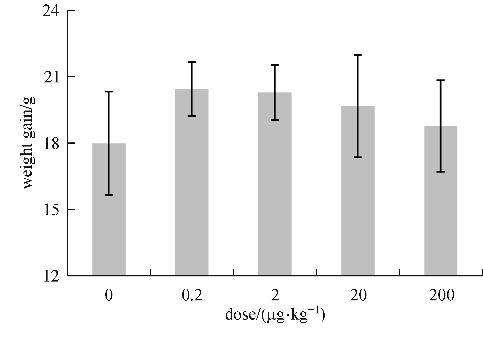
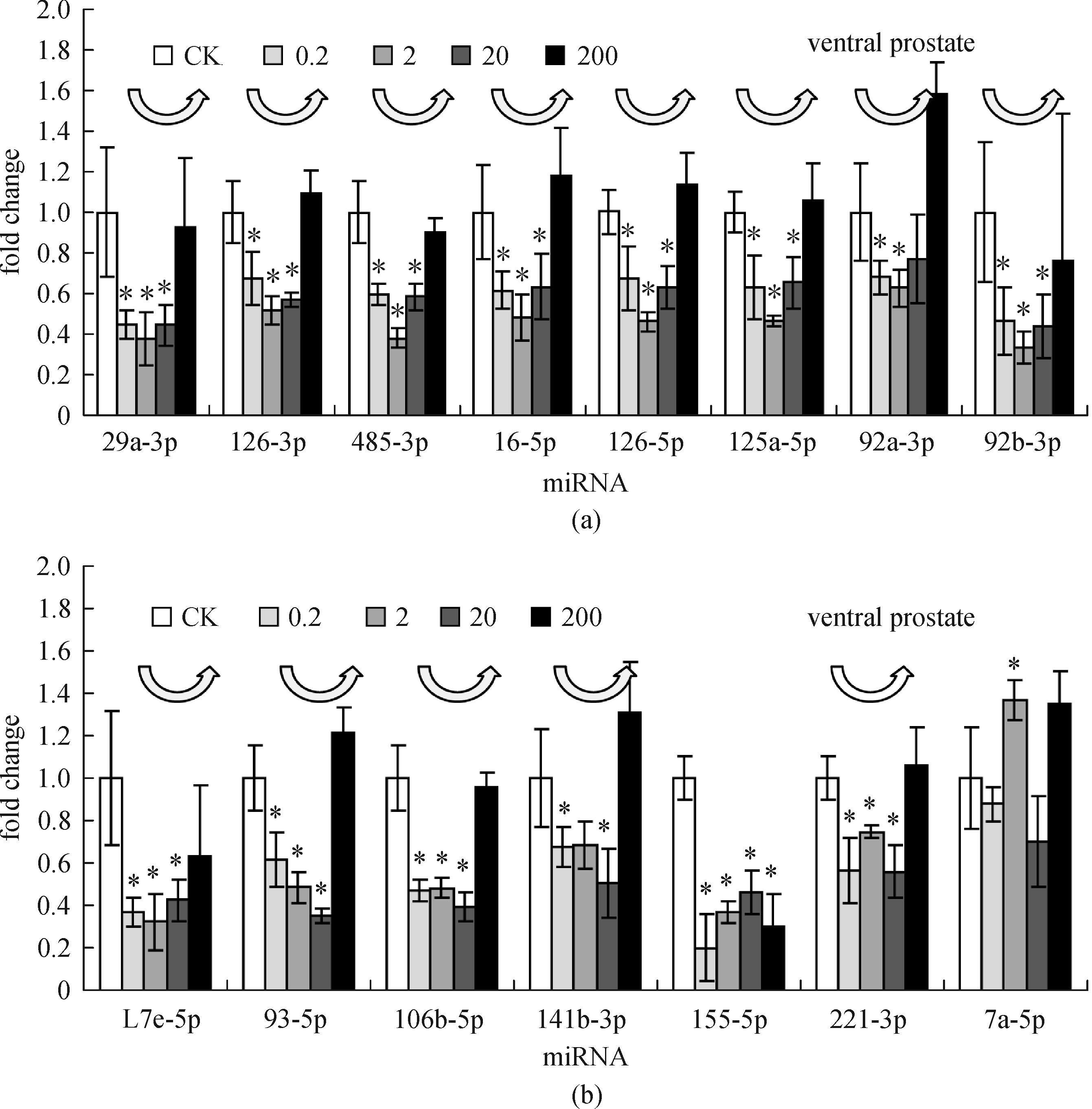
Abstract Estrogen regulates miRNA expression in a typical U-shaped dose-response pattern.
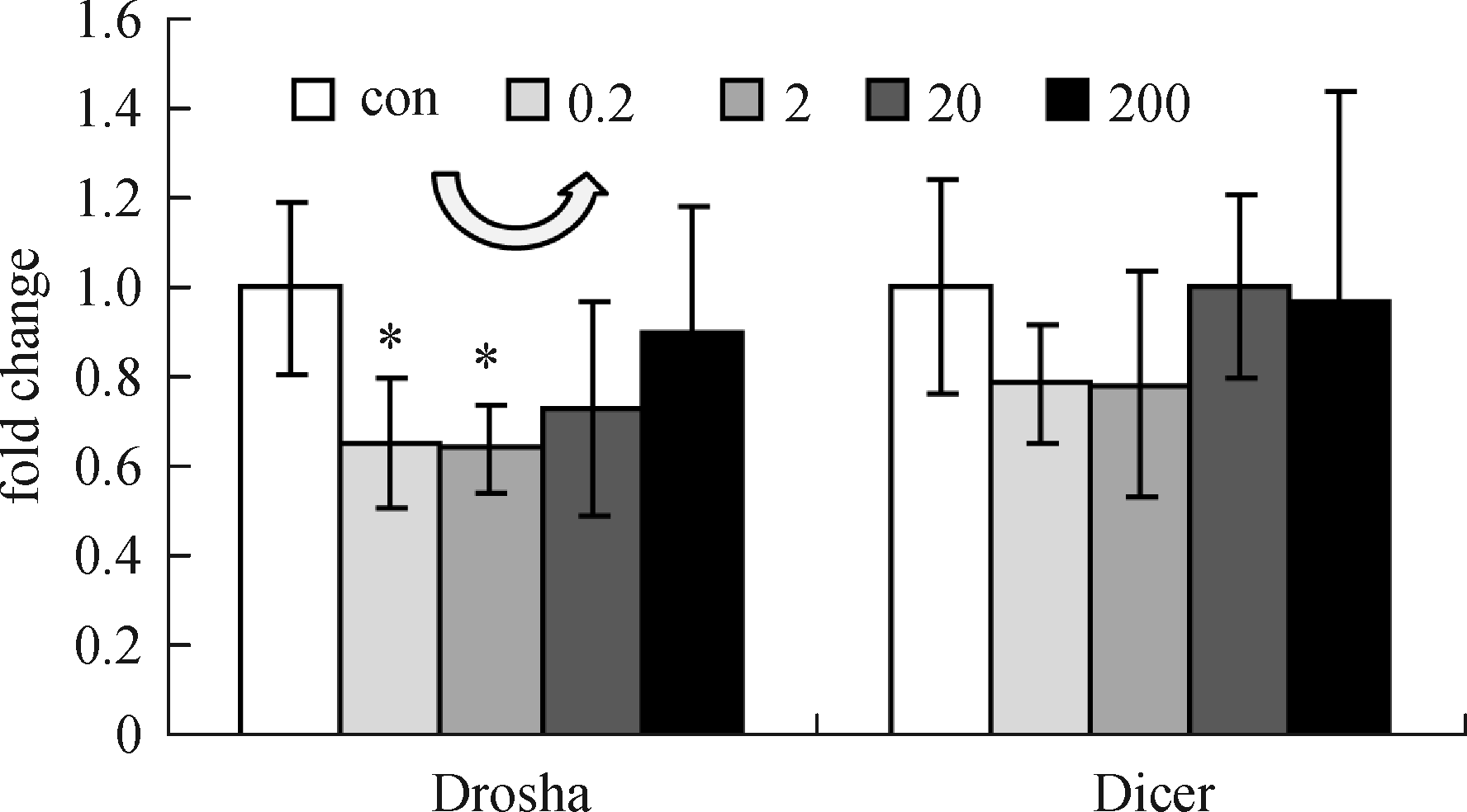
E2 can regulate drosha in the ventral prostate.
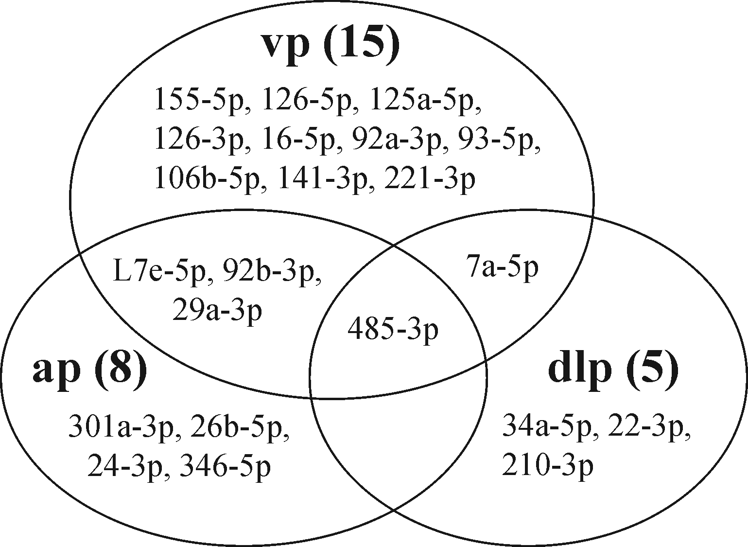
Mouse ventral prostate is most sensitive to estrogen.
Nonmonotonic dose-response in prostate could be a component of estrogen signature.
Many studies have focused on environmental estrogen-related diseases. However, no consistent gene markers or signatures for estrogenicity have been discovered in mammals. This study investigated the estrogenic effects of 17β-estradiol on the prostate in immature male mice. Consistent U-shaped responses were seen in bodyweight, ventral prostate epithelial morphology, and miRNA expression levels. Specifically, most estradiol regulated miRNAs were downregulated at low doses of estradiol (0.2 and 2 mg·kg−1), and whose expression returned to the control level at a larger dose (200 mg·kg−1). The function of these regulated miRNAs is related to the prostate cancer and PI3K-Akt signaling pathways, which is consistent with the function of estradiol. Furthermore, the miRNA-processing machinery, Drosha, in the prostate was also regulated in a similar pattern, which could be a part of the U-shaped miRNA expression mechanism. All of these data indicate that the prostate is a reliable organ for evaluating estrogenic activity and that the typical nonmonotonic dose-response relationship could be used as a novel biomarker for estrogenicity.
|
| Keywords
miRNA
Prostate
Estradiol
Nonmonotonic dose-response
Estrogenicity
Drosha
|
|
|
| Fund: |
|
Corresponding Author(s):
Yun Lu
|
|
Issue Date: 19 October 2016
|
|
| 1 |
Kerdivel G, Habauzit D, Pakdel F. Assessment and molecular actions of endocrine-disrupting chemicals that interfere with estrogen receptor pathways. International Journal of Endocrinology, 2013, 2013: 501851
https://doi.org/10.1155/2013/501851
pmid: 23737774
|
| 2 |
Pelekanou V, Leclercq G. Recent insights into the effect of natural and environmental estrogens on mammary development and carcinogenesis. The International Journal of Developmental Biology, 2011, 55(7-8-9): 869–878
https://doi.org/10.1387/ijdb.113369vp
pmid: 22161842
|
| 3 |
Shang G, Xue J, Li M, Hu H Y, Lu Y. Estrogen receptor affinity chromatography: a new method for characterization of novel estrogenic disinfection by-products. Chemosphere, 2014, 104: 251–257
https://doi.org/10.1016/j.chemosphere.2014.01.027
pmid: 24548648
|
| 4 |
Li M, Xu B, Liungai Z, Hu H Y, Chen C, Qiao J, Lu Y. The removal of estrogenic activity with UV/chlorine technology and identification of novel estrogenic disinfection by-products. Journal of Hazardous Materials, 2016, 307: 119–126
https://doi.org/10.1016/j.jhazmat.2016.01.003
pmid: 26780699
|
| 5 |
Williams G P. The role of oestrogen in the pathogenesis of obesity, type 2 diabetes, breast cancer and prostate disease. European Journal of Cancer Prevention, 2010, 19(4): 256–271
https://doi.org/10.1097/CEJ.0b013e328338f7d2
pmid: 20535861
|
| 6 |
Burns K A, Korach K S. Estrogen receptors and human disease: an update. Archives of Toxicology, 2012, 86(10): 1491–1504
https://doi.org/10.1007/s00204-012-0868-5
pmid: 22648069
|
| 7 |
vomSaal F S, Timms B G, Montano M M, Palanza P, Thayer K A, Nagel S C, Dhar M D, Ganjam V K, Parmigiani S, Welshons W V. Prostate enlargement in mice due to fetal exposure to low doses of estradiol or diethylstilbestrol and opposite effects at high doses. Proceedings of the National Academy of Sciences of the United States of America, 1997, 94(5): 2056–2061
https://doi.org/10.1073/pnas.94.5.2056
pmid: 9050904
|
| 8 |
Timms B G, Howdeshell K L, Barton L, Bradley S, Richter C A, vomSaal F S. Estrogenic chemicals in plastic and oral contraceptives disrupt development of the fetal mouse prostate and urethra. Proceedings of the National Academy of Sciences of the United States of America, 2005, 102(19): 7014–7019
https://doi.org/10.1073/pnas.0502544102
pmid: 15867144
|
| 9 |
Nelson W G, DeMarzo A M, Yegnasubramanian S. The diet as a cause of human prostate cancer. In: Zappia V, Panico S, Russo GL, Budillon A, DellaRagione F, eds. Advances in Nutrition and Cancer. Cancer Treatment and Research.Berlin: Springer Berlin Heidelberg,2014, 159:51–68
|
| 10 |
Sugimura Y, Cunha G R, Donjacour A A. Morphogenesis of ductal networks in the mouse prostate. Biology of Reproduction, 1986, 34(5): 961–971
https://doi.org/10.1095/biolreprod34.5.961
pmid: 3730488
|
| 11 |
Jarred R A, McPherson S J, Bianco J J, Couse J F, Korach K S, Risbridger G P. Prostate phenotypes in estrogen-modulated transgenic mice. Trends in Endocrinology and Metabolism, 2002, 13(4): 163–168
https://doi.org/10.1016/S1043-2760(02)00575-1
pmid: 11943560
|
| 12 |
Hewitt S C, Deroo B J, Hansen K, Collins J, Grissom S, Afshari C A, Korach K S. Estrogen receptor-dependent genomic responses in the uterus mirror the biphasic physiological response to estrogen. Molecular Endocrinology (Baltimore, Md.), 2003, 17(10): 2070–2083
https://doi.org/10.1210/me.2003-0146
pmid: 12893882
|
| 13 |
Watanabe H, Suzuki A, Kobayashi M, Takahashi E, Itamoto M, Lubahn D B, Handa H, Iguchi T. Analysis of temporal changes in the expression of estrogen-regulated genes in the uterus. Journal of Molecular Endocrinology, 2003, 30(3): 347–358
https://doi.org/10.1677/jme.0.0300347
pmid: 12790804
|
| 14 |
Hong S H, Nah H Y, Lee J Y, Gye M C, Kim C H, Kim M K. Analysis of estrogen-regulated genes in mouse uterus using cDNA microarray and laser capture microdissection. Journal of Endocrinology, 2004, 181(1): 157–167
https://doi.org/10.1677/joe.0.1810157
pmid: 15072576
|
| 15 |
An B S, Choi K C, Kang S K, Hwang W S, Jeung E B. Novel Calbindin-D(9k) protein as a useful biomarker for environmental estrogenic compounds in the uterus of immature rats. Reproductive Toxicology (Elmsford, N.Y.), 2003, 17(3): 311–319
https://doi.org/10.1016/S0890-6238(03)00003-0
pmid: 12759100
|
| 16 |
Calin G A, Croce C M. MicroRNA signatures in human cancers. Nature Reviews, Cancer, 2006, 6(11): 857–866
https://doi.org/10.1038/nrc1997
pmid: 17060945
|
| 17 |
Ha M, Kim V N. Regulation of microRNA biogenesis. Nature Reviews, Molecular Cell Biology, 2014, 15(8): 509–524
https://doi.org/10.1038/nrm3838
pmid: 25027649
|
| 18 |
Witwer K W. Circulating microRNA biomarker studies: pitfalls and potential solutions. Clinical Chemistry, 2015, 61(1): 56–63
https://doi.org/10.1373/clinchem.2014.221341
pmid: 25391989
|
| 19 |
Gore AC, Chappell VA, Fenton SE, Flaws JA, Nadal A, Prins GS, Toppari J, Zoeller RT.EDC-2: the endocrine society’s second scientific statement on endocrine-disrupting chemicals. Endocrine Reviews, 2015, 36(6): E1–E150
https://doi.org/10.1210/er.2015-1010
|
| 20 |
Vlachos I S, Kostoulas N, Vergoulis T, Georgakilas G, Reczko M, Maragkakis M, Paraskevopoulou M D, Prionidis K, Dalamagas T, Hatzigeorgiou A G. DIANA miRPath v.2.0: investigating the combinatorial effect of microRNAs in pathways. Nucleic Acids Research, 2012, 40(Web Server issueW1): W498–W504
https://doi.org/10.1093/nar/gks494
pmid: 22649059
|
| 21 |
Lee Y R, Park J, Yu H N, Kim J S, Youn H J, Jung S H. Up-regulation of PI3K/Akt signaling by 17beta-estradiol through activation of estrogen receptor-alpha, but not estrogen receptor-beta, and stimulates cell growth in breast cancer cells. Biochemical and Biophysical Research Communications, 2005, 336(4): 1221–1226
https://doi.org/10.1016/j.bbrc.2005.08.256
pmid: 16169518
|
| 22 |
Guo R X, Wei L H, Tu Z, Sun P M, Wang J L, Zhao D, Li X P, Tang J M. 17 beta-estradiol activates PI3K/Akt signaling pathway by estrogen receptor (ER)-dependent and ER-independent mechanisms in endometrial cancer cells. Journal of Steroid Biochemistry and Molecular Biology, 2006, 99(1): 9–18
https://doi.org/10.1016/j.jsbmb.2005.11.013
pmid: 16567092
|
| 23 |
Hua K, Feng W, Cao Q, Zhou X, Lu X, Feng Y. Estrogen and progestin regulate metastasis through the PI3K/AKT pathway in human ovarian cancer. International Journal of Oncology, 2008, 33(5): 959–967 doi:10.3892/ijo_00000083
pmid: 18949358
|
| 24 |
Sosa L, Gutiérrez S, Petiti J P, Palmeri C M, Mascanfroni I D, Soaje M, De Paul A L, Torres A I. 17b-Estradiol modulates the prolactin secretion induced by TRH through membrane estrogen receptors via PI3K/Akt in female rat anterior pituitary cell culture. American Journal of Physiology-Endocrinology and Metabolism,2012, 302(10): E1189–E1197
https://doi.org/10.1152/ajpendo.00408.2011
pmid: 22354782
|
| 25 |
Li Z, Yang S, Liu S. Estrogen protects MIN6 beta-cell from hypoxic cell death via PI3K/Aktpathway. Diabetes, 2013, 62(suppl. 1): A562–A562
|
| 26 |
Marker P C, Donjacour A A, Dahiya R, Cunha G R. Hormonal, cellular, and molecular control of prostatic development. Developmental Biology, 2003, 253(2): 165–174
https://doi.org/10.1016/S0012-1606(02)00031-3
pmid: 12645922
|
| 27 |
Huang L, Pu Y, Hu W Y, Birch L, Luccio-Camelo D, Yamaguchi T, Prins G S. The role of Wnt5a in prostate gland development. Developmental Biology, 2009, 328(2): 188–199
https://doi.org/10.1016/j.ydbio.2009.01.003
pmid: 19389372
|
| 28 |
Klinge C M. Estrogen receptor interaction with co-activators and co-repressors. Steroids, 2000, 65(5): 227–251
https://doi.org/10.1016/S0039-128X(99)00107-5
pmid: 10751636
|
| 29 |
Amara J F, Dannies P S. 17 beta-Estradiol has a biphasic effect on gh cell growth. Endocrinology, 1983, 112(3): 1141–1143
https://doi.org/10.1210/endo-112-3-1141
pmid: 6822206
|
| 30 |
Taylor J A, Grady L H, Engler K S, Welshons W V. Relationship of growth stimulated by lithium, estradiol, and EGF to phospholipase C activity in MCF-7 human breast cancer cells. Breast Cancer Research and Treatment, 1995, 34(3): 265–277
https://doi.org/10.1007/BF00689718
pmid: 7579491
|
| 31 |
Wetherill Y B, Petre C E, Monk K R, Puga A, Knudsen K E. The xenoestrogenbisphenol A induces inappropriate androgen receptor activation and mitogenesis in prostatic adenocarcinoma cells. Molecular Cancer Therapeutics, 2002, 1(7): 515–524
pmid: 12479269
|
| 32 |
Alworth L C, Howdeshell K L, Ruhlen R L, Day J K, Lubahn D B, Huang T H M, Besch-Williford C L, vomSaal F S. Uterine responsiveness to estradiol and DNA methylation are altered by fetal exposure to diethylstilbestrol and methoxychlor in CD-1 mice: effects of low versus high doses. Toxicology and Applied Pharmacology, 2002, 183(1): 10–22
https://doi.org/10.1006/taap.2002.9459
pmid: 12217638
|
| 33 |
Welshons W V, Thayer K A, Judy B M, Taylor J A, Curran E M, vomSaal F S. Large effects from small exposures. I. Mechanisms for endocrine-disrupting chemicals with estrogenic activity. Environmental Health Perspectives, 2003, 111(8): 994–1006
https://doi.org/10.1289/ehp.5494
pmid: 12826473
|
| 34 |
Chen T. The role of MicroRNA in chemical carcinogenesis. Journal of Environmental Science and Health. Part C, Environmental Carcinogenesis & Ecotoxicology Reviews, 2010, 28(2): 89–124
https://doi.org/10.1080/10590501.2010.481477
pmid: 20552498
|
| 35 |
Amiel J, de Pontual L, Henrion-Caude A. miRNA, development and disease. Advances in Genetics, 2012, 80: 1–36
https://doi.org/10.1016/B978-0-12-404742-6.00001-6
pmid: 23084872
|
| 36 |
Klinge C M. miRNAs and estrogen action. Trends in Endocrinology and Metabolism, 2012, 23(5): 223–233
https://doi.org/10.1016/j.tem.2012.03.002
pmid: 22503553
|
| 37 |
Nothnick W B, Healy C. Estrogen induces distinct patterns of microRNA expression within the mouse uterus. Reproductive Sciences (Thousand Oaks, Calif.), 2010, 17(11): 987–994
https://doi.org/10.1177/1933719110377472
pmid: 20720260
|
| 38 |
Hou L, Lu Y, Li Y, Li L. MiRNA-451 is a potential biomarker for estrogenicity in mouse uterus. Frontiers of Environmental Science &Engineering,2014, 8(1): 99–105
https://doi.org/10.1007/s11783-013-0490-7
|
| 39 |
Moggs J G, Tinwell H, Spurway T, Chang H S, Pate I, Lim F L, Moore D J, Soames A, Stuckey R, Currie R, Zhu T, Kimber I, Ashby J, Orphanides G. Phenotypic anchoring of gene expression changes during estrogen-induced uterine growth. Environmental Health Perspectives, 2004, 112(16): 1589–1606
https://doi.org/10.1289/ehp.7345
pmid: 15598610
|
|
Viewed |
|
|
|
Full text
|
|
|
|
|
Abstract
|
|
|
|
|
Cited |
|
|
|
|
| |
Shared |
|
|
|
|
| |
Discussed |
|
|
|
|