|
|
|
Bacterial inactivation, DNA damage, and faster ATP degradation induced by ultraviolet disinfection |
Chao Yang, Wenjun Sun( ), Xiuwei Ao ), Xiuwei Ao |
| School of Environment, Tsinghua University, Beijing 100084, China |
|
|
|
|
Abstract • Long amplicon is more effective to test DNA damage induced by UV. • ATP in bacteria does not degrade instantly but does eventually after UV exposure. • After medium pressure UV exposure, ATP degraded faster.
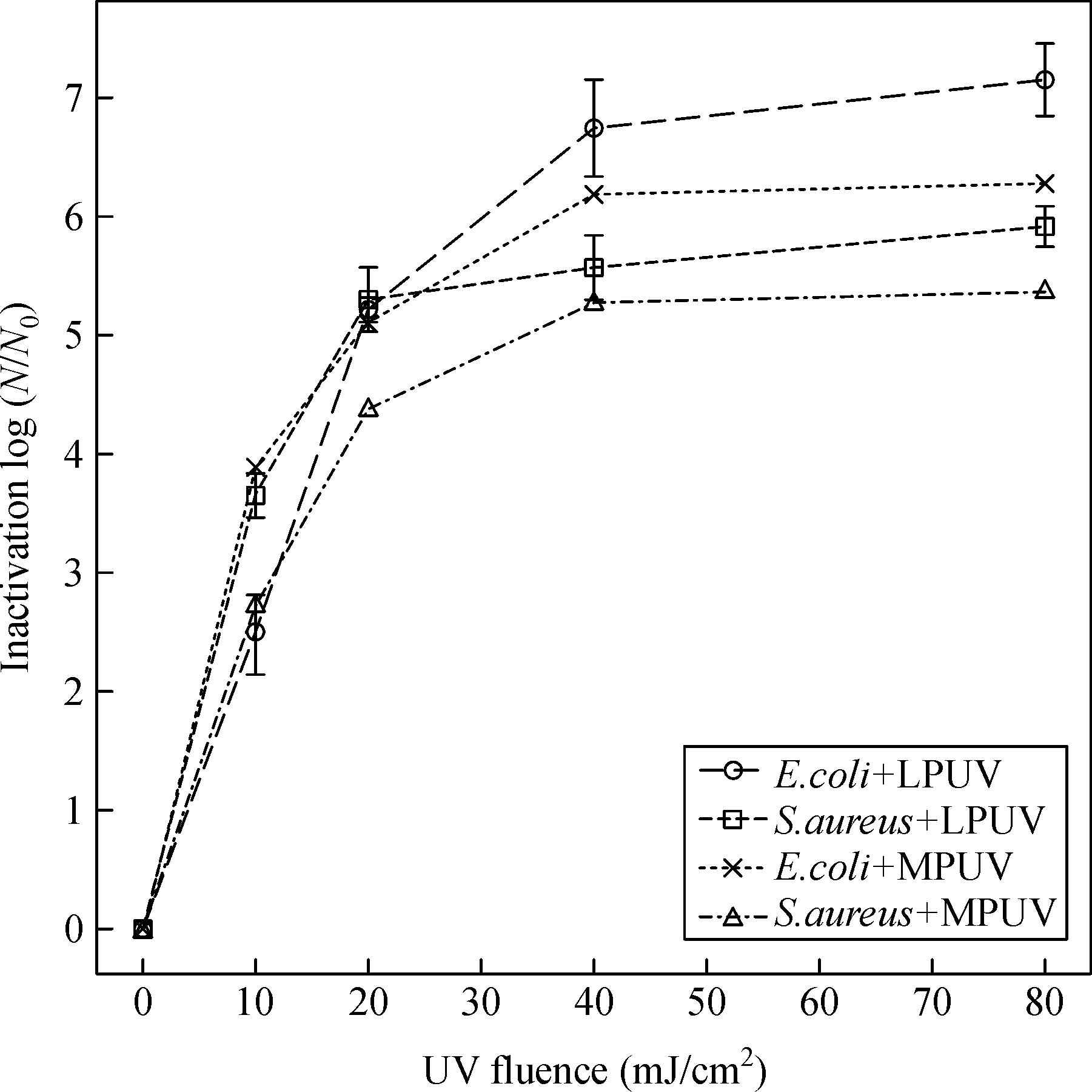
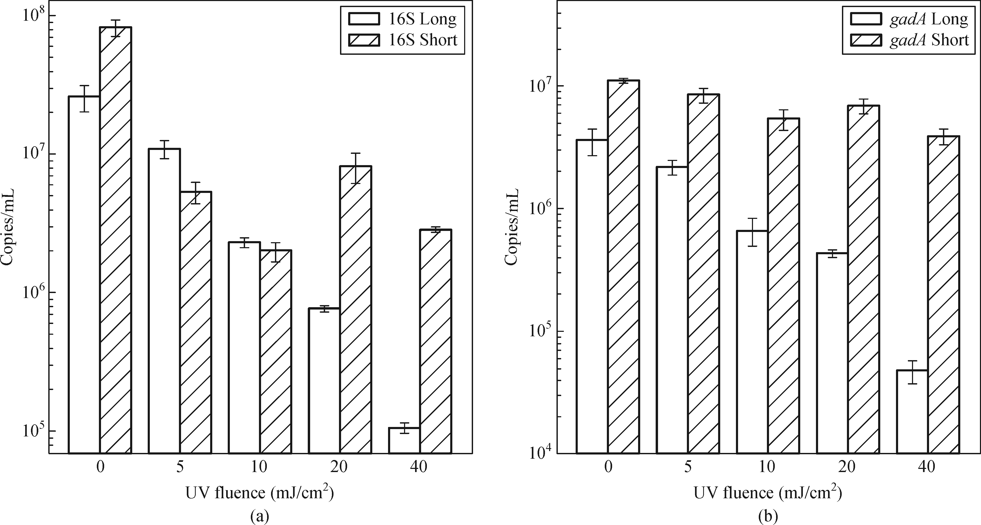
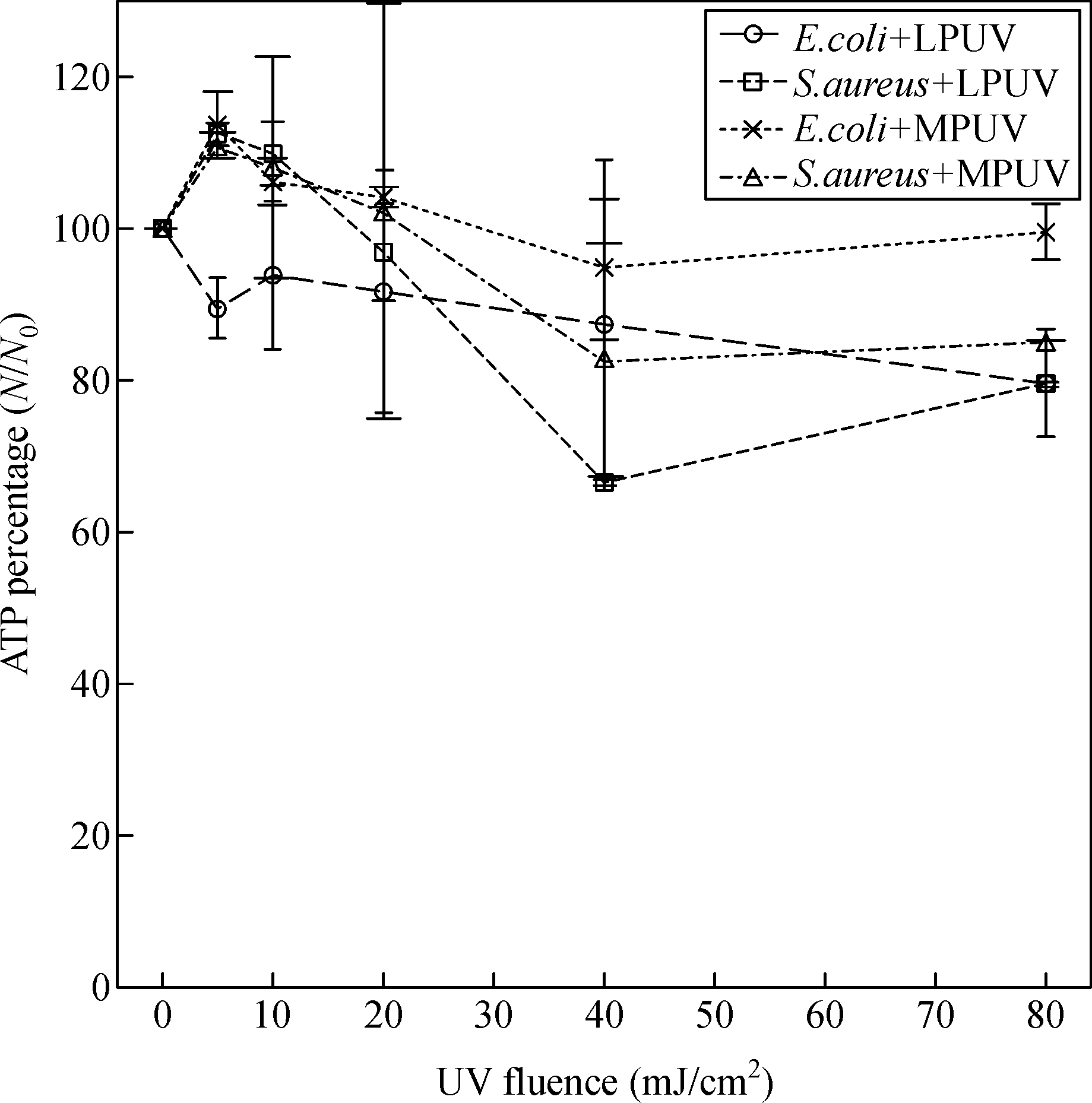
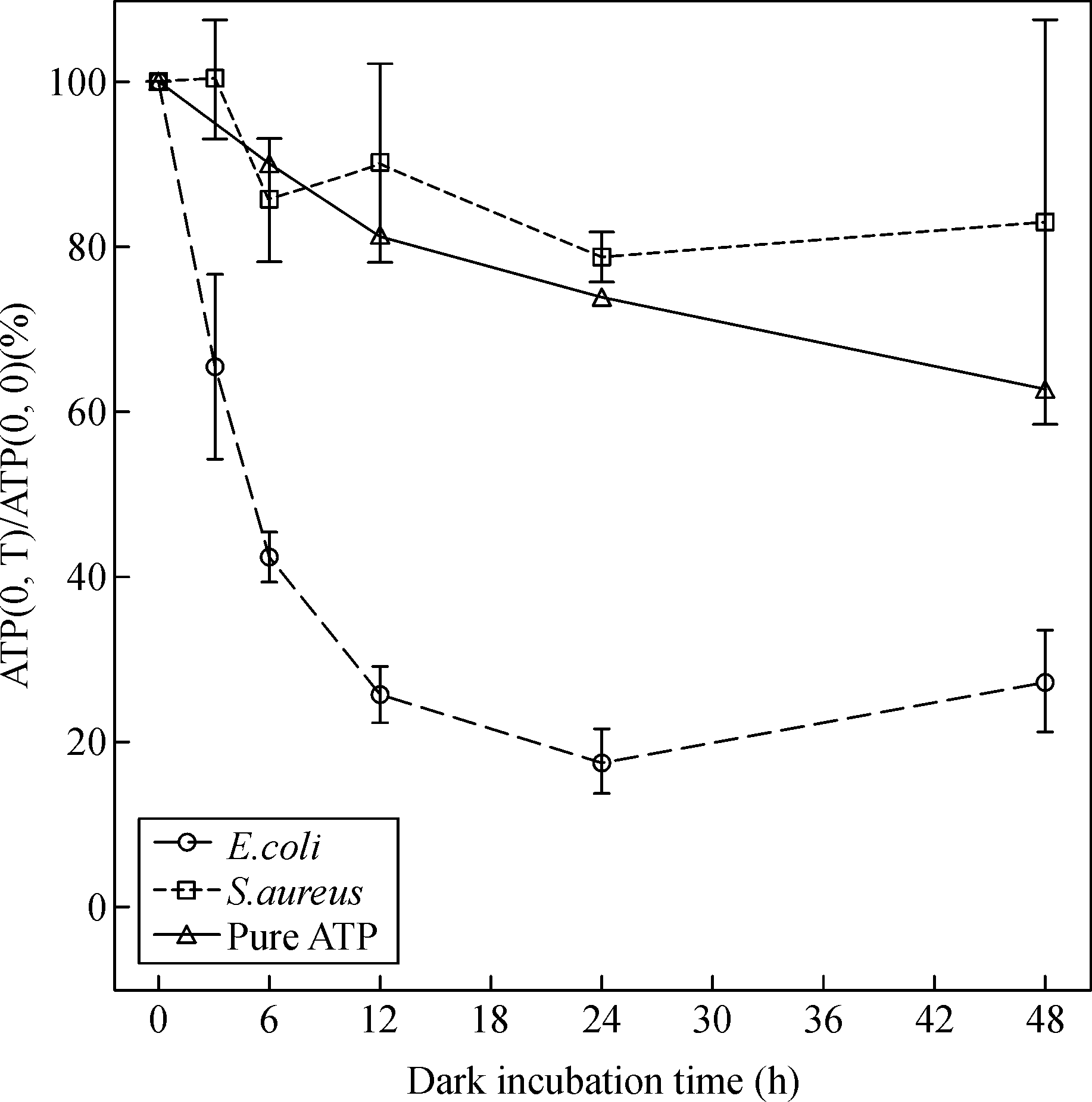
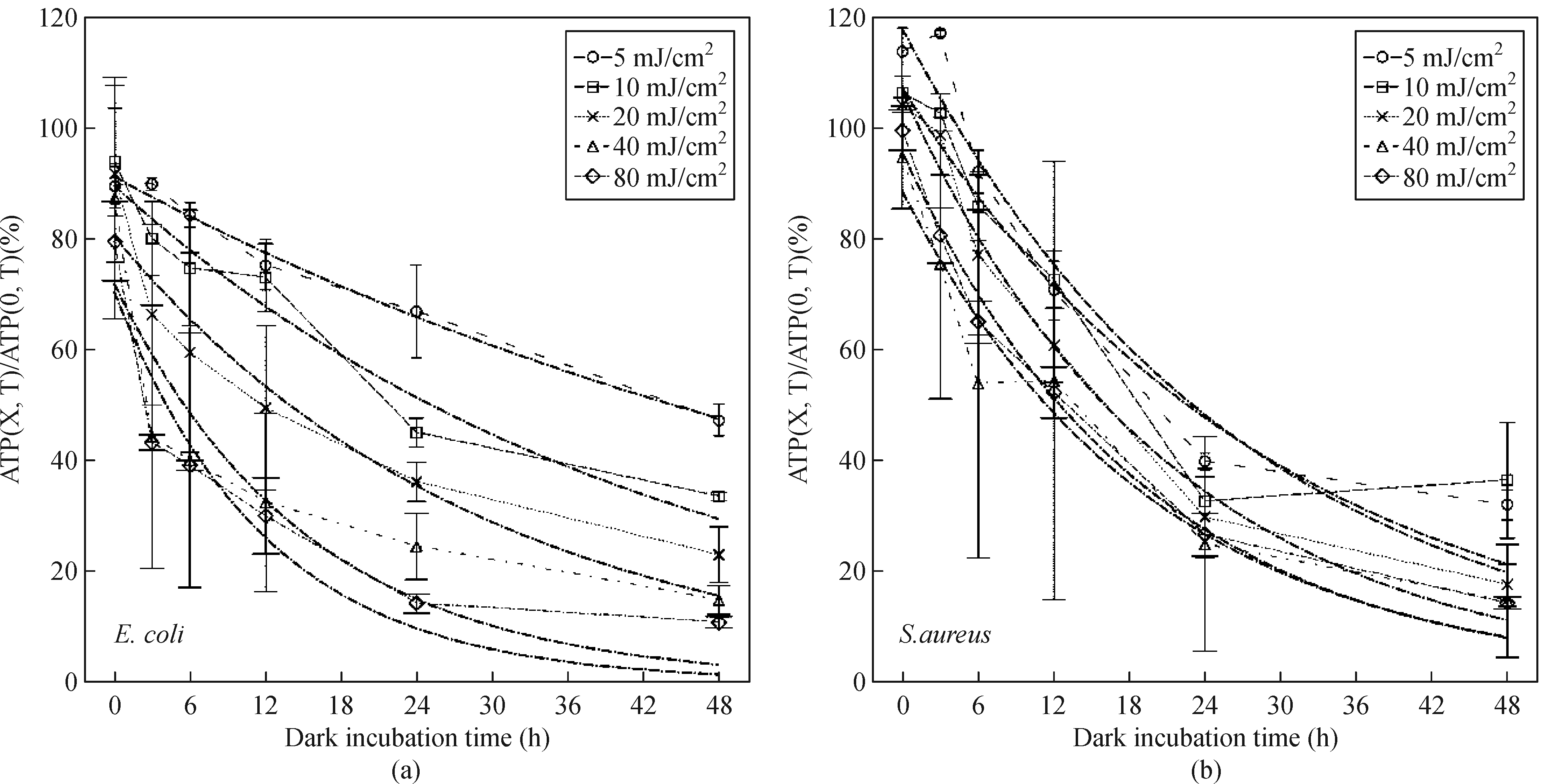
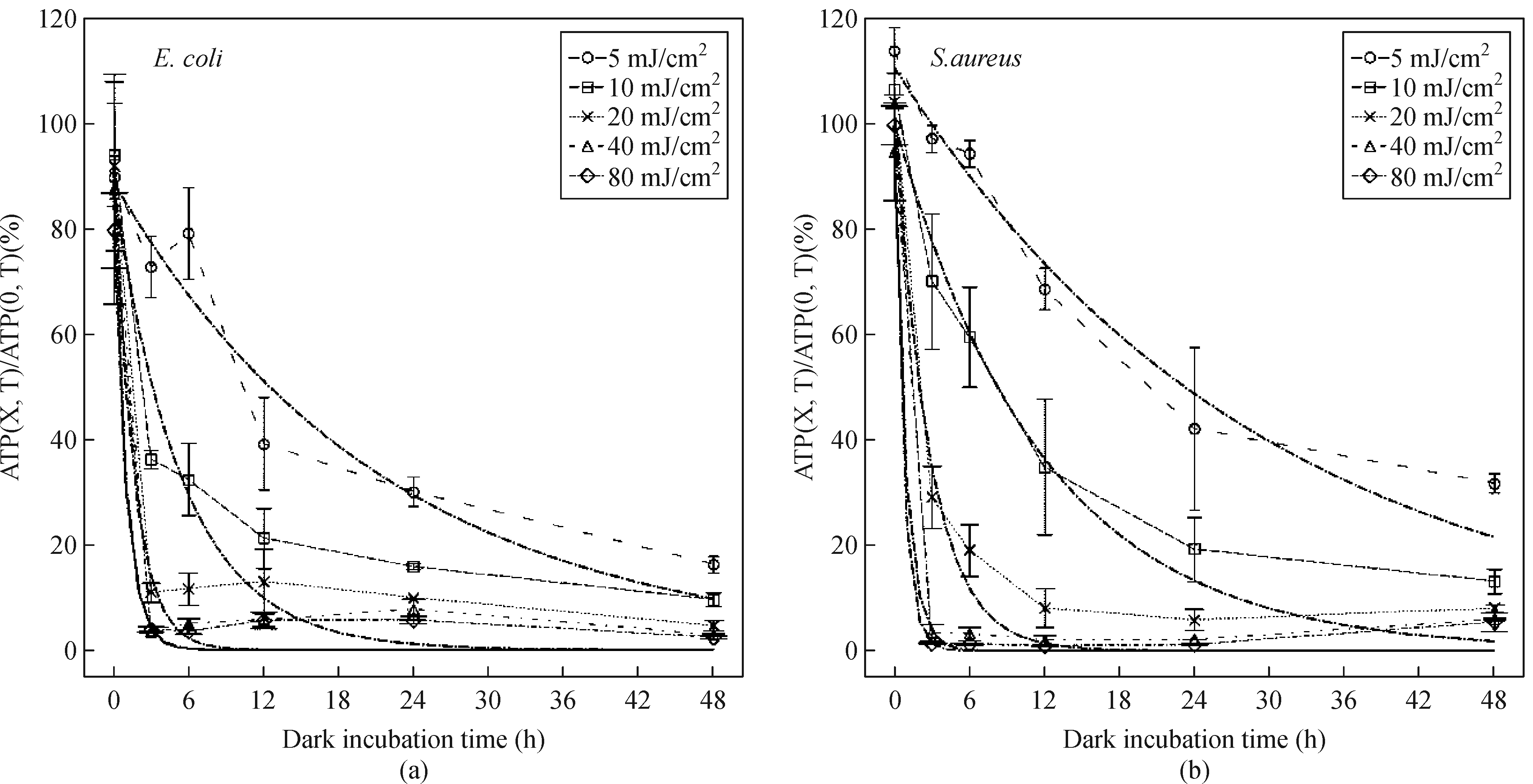
![]() The efficacy of ultraviolet (UV) disinfection has been validated in numerous studies by using culture-based methods. However, the discovery of viable but non-culturable bacteria has necessitated the investigation of UV disinfection based on bacterial viability parameters. We used quantitative polymerase chain reaction (qPCR) to investigate DNA damage and evaluated adenosine triphosphate (ATP) to indicate bacterial viability. The results of qPCR effectively showed the DNA damage induced by UV when using longer gene amplicons, in that sufficiently long amplicons of both 16S and gadA indicated that the UV induced DNA damages. The copy concentrations of the long amplicons of 16S and gadA decreased by 2.38 log/mL and 1.88 log/mL, respectively, after exposure to 40 mJ/cm2 low-pressure UV. After UV exposure, the ATP level in the bacteria did not decrease instantly. Instead it decreased gradually at a rate that was positively related to the UV fluence. For low-pressure UV, this rate of decrease was slow, but for medium pressure UV, this rate of decrease was relatively high when the UV fluence reached 40 mJ/cm2. At the same UV fluence, the ATP level in the bacteria decreased at a faster rate after exposure to medium-pressure UV.
|
| Keywords
UV disinfection
DNA damage
qPCR
ATP
|
|
Corresponding Author(s):
Wenjun Sun
|
|
Issue Date: 15 November 2019
|
|
| 1 |
S Ayala-Torres, Y Chen, T Svoboda, J Rosenblatt, B Van Houten (2000). Analysis of gene-specific DNA damage and repair using quantitative polymerase chain reaction. Methods (San Diego, Calif.), 22(2): 135–147
https://doi.org/10.1006/meth.2000.1054
pmid: 11020328
|
| 2 |
E R Blatchley 3rd, N Dumoutier, T N Halaby, Y Levi, J M Laîné (2001). Bacterial responses to ultraviolet irradiation. Water Science and Technology, 43(10): 179–186
https://doi.org/10.2166/wst.2001.0614
pmid: 11436779
|
| 3 |
E R Blatchley, K Oguma, R Sommer (2017). Comment on ‘UV disinfection induces a VBNC state in Escherichia coli and Pseudomonas aeruginosa’. IUVA News, 18(3): 12–16
|
| 4 |
J R Bolton, K G Linden (2003). Standardization of methods for fluence (UV dose) determination in bench-scale UV experiments. Journal of Environmental Engineering, 129(3): 209–215
https://doi.org/10.1061/(ASCE)0733-9372(2003)129:3(209)
|
| 5 |
A C, Eischeid J A Thurston, K G Linden (2011). UV disinfection of adenovirus: present state of the research and future directions. Critical Reviews in Environmental Science and Technology, 41(15): 1375–1396
https://doi.org/10.1080/10643381003608268
|
| 6 |
J Fang, H Liu, C Shang, M Zeng, M Ni, W Liu (2014). E. coli and bacteriophage MS2 disinfection by UV, ozone and the combined UV and ozone processes. Frontiers of Environmental Science & Engineering, 8(4): 547–552
https://doi.org/10.1007/s11783-013-0620-2
|
| 7 |
A F Gaudy Jr, F Abu-Niaaj, E T Gaudy (1963). Statistical study of the spot-plate technique for viable-cell counts. Applied Microbiology, 11(4): 305–309
pmid: 13946830
|
| 8 |
M Guo, H Hu, J R Bolton, M G El-Din (2009). Comparison of low- and medium-pressure ultraviolet lamps: Photoreactivation of Escherichia coli and total coliforms in secondary effluents of municipal wastewater treatment plants. Water Research, 43(3): 815–821
https://doi.org/10.1016/j.watres.2008.11.028
pmid: 19081599
|
| 9 |
H He, P Zhou, K K Shimabuku, X Fang, S Li, Y Lee, M C Dodd (2019). Degradation and deactivation of bacterial antibiotic resistance genes during exposure to free chlorine, monochloramine, chlorine dioxide, ozone, ultraviolet light, and hydroxyl radical. Environmental Science & Technology, 53(4): 2013–2026
https://doi.org/10.1021/acs.est.8b04393
pmid: 30712343
|
| 10 |
W A Hijnen, E F Beerendonk, G J Medema (2006). Inactivation credit of UV radiation for viruses, bacteria and protozoan (oo)cysts in water: A review. Water Research, 40(1): 3–22
https://doi.org/10.1016/j.watres.2005.10.030
pmid: 16386286
|
| 11 |
J Jagger (1967). Introduction to Research in Ultraviolet Photobiology. Englewood Cliffs, NJ: Prentice-Hall
|
| 12 |
X Kong, J Ma, G Wen, Y Wei (2016). Considerable discrepancies among HPC, ATP, and FCM detection methods in evaluating the disinfection efficiency of Gram-positive and-negative bacterium by ultraviolet radiation and chlorination. Desalination and Water Treatment, 57(37): 17537–17546
https://doi.org/10.1080/19443994.2015.1086693
|
| 13 |
M J Lehtola, I T Miettinen, T Vartiainen, P Rantakokko, A Hirvonen, P J Martikainen (2003). Impact of UV disinfection on microbially available phosphorus, organic carbon, and microbial growth in drinking water. Water Research, 37(5): 1064–1070
https://doi.org/10.1016/S0043-1354(02)00462-1
pmid: 12553981
|
| 14 |
K G Linden, J L Darby (1997). Estimating effective germicidal dose from medium pressure UV lamps. Journal of Environmental Engineering, 123(11): 1142–1149
https://doi.org/10.1061/(ASCE)0733-9372(1997)123:11(1142)
|
| 15 |
Y Liu, Q Zhang, Y Hong (2017). Formation of disinfection byproducts from accumulated soluble products of oleaginous microalga after chlorination. Frontiers of Environmental Science & Engineering, 11(6): 1
https://doi.org/10.1007/s11783-017-0938-2
|
| 16 |
S Lu, N Wang, C Wang (2018). Oxidation and biotoxicity assessment of microcystin-LR using different AOPs based on UV, O3 and H2O2. Frontiers of Environmental Science & Engineering, 12(3): 12
https://doi.org/10.1007/s11783-018-1030-2
|
| 17 |
K E Murray, E I Manitou-Alvarez, E C Inniss, F G Healy, A A Bodour (2015). Assessment of oxidative and UV-C treatments for inactivating bacterial biofilms from groundwater wells. Frontiers of Environmental Science & Engineering, 9(1): 39–49
https://doi.org/10.1007/s11783-014-0699-0
|
| 18 |
X Nie, W Liu, M Chen, M Liu, L Ao (2016). Flow cytometric assessment of the effects of chlorine, chloramine, and UV on bacteria by using nucleic acid stains and 5-cyano-2,3-ditolyltetrazolium chloride. Frontiers of Environmental Science & Engineering, 10(6): 12
https://doi.org/10.1007/s11783-016-0884-4
|
| 19 |
X Nie, W Liu, L Zhang, Q Liu (2017). Genotoxicity of drinking water treated with different disinfectants and effects of disinfection conditions detected by umu-test. Journal of Environmental Sciences-China, 56: 36–44
https://doi.org/10.1016/j.jes.2016.07.016
pmid: 28571868
|
| 20 |
K Oguma, H Katayama, H Mitani, S Morita, T Hirata, S Ohgaki (2001). Determination of pyrimidine dimers in Escherichia coli and Cryptosporidium parvum during UV light inactivation, photoreactivation, and dark repair. Applied and Environmental Microbiology, 67(10): 4630–4637
https://doi.org/10.1128/AEM.67.10.4630-4637.2001
pmid: 11571166
|
| 21 |
J D Oliver (2000). The Public Health Significance of Viable but Nonculturable Bacteria. Boston: Springer, 277–300
|
| 22 |
B M Pecson, M Ackermann, T Kohn (2011). Framework for using quantitative PCR as a nonculture based method to estimate virus infectivity. Environmental Science & Technology, 45(6): 2257–2263
https://doi.org/10.1021/es103488e
pmid: 21322644
|
| 23 |
D Pinto, M A Santos, L Chambel (2015). Thirty years of viable but nonculturable state research: Unsolved molecular mechanisms. Critical Reviews in Microbiology, 41(1): 61–76
https://doi.org/10.3109/1040841X.2013.794127
pmid: 23848175
|
| 24 |
M Pirnie, K G Linden, J P Malley, D Schmelling, O O W Usa (2006). Ultraviolet Disinfection Guidance Manual for the Final Long Term 2 Enhanced Surface Water Treatment Rule: EPA 815-R-06–007. Washington, DC: EPA, 2–8
|
| 25 |
A M Rauth (1965). The physical state of viral nucleic acid and the sensitivity of viruses to ultraviolet light. Biophysical Journal, 5(3): 257–273
https://doi.org/10.1016/S0006-3495(65)86715-7
pmid: 19431332
|
| 26 |
D A Reckhow, K G Linden, J Kim, H Shemer, G Makdissy (2010). Effect of UV treatment on DBP formation. Journal-American Water Works Association, 102(6): 100–113
|
| 27 |
C A Smith, J Baeten, J S Taylor (1998). The ability of a variety of polymerases to synthesize past site-specific cis-syn, trans-syn-II, (6-4), and Dewar photoproducts of thymidylyl-(3′→5′)-thymidine. Journal of Biological Chemistry, 273(34): 21933–21940
https://doi.org/10.1074/jbc.273.34.21933
pmid: 9705333
|
| 28 |
M R Snowball, I S Hornsey (1988). Purification of water supplies using ultraviolet light. Developments in Food Microbiology, 3: 171–191
|
| 29 |
R Sommer, T Haider, A Cabaj, W Pribil, M Lhotsky (1998). Time dose reciprocity in UV disinfection of water. Water Science and Technology, 38(12): 145–150
https://doi.org/10.2166/wst.1998.0526
|
| 30 |
R Sommer, M Lhotsky, T Haider, A Cabaj (2000). UV inactivation, liquid-holding recovery, and photoreactivation of Escherichia coli O157 and other pathogenic Escherichia coli strains in water. Journal of Food Protection, 63(8): 1015–1020
https://doi.org/10.4315/0362-028X-63.8.1015
pmid: 10945573
|
| 31 |
F Wang, W Li, Y Li, J Zhang, J Chen, W Zhang, X Wu (2018). Molecular analysis of bacterial community in the tap water with different water ages of a drinking water distribution system. Frontiers of Environmental Science & Engineering, 12(3): 6
https://doi.org/10.1007/s11783-018-1020-4
|
| 32 |
N Y Wang, K Wang, C Wang (2017). Comparison of different algicides on growth of Microcystis aeruginosa and microcystin release, as well as its removal pathway in riverways. Frontiers of Environmental Science & Engineering, 11(6): 3
|
| 33 |
H S Xu, N Roberts, F L Singleton, R W Attwell, D J Grimes, R R Colwell (1982). Survival and viability of nonculturable Escherichia coli and Vibrio cholerae in the estuarine and marine environment. Microbial Ecology, 8(4): 313–323
https://doi.org/10.1007/BF02010671
pmid: 24226049
|
| 34 |
L Xu, C Zhang, P Xu, X C Wang (2018). Mechanisms of ultraviolet disinfection and chlorination of Escherichia coli: Culturability, membrane permeability, metabolism, and genetic damage. Journal of Environmental Sciences-China, 65: 356–366
https://doi.org/10.1016/j.jes.2017.07.006
pmid: 29548407
|
| 35 |
C Yang, W Sun, X Ao (2019). Using mRNA to investigate the effect of low-pressure ultraviolet disinfection on the viability of E. coli. Frontiers of Environmental Science & Engineering, 13(2): 26
https://doi.org/10.1007/s11783-019-1111-x
|
| 36 |
S Zhang, C Ye, H Lin, L Lv, X Yu (2015). UV disinfection induces a VBNC state in Escherichia coli and Pseudomonas aeruginosa. Environmental Science & Technology, 49(3): 1721–1728
https://doi.org/10.1021/es505211e
pmid: 25584685
|
| 37 |
J L Zimmer, R M Slawson (2002). Potential repair of Escherichia coli DNA following exposure to UV radiation from both medium- and low-pressure UV sources used in drinking water treatment. Applied and Environmental Microbiology, 68(7): 3293–3299
https://doi.org/10.1128/AEM.68.7.3293-3299.2002
pmid: 12089006
|
|
Viewed |
|
|
|
Full text
|
|
|
|
|
Abstract
|
|
|
|
|
Cited |
|
|
|
|
| |
Shared |
|
|
|
|
| |
Discussed |
|
|
|
|