|
|
|
Acetyl salicylic acid attenuates cardiac hypertrophy through Wnt signaling |
Samuel Chege Gitau1,3,Xuelian Li1,Dandan Zhao1,Zhenfeng Guo1,Haihai Liang1,Ming Qian1,Lifang Lv1,Tianshi Li1,Bozhi Xu1,Zhiguo Wang2,Yong Zhang1,Chaoqian Xu1,Yanjie Lu1,2,Zhiming Du4,Hongli Shan1,*( ),Baofeng Yang1,2,*() ),Baofeng Yang1,2,*() |
1. Department of Pharmacology (State-Province Key Laboratories of Biomedicine-Pharmaceutics of China, Key Laboratory of Cardiovascular Research, Ministry of Education), Harbin Medical University, Harbin 150081, China
2. Institute of Cardiovascular Research, Harbin Medical University, Harbin 150081, China
3. Department of Pharmacy and Complementary Medicine, School of Health Sciences, Kenyatta University, P.O. BOX 43844-00100, Nairobi, Kenya
4. Institute of Clinical Pharmacy, the Second Affiliated Hospital, Harbin Medical University, Harbin 150081, China |
|
|
|
|
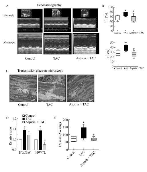
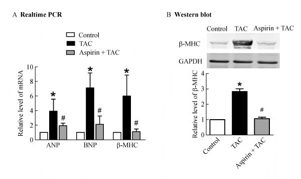
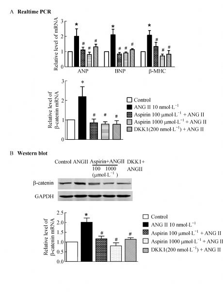
Abstract Ventricular hypertrophy is a powerful and independent predictor of cardiovascular morbid events. The vascular properties of low-dose acetyl salicylic acid (aspirin) provide cardiovascular benefits through the irreversible inhibition of platelet cyclooxygenase 1; however, the possible anti-hypertrophic properties and potential mechanism of aspirin have not been investigated in detail. In this study, healthy wild-type male mice were randomly divided into three groups and subjected to transverse aortic constriction (TAC) or sham operation. The TAC-operated mice were treated with the human equivalent of low-dose aspirin (10 mg·kg−1·d−1); the remaining mice received an equal amount of phosphate buffered saline with 0.65% ethanol, which was used as a vehicle. A cardiomyocyte hypertrophy model induced by angiotensin II (10 nmol·L−1) was treated with the human equivalent of low (10 or 100 µmol·L−1) and high (1000 µmol·L−1) aspirin concentrations in plasma. Changes in the cardiac structure and function were assessed through echocardiography and transmission electron microscopy. Gene expression was determined through RT-PCR and western blot analysis. Results indicated that aspirin treatment abrogated the increased thickness of the left ventricular anterior and posterior walls, the swelling of mitochondria, and the increased surface area in in vivo and in vitro hypertrophy models. Aspirin also normalized the upregulated hypertrophic biomarkers, β-myosin heavy chain (β-MHC), atrial natriuretic peptide (ANP), and b-type natriuretic peptide (BNP). Aspirin efficiently reversed the upregulation of β-catenin and P-Akt expression and the TAC- or ANG II-induced downregulation of GSK-3β. Therefore, low-dose aspirin possesses significant anti-hypertrophic properties at clinically relevant concentrations for anti-thrombotic therapy. The downregulation of β-catenin and Akt may be the underlying signaling mechanism of the effects of aspirin.
|
| Keywords
aspirin
Akt
cardiac hypertrophy
GSK-3β
Wnt/β-catenin
|
|
Corresponding Author(s):
Hongli Shan,Baofeng Yang
|
|
Just Accepted Date: 28 October 2015
Online First Date: 17 November 2015
Issue Date: 26 November 2015
|
|
| 1 |
Yusuf S, McKee M. Documenting the global burden of cardiovascular disease: a major achievement but still a work in progress. Circulation 2014; 129(14): 1459–1462
https://doi.org/10.1161/CIRCULATIONAHA.114.008302
pmid: 24573353
|
| 2 |
Gjesdal O, Bluemke DA, Lima JA. Cardiac remodeling at the population level—risk factors, screening, and outcomes. Nat Rev Cardiol 2011; 8(12): 673–685
https://doi.org/10.1038/nrcardio.2011.154
pmid: 22027657
|
| 3 |
Lavie CJ, Patel DA, Milani RV, Ventura HO, Shah S, Gilliland Y. Impact of echocardiographic left ventricular geometry on clinical prognosis. Prog Cardiovasc Dis 2014; 57(1): 3–9
https://doi.org/10.1016/j.pcad.2014.05.003
pmid: 25081397
|
| 4 |
Antithrombotic Trialists (ATT)’ Collaboration, Baigent C, Blackwell L, Collins R, Emberson J, Godwin J, Peto R, Buring J, Hennekens C, Kearney P, Meade T, Patrono C, Roncaglioni MC, Zanchetti A. Aspirin in the primary and secondary prevention of vascular disease: collaborative meta-analysis of individual participant data from randomised trials. Lancet 2009; 373(9678): 1849–1860
https://doi.org/10.1016/S0140-6736(09)60503-1
pmid: 19482214
|
| 5 |
Gaglia MA Jr, Clavijo L. Cardiovascular pharmacology core reviews: aspirin. J Cardiovasc Pharmacol Ther 2013; 18(6): 505–513
https://doi.org/10.1177/1074248413503043
pmid: 24057863
|
| 6 |
Eikelboom J, Hirsh J, Spencer F, Baglin T, Weitz J. Antiplatelet drugs: antithrombotic therapy and prevention of thrombosis. 9th ed. American College of Chest Physicians Evidence-Based Clinical Practice Guidelines. Chest 2012; 141(Suppl 2): e89S–e119S
|
| 7 |
Patrono C. Low-dose aspirin in primary prevention: cardioprotection, chemoprevention, both, or neither? Eur Heart J 2013; 34(44): 3403–3411
https://doi.org/10.1093/eurheartj/eht058
pmid: 23771843
|
| 8 |
Levy D, Garrison RJ, Savage DD, Kannel WB, Castelli WP. Prognostic implications of echocardiographically determined left ventricular mass in the Framingham Heart Study. N Engl J Med 1990; 322(22): 1561–1566
https://doi.org/10.1056/NEJM199005313222203
pmid: 2139921
|
| 9 |
Dovizio M, Tacconelli S, Sostres C, Ricciotti E, Patrignani P. Mechanistic and pharmacological issues of aspirin as an anticancer agent. Pharmaceuticals (Basel) 2012; 5(12): 1346–1371
https://doi.org/10.3390/ph5121346
pmid: 24281340
|
| 10 |
Yang Z, Guo L, Liu D, Sun L, Chen H, Deng Q, Liu Y, Yu M, Ma Y, Guo N, Shi M. Acquisition of resistance to trastuzumab in gastric cancer cells is associated with activation of IL-6/STAT3/Jagged-1/Notch positive feedback loop. Oncotarget 2015; 6(7): 5072–5087
pmid: 25669984
|
| 11 |
Farag M. Can aspirin and cancer prevention be ageless companions? J Clin Diagn Res 2015; 9(1): XE01–XE03
pmid: 25738074
|
| 12 |
Bergmann MW. WNT signaling in adult cardiac hypertrophy and remodeling: lessons learned from cardiac development. Circ Res 2010; 107(10): 1198–1208
https://doi.org/10.1161/CIRCRESAHA.110.223768
pmid: 21071717
|
| 13 |
Lu Z, Hunter T. Wnt-independent β-catenin transactivation in tumor development. Cell Cycle 2004; 3(5): 571–573
https://doi.org/10.4161/cc.3.5.885
pmid: 15107603
|
| 14 |
Paikin JS, Eikelboom JW. Aspirin. Circulation 2012; 125(10): e439–e442
https://doi.org/10.1161/CIRCULATIONAHA.111.046243
pmid: 22412097
|
| 15 |
Mehta SR, Tanguay JF, Eikelboom JW, Jolly SS, Joyner CD, Granger CB, Faxon DP, Rupprecht HJ, Budaj A, Avezum A, Widimsky P, Steg PG, Bassand JP, Montalescot G, Macaya C, Di Pasquale G, Niemela K, Ajani AE, White HD, Chrolavicius S, Gao P, Fox KA, Yusuf S; CURRENT-OASIS 7 trial investigators. Double-dose versus standard-dose clopidogrel and high-dose versus low-dose aspirin in individuals undergoing percutaneous coronary intervention for acute coronary syndromes (CURRENT-OASIS 7): a randomised factorial trial. Lancet 2010; 376(9748): 1233–1243
https://doi.org/10.1016/S0140-6736(10)61088-4
pmid: 20817281
|
| 16 |
Reagan-Shaw S, Nihal M, Ahmad N. Dose translation from animal to human studies revisited. FASEB J 2008; 22(3): 659–661
https://doi.org/10.1096/fj.07-9574LSF
pmid: 17942826
|
| 17 |
Houser SR, Margulies KB, Murphy AM, Spinale FG, Francis GS, Prabhu SD, Rockman HA, Kass DA, Molkentin JD, Sussman MA, Koch WJ; American Heart Association Council on Basic Cardiovascular Sciences, Council on Clinical Cardiology, and Council on Functional Genomics and Translational Biology. Animal models of heart failure: a scientific statement from the American Heart Association. Circ Res 2012; 111(1): 131–150
https://doi.org/10.1161/RES.0b013e3182582523
pmid: 22595296
|
| 18 |
Lichte P, Pfeifer R, Kobbe P, Tohidnezhad M, Pufe T, Almahmoud K, Hildebrand F, Pape HC. Inhalative IL-10 treatment after bilateral femoral fractures affect pulmonary inflammation in mice. Ann Anat 2015; 200(5): 73–78
https://doi.org/10.1016/j.aanat.2015.02.005
pmid: 25801583
|
| 19 |
Li C, Li X, Gao X, Zhang R, Zhang Y, Liang H, Xu C, Du W, Zhang Y, Liu X, Ma N, Xu Z, Wang L, Chen X, Lu Y, Ju J, Yang B, Shan H. MicroRNA-328 as a regulator of cardiac hypertrophy. Int J Cardiol 2014; 173(2): 268–276
https://doi.org/10.1016/j.ijcard.2014.02.035
pmid: 24631114
|
| 20 |
Sun B, Huo R, Sheng Y, Li Y, Xie X, Chen C, Liu HB, Li N, Li CB, Guo WT, Zhu JX, Yang BF, Dong DL. Bone morphogenetic protein-4 mediates cardiac hypertrophy, apoptosis, and fibrosis in experimentally pathological cardiac hypertrophy. Hypertension 2013; 61(2): 352–360
https://doi.org/10.1161/HYPERTENSIONAHA.111.00562
pmid: 23248151
|
| 21 |
Rothstein S, Simkins T, Nuñez JL. Response to neonatal anesthesia: effect of sex on anatomical and behavioral outcome. Neuroscience 2008; 152(4): 959–969
https://doi.org/10.1016/j.neuroscience.2008.01.027
pmid: 18329814
|
| 22 |
Bochner F, Somogyi AA, Wilson KM. Bioinequivalence of four 100 mg oral aspirin formulations in healthy volunteers. Clin Pharmacokinet 1991; 21(5): 394–399
https://doi.org/10.2165/00003088-199121050-00006
pmid: 1773551
|
| 23 |
Rosenkranz B, Frölich JC. Plasma concentrations and anti-platelet effects after low dose acetylsalicylic acid. Prostaglandins Leukot Med 1985; 19(3): 289–300
https://doi.org/10.1016/0262-1746(85)90142-8
pmid: 3864170
|
| 24 |
Hermans H, Swinnen M, Pokreisz P, Caluwe E, Dymarkowski S, Herregods MC, Janssens S, Herijgers P. Murine pressure overload models: a 30-MHz look brings a whole new “sound” into data interpretation. J Appl Physiol (Bethesda, MD: 1985) 2014; 117(5): 563–571
|
| 25 |
Bode-Böger SM, Böger RH, Schubert M, Frölich JC. Effects of very low dose and enteric-coated acetylsalicylic acid on prostacyclin and thromboxane formation and on bleeding time in healthy subjects. Eur J Clin Pharmacol 1998; 54(9–10): 707–714
https://doi.org/10.1007/s002280050539
pmid: 9923572
|
| 26 |
Zheng Q, Chen P, Xu Z, Li F, Yi XP. Expression and redistribution of β-catenin in the cardiac myocytes of left ventricle of spontaneously hypertensive rat. J Mol Histol 2013; 44(5): 565–573
https://doi.org/10.1007/s10735-013-9507-6
pmid: 23591738
|
| 27 |
Shanmugam P, Valente AJ, Prabhu SD, Venkatesan B, Yoshida T, Delafontaine P, Chandrasekar B. Angiotensin-II type 1 receptor and NOX2 mediate TCF/LEF and CREB dependent WISP1 induction and cardiomyocyte hypertrophy. J Mol Cell Cardiol 2011; 50(6): 928–938
https://doi.org/10.1016/j.yjmcc.2011.02.012
pmid: 21376054
|
| 28 |
Fang D, Hawke D, Zheng Y, Xia Y, Meisenhelder J, Nika H, Mills GB, Kobayashi R, Hunter T, Lu Z. Phosphorylation of β-catenin by AKT promotes β-catenin transcriptional activity. J Biol Chem 2007; 282(15): 11221–11229
https://doi.org/10.1074/jbc.M611871200
pmid: 17287208
|
| 29 |
Lincová E, Hampl A, Pernicová Z, Starsíchová A, Krcmár P, Machala M, Kozubík A, Soucek K. Multiple defects in negative regulation of the PKB/Akt pathway sensitise human cancer cells to the antiproliferative effect of non-steroidal anti-inflammatory drugs. Biochem Pharmacol 2009; 78(6): 561–572
https://doi.org/10.1016/j.bcp.2009.05.001
pmid: 19433066
|
| 30 |
Ferreira Filho C, Abreu LC, Valenti VE, Ferreira M, Meneghini A, Silveira JA, Riera AR, Colombari E, Murad N, Santos-Silva PR, Silva LJ, Vanderlei LC, Carvalho TD, Ferreira C. Anti-hypertensive drugs have different effects on ventricular hypertrophy regression. Clinics (Sao Paulo) 2010; 65(7): 723–728
https://doi.org/10.1590/S1807-59322010000700012
pmid: 20668631
|
| 31 |
Bisping E, Wakula P, Poteser M, Heinzel FR. Targeting cardiac hypertrophy: toward a causal heart failure therapy. J Cardiovasc Pharmacol 2014; 64(4): 293–305
https://doi.org/10.1097/FJC.0000000000000126
pmid: 25286359
|
| 32 |
Levy D. Left ventricular hypertrophy. Epidemiological insights from the Framingham Heart Study. Drugs 1988; 35(Suppl 5): 1–5
https://doi.org/10.2165/00003495-198800355-00002
pmid: 2975214
|
| 33 |
Nagelschmitz J, Blunck M, Kraetzschmar J, Ludwig M, Wensing G, Hohlfeld T. Pharmacokinetics and pharmacodynamics of acetylsalicylic acid after intravenous and oral administration to healthy volunteers. Clin Pharmacol 2014; 6: 51–59
https://doi.org/ 10.2147/CPAA.S47895
pmid: 24672263
|
| 34 |
Wu R, Yin D, Sadekova N, Deschepper CF, de Champlain J, Girouard H. Protective effects of aspirin from cardiac hypertrophy and oxidative stress in cardiomyopathic hamsters. Oxid Med Cell Longev 2012; 2012: 761710
https://doi.org/10.1155/2012/761710
pmid: 22829962
|
| 35 |
Wu R, Laplante MA, De Champlain J. Prevention of angiotensin II-induced hypertension, cardiovascular hypertrophy and oxidative stress by acetylsalicylic acid in rats. J Hypertens 2004; 22(4): 793–801
https://doi.org/10.1097/00004872-200404000-00023
pmid: 15126922
|
| 36 |
Halvorsen S, Andreotti F, ten Berg JM, Cattaneo M, Coccheri S, Marchioli R, Morais J, Verheugt FW, De Caterina R. Aspirin therapy in primary cardiovascular disease prevention: a position paper of the European Society of Cardiology working group on thrombosis. J Am Coll Cardiol 2014; 64(3): 319–327
https://doi.org/10.1016/j.jacc.2014.03.049
pmid: 25034070
|
| 37 |
Bae SK, Seo KA, Jung EJ, Kim HS, Yeo CW, Shon JH, Park KM, Liu KH, Shin JG. Determination of acetylsalicylic acid and its major metabolite, salicylic acid, in human plasma using liquid chromatography-tandem mass spectrometry: application to pharmacokinetic study of Astrix in Korean healthy volunteers. Biomed Chromatogr 2008; 22(6): 590–595
https://doi.org/10.1002/bmc.973
pmid: 18254152
|
| 38 |
Clarke RJ, Mayo G, Price P, FitzGerald GA. Suppression of thromboxane A2 but not of systemic prostacyclin by controlled-release aspirin. N Engl J Med 1991; 325(16): 1137–1141
https://doi.org/10.1056/NEJM199110173251605
pmid: 1891022
|
| 39 |
Rubak P, Hardlei TF, Würtz M, Kristensen SD, Hvas AM. Low-dose acetylsalicylic acid therapy monitored with ultra high performance liquid chromatography. Clin Biochem 2013; 46(12): 988–992
https://doi.org/10.1016/j.clinbiochem.2013.04.007
pmid: 23608356
|
| 40 |
Sklepkiewicz P, Shiomi T, Kaur R, Sun J, Kwon S, Mercer B, Bodine P, Schermuly RT, George I, Schulze PC, D'Armiento JM. Loss of secreted frizzled-related protein-1 leads to deterioration of cardiac function in mice and plays a role in human cardiomyopathy. Circ Heart Fail 2015; 8(2): 362–372
https://doi.org/10.1161/CIRCHEARTFAILURE.114.001274
pmid: 25669938
|
| 41 |
Fujishima Y, Maeda N, Matsuda K, Komura N, Hirata A, Mori T, Sekimoto R, Tsushima Y, Nishizawa H, Funahashi T, Shimomura I. Effect of adiponectin on cardiac β-catenin signaling pathway under angiotensin II infusion. Biochem Biophys Res Commun 2014; 444(2): 224–229
https://doi.org/10.1016/j.bbrc.2014.01.043
pmid: 24462873
|
| 42 |
Aoyagi T, Matsui T. Phosphoinositide-3 kinase signaling in cardiac hypertrophy and heart failure. Curr Pharm Des 2011; 17(18): 1818–1824
https://doi.org/10.2174/138161211796390976
pmid: 21631421
|
| 43 |
Ellison GM, Waring CD, Vicinanza C, Torella D. Physiological cardiac remodelling in response to endurance exercise training: cellular and molecular mechanisms. Heart 2012; 98(1): 5–10
https://doi.org/10.1136/heartjnl-2011-300639
pmid: 21880653
|
| 44 |
Yoshida T, Friehs I, Mummidi S, del Nido PJ, Addulnour-Nakhoul S, Delafontaine P, Valente AJ, Chandrasekar B. Pressure overload induces IL-18 and IL-18R expression, but markedly suppresses IL-18BP expression in a rabbit model. IL-18 potentiates TNF-α-induced cardiomyocyte death. J Mol Cell Cardiol 2014; 75(10): 141–151
https://doi.org/10.1016/j.yjmcc.2014.07.007
pmid: 25108227
|
| 45 |
Ishida H, Kogaki S, Narita J, Ichimori H, Nawa N, Okada Y, Takahashi K, Ozono K. LEOPARD-type SHP2 mutant Gln510Glu attenuates cardiomyocyte differentiation and promotes cardiac hypertrophy via dysregulation of Akt/GSK-3β/β-catenin signaling. Am J Physiol Heart Circ Physiol 2011; 301(4): H1531–H1539
https://doi.org/10.1152/ajpheart.00216.2011
pmid: 21803945
|
| 46 |
Askevold ET, Aukrust P, Nymo SH, Lunde IG, Kaasbøll OJ, Aakhus S, Florholmen G, Ohm IK, Strand ME, Attramadal H, Fiane A, Dahl CP, Finsen AV, Vinge LE, Christensen G, Yndestad A, Gullestad L, Latini R, Masson S, Tavazzi L, Ueland T; GISSI-HF Investigators, Ueland T. The cardiokine secreted Frizzled-related protein 3, a modulator of Wnt signalling, in clinical and experimental heart failure. J Intern Med 2014; 275(6): 621–630
https://doi.org/10.1111/joim.12175
pmid: 24330105
|
|
Viewed |
|
|
|
Full text
|
|
|
|
|
Abstract
|
|
|
|
|
Cited |
|
|
|
|
| |
Shared |
|
|
|
|
| |
Discussed |
|
|
|
|