| LETTER TO FRONTIERS OF MEDICINE |
|
|
|
Determining “abnormal” levator hiatus distensibility using three-dimensional transperineal ultrasound in Chinese women |
Chaoran Dou, Qin Li, Tao Ying( ), Yulin Yan, Xia Wang, Bing Hu ), Yulin Yan, Xia Wang, Bing Hu |
| Department of Ultrasound in Medicine, Shanghai Institute of Ultrasound in Medicine, Shanghai Jiao Tong University Affiliated Sixth People’s Hospital, Shanghai 200233, China |
|
|
|
|
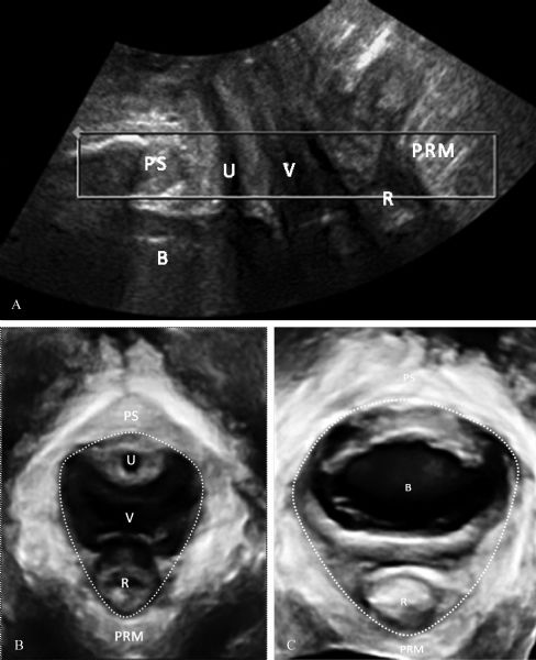
Abstract The dimension of the levator hiatus is a possible predictor of pelvic organ prolapse (POP). This retrospective study investigated 360 women who went to urogynecological clinic for pelvic floor discomfort. Levator hiatus dimensions were obtained by three-dimensional transperineal ultrasound and results were compared between women with and without significantly objective prolapse (International Continence Society POP quantification, grade 2 or higher). Receiver operating characteristic (ROC) curve analyses were performed to determine valid screening index for detecting abnormal levator hiatus distensibility. Women with significantly objective prolapse had significantly higher levator hiatus dimensions than those without (all P <0.001). ROC curve analyses confirmed that hiatal area (HA) of 19.5 cm2 during Valsalva maneuver can be used as single-screening index for abnormal levator hiatus distensibility with sensitivity of 0.80 and specificity of 0.70. In this study, we used a two-step method and achieved higher sensibility (0.80 vs. 0.87) without reducing specificity (0.70 vs. 0.71) compared with a single-screening index method. As a result, we suggest that HA≥19.5 cm2 during Valsalva maneuver is an indicator of abnormal levator hiatus distensibility in Chinese women and that the two-step method has higher sensitivity in detecting abnormal distensibility.
|
| Keywords
three-dimensional transperineal ultrasound
levator hiatus
levator ani muscle
pelvic organ prolapse
|
|
Corresponding Author(s):
Tao Ying
|
|
Just Accepted Date: 25 September 2017
Online First Date: 22 November 2017
Issue Date: 29 September 2018
|
|
| 1 |
Olsen AL, Smith VJ, Bergstrom JO, Colling JC, Clark AL. Epidemiology of surgically managed pelvic organ prolapse and urinary incontinence. Obstet Gynecol 1997; 89(4): 501–506
https://doi.org/10.1016/S0029-7844(97)00058-6
pmid: 9083302
|
| 2 |
DeLancey JO. The hidden epidemic of pelvic floor dysfunction: achievable goals for improved prevention and treatment. Am J Obstet Gynecol 2005; 192(5): 1488–1495
https://doi.org/10.1016/j.ajog.2005.02.028
pmid: 15902147
|
| 3 |
Mant J, Painter R, Vessey M. Epidemiology of genital prolapse: observations from the Oxford Family Planning Association Study. Br J Obstet Gynaecol 1997; 104(5): 579–585
https://doi.org/10.1111/j.1471-0528.1997.tb11536.x
pmid: 9166201
|
| 4 |
Walker GJ, Gunasekera P. Pelvic organ prolapse and incontinence in developing countries: review of prevalence and risk factors. Int Urogynecol J Pelvic Floor Dysfunct 2011; 22(2): 127–135
https://doi.org/10.1007/s00192-010-1215-0
pmid: 20617303
|
| 5 |
Kolberg Tennfjord M, Hilde G, Staer-Jensen J, Siafarikas F, Engh ME, Bø K. Effect of postpartum pelvic floor muscle training on vaginal symptoms and sexual dysfunction-secondary analysis of a randomised trial. BJOG 2016; 123(4): 634–642
https://doi.org/10.1111/1471-0528.13823
pmid: 26691895
|
| 6 |
Wiegersma M, Panman CM, Kollen BJ, Berger MY, Lisman-Van Leeuwen Y, Dekker JH. Effect of pelvic floor muscle training compared with watchful waiting in older women with symptomatic mild pelvic organ prolapse: randomised controlled trial in primary care. BMJ 2014; 349(dec22 1): g7378
https://doi.org/10.1136/bmj.g7378
pmid: 25533442
|
| 7 |
Bump RC, Mattiasson A, Bø K, Brubaker LP, DeLancey JO, Klarskov P, Shull BL, Smith AR. The standardization of terminology of female pelvic organ prolapse and pelvic floor dysfunction. Am J Obstet Gynecol 1996; 175(1): 10–17
https://doi.org/10.1016/S0002-9378(96)70243-0
pmid: 8694033
|
| 8 |
Notten KJ, Weemhoff M, Kluivers KB, Schweitzer KJ, Mulder F, Stoker J, Beets-Tan RG, Futterer JJ, Vliegen RF, Evers JL, Link G, Bergmans MG, Kampschöer PH, Gondrie ET, van Gestel I, van Dooren I, Dirksen C, Smits LJ, Bossuyt PM, Roovers JP. Protocol for translabial 3D-ultrasonography for diagnosing levator defects (TRUDIL): a multicentre cohort study for estimating the diagnostic accuracy of translabial 3D-ultrasonography of the pelvic floor as compared to MR imaging. BMC Womens Health 2011; 11(1): 23
https://doi.org/10.1186/1472-6874-11-23
pmid: 21639876
|
| 9 |
Berger MB, Morgan DM, DeLancey JO. Levator ani defect scores and pelvic organ prolapse: is there a threshold effect? Int Urogynecol J Pelvic Floor Dysfunct 2014; 25(10): 1375–1379
https://doi.org/10.1007/s00192-014-2388-8
pmid: 24788366
|
| 10 |
DeLancey JO, Sørensen HC, Lewicky-Gaupp C, Smith TM. Comparison of the puborectal muscle on MRI in women with POP and levator ani defects with those with normal support and no defect. Int Urogynecol J Pelvic Floor Dysfunct 2012; 23(1): 73–77
https://doi.org/10.1007/s00192-011-1527-8
pmid: 21822711
|
| 11 |
Athanasiou S, Chaliha C, Toozs-Hobson P, Salvatore S, Khullar V, Cardozo L. Direct imaging of the pelvic floor muscles using two-dimensional ultrasound: a comparison of women with urogenital prolapse versus controls. BJOG 2007; 114(7): 882–888
https://doi.org/10.1111/j.1471-0528.2007.01322.x
pmid: 17501961
|
| 12 |
Delancey JO, Hurd WW. Size of the urogenital hiatus in the levator ani muscles in normal women and women with pelvic organ prolapse. Obstet Gynecol 1998; 91(3): 364–368
https://doi.org/10.1016/S0029-7844(97)00682-0
pmid: 9491861
|
| 13 |
Andrew BP, Shek KL, Chantarasorn V, Dietz HP. Enlargement of the levator hiatus in female pelvic organ prolapse: cause or effect? Aust N Z J Obstet Gynaecol 2013; 53(1): 74–78
https://doi.org/10.1111/ajo.12026
pmid: 23278472
|
| 14 |
Dietz HP, Shek C, De Leon J, Steensma AB. Ballooning of the levator hiatus. Ultrasound Obstet Gynecol 2008; 31(6): 676–680
https://doi.org/10.1002/uog.5355
pmid: 18470963
|
| 15 |
Cheung RY, Shek KL, Chan SS, Chung TK, Dietz HP. Pelvic floor muscle biometry and pelvic organ mobility in East Asian and Caucasian nulliparae. Ultrasound Obstet Gynecol 2015; 45(5): 599–604
https://doi.org/10.1002/uog.14656
pmid: 25175901
|
| 16 |
van Veelen GA, Schweitzer KJ, van der Vaart CH. Ultrasound imaging of the pelvic floor: changes in anatomy during and after first pregnancy. Ultrasound Obstet Gynecol 2014; 44(4): 476–480
https://doi.org/10.1002/uog.13301
pmid: 24436146
|
| 17 |
Chan SS, Cheung RY, Yiu KW, Lee LL, Chung TK. Pelvic floor biometry in Chinese primiparous women 1 year after delivery: a prospective observational study. Ultrasound Obstet Gynecol 2014; 43(4): 466–474
https://doi.org/10.1002/uog.13249
pmid: 24254134
|
| 18 |
Dietz HP, Shek C, Clarke B. Biometry of the pubovisceral muscle and levator hiatus by three-dimensional pelvic floor ultrasound. Ultrasound Obstet Gynecol 2005; 25(6): 580–585
https://doi.org/10.1002/uog.1899
pmid: 15883982
|
| 19 |
Kruger JA, Heap SW, Murphy BA, Dietz HP. How best to measure the levator hiatus: evidence for the non-Euclidean nature of the ‘plane of minimal dimensions’. Ultrasound Obstet Gynecol 2010; 36(6): 755–758
https://doi.org/10.1002/uog.7750
pmid: 20645397
|
| 20 |
Balmforth J, Toozs-Hobson P, Cardozo L. Ask not what childbirth can do to your pelvic floor but what your pelvic floor can do in childbirth. Neurourol Urodyn 2003; 22(5): 540–542
|
| 21 |
Lanzarone V, Dietz HP. Three-dimensional ultrasound imaging of the levator hiatus in late pregnancy and associations with delivery outcomes. Aust N Z J Obstet Gynaecol 2007; 47(3): 176–180
https://doi.org/10.1111/j.1479-828X.2007.00714.x
pmid: 17550482
|
| 22 |
Dietz HP, Wong V, Shek KL. A simplified method for determining hiatal biometry. Aust N Z J Obstet Gynaecol 2011; 51(6): 540–543
https://doi.org/10.1111/j.1479-828X.2011.01352.x
pmid: 21951068
|
| 23 |
Pineda M, Shek K, Wong V, Dietz HP. Can hiatal ballooning be determined by two-dimensional translabial ultrasound? Aust N Z J Obstet Gynaecol 2013; 53(5): 489–493
pmid: 23909797
|
| 24 |
Smith FJ, Holman CD, Moorin RE, Tsokos N. Lifetime risk of undergoing surgery for pelvic organ prolapse. Obstet Gynecol 2010; 116(5): 1096–1100
https://doi.org/10.1097/AOG.0b013e3181f73729
pmid: 20966694
|
| 25 |
Chen L, Ashton-Miller JA, Hsu Y, DeLancey JO. Interaction among apical support, levator ani impairment, and anterior vaginal wall prolapse. Obstet Gynecol 2006; 108(2): 324–332
https://doi.org/10.1097/01.AOG.0000227786.69257.a8
pmid: 16880302
|
| 26 |
Ghetti C, Gregory WT, Edwards SR, Otto LN, Clark AL. Severity of pelvic organ prolapse is associated with measurements of genital hiatus. Int Urogynecol J Pelvic Floor Dysfunct 2005; 16(6): 432–436
https://doi.org/10.1007/s00192-004-1274-1
pmid: 15660182
|
| 27 |
Haylen BT, Maher CF, Barber MD, Camargo S, Dandolu V, Digesu A, Goldman HB, Huser M, Milani AL, Moran PA, Schaer GN, Withagen MI. An International Urogynecological Association (IUGA)/International Continence Society (ICS) joint report on the terminology for female pelvic organ prolapse (POP). Int Urogynecol J Pelvic Floor Dysfunct 2016; 27(4): 655–684
https://doi.org/10.1007/s00192-016-3003-y
pmid: 26984443
|
| 28 |
Dietz HP. Quantification of major morphological abnormalities of the levator ani. Ultrasound Obstet Gynecol 2007; 29(3): 329–334
https://doi.org/10.1002/uog.3951
pmid: 17323308
|
| 29 |
Majida M, Brækken IH, Bø K, Engh ME. Levator hiatus dimensions and pelvic floor function in women with and without major defects of the pubovisceral muscle. Int Urogynecol J Pelvic Floor Dysfunct 2012; 23(6): 707–714
https://doi.org/10.1007/s00192-011-1652-4
pmid: 22246577
|
| 30 |
Braekken IH, Majida M, Engh ME, Bø K. Morphological changes after pelvic floor muscle training measured by 3-dimensional ultrasonography: a randomized controlled trial. Obstet Gynecol 2010; 115(2 Pt 1): 317–324
https://doi.org/10.1097/AOG.0b013e3181cbd35f
pmid: 20093905
|
|
Viewed |
|
|
|
Full text
|
|
|
|
|
Abstract
|
|
|
|
|
Cited |
|
|
|
|
| |
Shared |
|
|
|
|
| |
Discussed |
|
|
|
|