|
|
|
Effect of surgical factors on the augmentation of cement- injectable cannulated pedicle screw fixation by a novel calcium phosphate-based nanocomposite |
Haolin Sun1,4, Chun Liu2, Shunlun Chen1, Yanjie Bai3, Huilin Yang2,4, Chunde Li1( ), Lei Yang2,4,5() ), Lei Yang2,4,5() |
1. Department of Orthopedics, Peking University First Hospital, Beijing 100034, China
2. Orthopedic Institute, Department of Orthopedics, First Affiliated Hospital, Soochow University, Suzhou 215006, China
3. School of Public Health, Medical College, Soochow University, Suzhou 215100, China
4. International Research Center for Translational Orthopedics, Suzhou 215006, China
5. School of Materials Science and Engineering, Hebei University of Technology, Tianjin 300130, China |
|
|
|
|
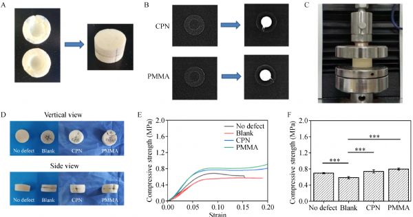
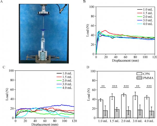
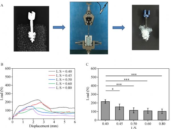
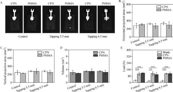
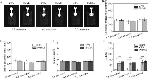
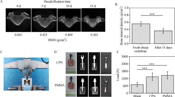
Abstract Bone cement-augmented pedicle screw system demonstrates great efficacy in spinal disease treatments. However, the intrinsic drawbacks associated with clinically used polymethylmethacrylate (PMMA) cement demands for new bone cement formulations. On the basis of our previous studies, a novel injectable and biodegradable calcium phosphate-based nanocomposite (CPN) for the augmentation of pedicle screw fixation was systematically evaluated for its surgical feasibility and biomechanical performance by simulated and animal osteoporotic bone models, and the results were compared with those of clinical PMMA cement. ASTM-standard solid foam and open-cell foam models and decalcified sheep vertebra models were employed to evaluate the augmentation effects of CPN on bone tissue and on the cement-injected cannulated pedicle screws (CICPs) placed in osteoporotic bone. Surgical factors in CICPs application, such as injection force, tapping technique, screw diameter, and pedicle screw loosening scenarios, were studied in comparison with those in PMMA. When directly injected to the solid foam model, CPN revealed an identical augmentation effect to that of PMMA, as shown by the similar compressive strengths (0.73±0.04 MPa for CPN group vs. 0.79±0.02 MPa for PMMA group). The average injection force of CPN at approximately 40–50 N was higher than that of PMMA at approximately 20 N. Although both values are acceptable to surgeons, CPN revealed a more consistent injection force pattern than did PMMA. The dispersing and anti-pullout ability of CPN were not affected by the surgical factors of tapping technique and screw diameter. The axial pullout strength of CPN evaluated by the decalcified sheep vertebra model revealed a similar augmentation level as that of PMMA (1351.6±324.2 N for CPN vs. 1459.7±304.4 N for PMMA). The promising results of CPN clearly suggest its potential for replacing PMMA in CICPs augmentation application and the benefits of further study and development for clinical uses.
|
| Keywords
bone cement
pedicle screw
degenerative spinal diseases
calcium phosphate
injectable
|
|
Corresponding Author(s):
Chunde Li,Lei Yang
|
|
Just Accepted Date: 01 August 2019
Online First Date: 23 September 2019
Issue Date: 14 October 2019
|
|
| 1 |
PW Hitchon, MD Brenton, JK Coppes, AM From, JC Torner. Factors affecting the pullout strength of self-drilling and self-tapping anterior cervical screws. Spine 2003; 28(1): 9–13
https://doi.org/10.1097/00007632-200301010-00004
pmid: 12544947
|
| 2 |
DM Lai, YT Shih, YH Chen, A Chien, JL Wang. Effect of pedicle screw diameter on screw fixation efficacy in human osteoporotic thoracic vertebrae. J Biomech 2018; 70: 196–203
https://doi.org/10.1016/j.jbiomech.2017.10.009
pmid: 29126607
|
| 3 |
L Weiser, G Huber, K Sellenschloh, L Viezens, K Püschel, MM Morlock, W Lehmann. Time to augment?! Impact of cement augmentation on pedicle screw fixation strength depending on bone mineral density. Eur Spine J 2018; 27(8): 1964–1971
https://doi.org/10.1007/s00586-018-5660-7
pmid: 29948322
|
| 4 |
M Pishnamaz, H Lange, C Herren, HS Na, P Lichte, F Hildebrand, HC Pape, P Kobbe. The quantity of bone cement influences the anchorage of augmented pedicle screws in the osteoporotic spine: a biomechanical human cadaveric study. Clin Biomech (Bristol, Avon) 2018; 52: 14–19
https://doi.org/10.1016/j.clinbiomech.2017.12.012
pmid: 29309925
|
| 5 |
PE Paré, JL Chappuis, R Rampersaud, AO Agarwala, JH Perra, S Erkan, C Wu. Biomechanical evaluation of a novel fenestrated pedicle screw augmented with bone cement in osteoporotic spines. Spine 2011; 36(18): E1210–E1214
https://doi.org/10.1097/BRS.0b013e318205e3af
pmid: 21325986
|
| 6 |
TJ Choma, WF Frevert, WL Carson, NP Waters, FM Pfeiffer. Biomechanical analysis of pedicle screws in osteoporotic bone with bioactive cement augmentation using simulated in vivo multicomponent loading. Spine 2011; 36(6): 454–462
https://doi.org/10.1097/BRS.0b013e3181d449ec
pmid: 20881517
|
| 7 |
I Janssen, YM Ryang, J Gempt, S Bette, J Gerhardt, JS Kirschke, B Meyer. Risk of cement leakage and pulmonary embolism by bone cement-augmented pedicle screw fixation of the thoracolumbar spine. Spine J 2017; 17(6): 837–844
https://doi.org/10.1016/j.spinee.2017.01.009
pmid: 28108403
|
| 8 |
Csaba Padányi, F Misik, Z Papp, D Vitanovics, A Balogh, R Veres, L Lipóth, P Banczerowski. Treatment of osteoporotic vertebral compression fractures with PMMA-augmented pedicle screw fixation. Ideggyogy Sz 2015; 68(1-2): 52–58 (in Hungarian)
pmid: 25842917
|
| 9 |
BJ Moon, BY Cho, EY Choi, HY Zhang. Polymethylmethacrylate-augmented screw fixation for stabilization of the osteoporotic spine: a three-year follow-up of 37 patients. J Korean Neurosurg Soc 2009; 46(4): 305–311
https://doi.org/10.3340/jkns.2009.46.4.305
pmid: 19893717
|
| 10 |
D Liu, J Sheng, HH Wu, X Kang, QY Xie, Y Luo, JJ Zhou, W Zheng. Biomechanical study of injectable hollow pedicle screws for PMMA augmentation in severely osteoporotic lumbar vertebrae: effect of PMMA distribution and volume on screw stability. J Neurosurg Spine 2018; 29(6): 639–646
https://doi.org/10.3171/2018.4.SPINE171225
pmid: 30192220
|
| 11 |
F Galbusera, D Volkheimer, S Reitmaier, N Berger-Roscher, A Kienle, HJ Wilke. Pedicle screw loosening: a clinically relevant complication? Eur Spine J 2015; 24(5): 1005–1016
https://doi.org/10.1007/s00586-015-3768-6
pmid: 25616349
|
| 12 |
SF María, T Claudia, P Victor Hugo, V Marcela, V Luis, F María Luisa, C Luis, GJ Valenzuela. Perinatal neuroendocrine regulation. Development of the circadian time-keeping system. J Biomech 2004; 186(2): 169–173
|
| 13 |
C Fukuda, K Goto, M Imamura, M Neo, T Nakamura. Bone bonding ability and handling properties of a titania-polymethylmethacrylate (PMMA) composite bioactive bone cement modified with a unique PMMA powder. Acta Biomater 2011; 7(10): 3595–3600
https://doi.org/10.1016/j.actbio.2011.06.006
pmid: 21704200
|
| 14 |
AR Piñera, C Duran, B Lopez, I Saez, E Correia, L Alvarez. Instrumented lumbar arthrodesis in elderly patients: prospective study using cannulated cemented pedicle screw instrumentation. Eur Spine J 2011; 20 (Suppl 3):408–414 PMID: 21850421 DOI: 10.1007/s00586-011-1907-2
|
| 15 |
SJ Breusch, KD Kühn. Bone cements based on polymethylmethacrylate. Orthopade 2003; 32(1): 41–50 (in German)
https://doi.org/10.1007/s00132-002-0411-0
pmid: 12557085
|
| 16 |
DE Mar, SJ Clary, DC Burton, TE McIff. Effect of spinous process tether tension for prophylactic treatment of proximal junctional kyphosis in adult spinal deformity surgery. Spine J 2017; 17(10): S190
https://doi.org/10.1016/j.spinee.2017.07.275
|
| 17 |
H Sun, C Liu, H Liu, Y Bai, Z Zhang, X Li, C Li, H Yang, L Yang. A novel injectable calcium phosphate-based nanocomposite for the augmentation of cannulated pedicle-screw fixation. Int J Nanomedicine 2017; 12: 3395–3406
https://doi.org/10.2147/IJN.S131962
pmid: 28490878
|
| 18 |
L Chun, L Huiling, S Haolin, Y Huilin, L Chunde, Y Lei. Development and optimization of biodegradable calcium phosphate-based nanocomposite for the application of spinal fixation. Ferroelectrics 2018; 527(1): 162–169
https://doi.org/10.1080/00150193.2018.1450566
|
| 19 |
H Liu, B Liu, C Gao, B Meng, H Yang, H Yu, L Yang. Injectable, biomechanically robust, biodegradable and osseointegrative bone cement for percutaneous kyphoplasty and vertebroplasty. Int Orthop 2018; 42(1): 125–132
https://doi.org/10.1007/s00264-017-3674-0
pmid: 29116357
|
| 20 |
HT Fan, RJ Zhang, CL Shen, FL Dong, Y Li, PW Song, C Gong, YJ Wang. The biomechanical properties of pedicle screw fixation combined with trajectory bone cement augmentation in osteoporotic vertebrae. Clin Spine Surg 2016; 29(2): 78–85
https://doi.org/10.1097/BSD.0b013e3182a14870
pmid: 26889991
|
| 21 |
SI Chen, RM Lin, CH Chang. Biomechanical investigation of pedicle screw-vertebrae complex: a finite element approach using bonded and contact interface conditions. Med Eng Phys 2003; 25(4): 275–282
https://doi.org/10.1016/S1350-4533(02)00219-9
pmid: 12649011
|
| 22 |
C Gao, H Liu, H Yang, L Yang. Fabrication and characterization of injectable calcium phosphate-based cements for kyphoplasty. Mater Technol 2015; 30(sup8): B256–B263
https://doi.org/10.1080/10667857.2015.1104828
|
| 23 |
LH Chen, CL Tai, DM Lee, PL Lai, YC Lee, CC Niu, WJ Chen. Pullout strength of pedicle screws with cement augmentation in severe osteoporosis: a comparative study between cannulated screws with cement injection and solid screws with cement pre-filling. BMC Musculoskelet Disord 2011; 12(1): 33
https://doi.org/10.1186/1471-2474-12-33
pmid: 21284883
|
| 24 |
AE Johnson, TS Keller. Mechanical properties of open-cell foam synthetic thoracic vertebrae. J Mater Sci Mater Med 2008; 19(3): 1317–1323
https://doi.org/10.1007/s10856-007-3158-7
pmid: 17882383
|
| 25 |
TS Keller, M Nathan. Height change caused by creep in intervertebral discs: a sagittal plane model. J Spinal Disord 1999; 12(4): 313–324
https://doi.org/10.1097/00002517-199908000-00008
pmid: 10451048
|
| 26 |
S Muthukrishnan, S Mitra, P Ranjith, S Gopalakrishnan, P Parthipan, B Al-Farhood. High heat resistant blends of poly (methyl methacrylate) and styrenic copolymers via post reactor modification. J Appl Polym Sci 2018; 135(18): 46220
https://doi.org/10.1002/app.46220
|
| 27 |
CC Hsu, CK Chao, JL Wang, SM Hou, YT Tsai, J Lin. Increase of pullout strength of spinal pedicle screws with conical core: biomechanical tests and finite element analyses. J Orthop Res 2005; 23(4): 788–794
https://doi.org/10.1016/j.orthres.2004.11.002
pmid: 16022991
|
| 28 |
FM Pfeiffer, DL Abernathie, DE Smith. A comparison of pullout strength for pedicle screws of different designs: a study using tapped and untapped pilot holes. Spine 2006; 31(23): E867–E870
https://doi.org/10.1097/01.brs.0000244658.35865.59
pmid: 17077722
|
| 29 |
JJ Carmouche, RW Molinari, T Gerlinger, J Devine, T Patience. Effects of pilot hole preparation technique on pedicle screw fixation in different regions of the osteoporotic thoracic and lumbar spine. J Neurosurg Spine 2005; 3(5): 364–370
https://doi.org/10.3171/spi.2005.3.5.0364
pmid: 16302630
|
| 30 |
S Fujibayashi, M Takemoto, M Neo, S Matsuda. Strategy for salvage pedicle screw placement: a technical note. Int J Spine Surg 2013; 7(1): e67–e71
https://doi.org/10.1016/j.ijsp.2013.03.002
pmid: 25694906
|
|
Viewed |
|
|
|
Full text
|
|
|
|
|
Abstract
|
|
|
|
|
Cited |
|
|
|
|
| |
Shared |
|
|
|
|
| |
Discussed |
|
|
|
|