|
|
|
Microfluidics for cell-cell interactions: A review |
Rui Li,Xuefei Lv,Xingjian Zhang,Omer Saeed,Yulin Deng( ) ) |
| School of Life Science, Beijing Institute of Technology, Beijing 100081, China |
|
|
|
|
Abstract Microfluidic chip has been applied in various biological fields owing to its low-consumption of reagents, high throughput, fluidic controllability and integrity. The well-designed microscale intermediary is also ideal for the study of cell biology. Particularly, microfluidic chip is helpful for better understanding cell-cell interactions. A general survey of recent publications would help to generalize the designs of the co-culture chips with different features. With ingenious and combinational utilization, the chips facilitate the implementation of some special co-culture models that are highly concerned in a different spatial and temporal way.
|
| Keywords
microfluidic chip
co-culture
cell-cell interactions
review
|
|
Corresponding Author(s):
Yulin Deng
|
|
Online First Date: 29 December 2015
Issue Date: 29 February 2016
|
|
| 1 |
Whitesides G M. The origins and the future of microfluidics. Nature, 2006, 442(7101): 368–373
https://doi.org/10.1038/nature05058
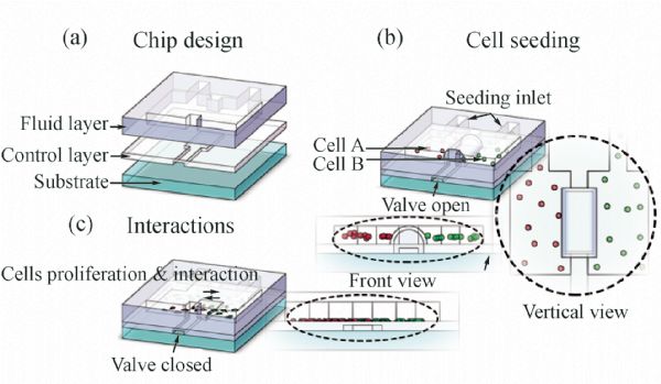
|
| 2 |
Haeberle S, Zengerle R. Microfluidic platforms for lab-on-a-chip applications. Lab on a Chip, 2007, 7(9): 1094–1110
https://doi.org/10.1039/b706364b
|
| 3 |
Han K N, Li C A, Seong G H. Microfluidic chips for immunoassays. Annual Review of Analytical Chemistry (Palo Alto, Calif.), 2013, 6(1): 119–141
https://doi.org/10.1146/annurev-anchem-062012-092616
|
| 4 |
Nan L, Jiang Z, Wei X. Emerging microfluidic devices for cell lysis: A review. Lab on a Chip, 2014, 14(6): 1060–1073
https://doi.org/10.1039/c3lc51133b
|
| 5 |
Smejkal P, Bottenus D, Breadmore M C, Guijt R M, Ivory C F, Foret F, Macka M. Microfluidic isotachophoresis: A review. Electrophoresis, 2013, 34(11): 1493–1509
https://doi.org/10.1002/elps.201300021
|
| 6 |
Ma S, Loufakis D N, Cao Z, Chang Y, Achenie L, Lu C. Diffusion-based microfluidic PCR for “one-pot” analysis of cells. Lab on a Chip, 2014, 14(16): 2905–2909
https://doi.org/10.1039/C4LC00498A
|
| 7 |
Sommer G J, Hatch A V, Singh A K, Wang Y C. Microfluidic device having an immobilized pH gradient and page gels for protein separation and analysis: US Patent 8728290,2014-5-20
|
| 8 |
Jebrail M J, Renzi R F, Sinha A, Van De Vreugde J, Gondhalekar C, Ambriz C, Meagher R J, Branda S S. A solvent replenishment solution for managing evaporation of biochemical reactions in air-matrix digital microfluidics devices. Lab on a Chip, 2015, 15(1): 151–158
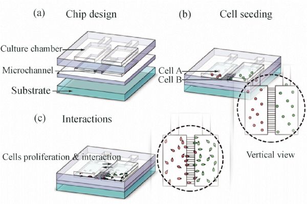
https://doi.org/10.1039/C4LC00703D
|
| 9 |
Ren K, Chen Y, Wu H. New materials for microfluidics in biology. Current Opinion in Biotechnology, 2014, 25: 78–85
https://doi.org/10.1016/j.copbio.2013.09.004
|
| 10 |
Sia S K, Whitesides G M. Microfluidic devices fabricated in poly(dimethylsiloxane) for biological studies. Electrophoresis, 2003, 24(21): 3563–3576
https://doi.org/10.1002/elps.200305584
|
| 11 |
Giulitti S, Magrofuoco E, Prevedello L, Elvassore N. Optimal periodic perfusion strategy for robust long-term microfluidic cell culture. Lab on a Chip, 2013, 13(22): 4430–4441
https://doi.org/10.1039/c3lc50643f
|
| 12 |
Zhang Q, Liu T, Qin J. A microfluidic-based device for study of transendothelial invasion of tumor aggregates in realtime. Lab on a Chip, 2012, 12(16): 2837–2842
https://doi.org/10.1039/c2lc00030j
|
| 13 |
Ziolkowska K, Jedrych E, Kwapiszewski R, Lopacinska J, Skolimowski M, Chudy M. PDMS/glass microfluidic cell culture system for cytotoxicity tests and cells passage. Sensors and Actuators. B, Chemical, 2010, 145(1): 533–542
https://doi.org/10.1016/j.snb.2009.11.010
|
| 14 |
El-Ali J, Sorger P K, Jensen K F. Cells on chips. Nature, 2006, 442(7101): 403–411
https://doi.org/10.1038/nature05063
|
| 15 |
Mehling M, Tay S. Microfluidic cell culture. Current Opinion in Biotechnology, 2014, 25: 95–102
https://doi.org/10.1016/j.copbio.2013.10.005
|
| 16 |
Xiong B, Ren K, Shu Y, Chen Y, Shen B, Wu H. Recent developments in microfluidics for cell studies. Advanced Materials, 2014, 26(31): 5525–5532
https://doi.org/10.1002/adma.201305348
|
| 17 |
Nge P N, Rogers C I, Woolley A T. Advances in microfluidic materials, functions, integration, and applications. Chemical Reviews, 2013, 113(4): 2550–2583
https://doi.org/10.1021/cr300337x
|
| 18 |
Torisawa Y S, Mosadegh B, Luker G D, Morell M, O’Shea K S, Takayama S. Microfluidic hydrodynamic cellular patterning for systematic formation of co-culture spheroids. Integrative Biology, 2009, 1(11-12): 649–654
https://doi.org/10.1039/b915965g
|
| 19 |
Skafte-Pedersen P, Hemmingsen M, Sabourin D, Blaga F S, Bruus H, Dufva M. A self-contained, programmable microfluidic cell culture system with real-time microscopy access. Biomedical Microdevices, 2012, 14(2): 385–399
https://doi.org/10.1007/s10544-011-9615-6
|
| 20 |
Wang D Y, Wu S C, Lin S P, Hsiao S H, Chung T W, Huang Y Y. Evaluation of transdifferentiation from mesenchymal stem cells to neuron-like cells using microfluidic patterned co-culture system. Biomedical Microdevices, 2011, 13(3): 517–526
https://doi.org/10.1007/s10544-011-9520-z
|
| 21 |
Wei C W, Cheng J Y, Young T H. Elucidating in vitro cell-cell interaction using a microfluidic coculture system. Biomedical Microdevices, 2006, 8(1): 65–71
https://doi.org/10.1007/s10544-006-6384-8
|
| 22 |
Kobel S, Valero A, Latt J, Renaud P, Lutolf M. Optimization of microfluidic single cell trapping for long-term on-chip culture. Lab on a Chip, 2010, 10(7): 857–863
https://doi.org/10.1039/b918055a
|
| 23 |
Mazutis L, Gilbert J, Ung W L, Weitz D A, Griffiths A D, Heyman J A. Single-cell analysis and sorting using droplet-based microfluidics. Nature Protocols, 2013, 8(5): 870–891
https://doi.org/10.1038/nprot.2013.046
|
| 24 |
Yin H, Marshall D. Microfluidics for single cell analysis. Current Opinion in Biotechnology, 2012, 23(1): 110–119
https://doi.org/10.1016/j.copbio.2011.11.002
|
| 25 |
Kim L, Toh Y C, Voldman J, Yu H. A practical guide to microfluidic perfusion culture of adherent mammalian cells. Lab on a Chip, 2007, 7(6): 681–694
https://doi.org/10.1039/b704602b
|
| 26 |
Huang C P, Lu J, Seon H, Lee A P, Flanagan L A, Kim H Y, Putnam A J, Jeon N L. Engineering microscale cellular niches for three-dimensional multicellular co-cultures. Lab on a Chip, 2009, 9(12): 1740–1748
https://doi.org/10.1039/b818401a
|
| 27 |
Liu T, Lin B, Qin J. Carcinoma-associated fibroblasts promoted tumor spheroid invasion on a microfluidic 3D co-culture device. Lab on a Chip, 2010, 10(13): 1671–1677
https://doi.org/10.1039/c000022a
|
| 28 |
Zhou M, Ma H, Lin H, Qin J. Induction of epithelial-to-mesenchymal transition in proximal tubular epithelial cells on microfluidic devices. Biomaterials, 2014, 35(5): 1390–1401
https://doi.org/10.1016/j.biomaterials.2013.10.070
|
| 29 |
Unger M A, Chou H P, Thorsen T, Scherer A, Quake S R. Monolithic microfabricated valves and pumps by multilayer soft lithography. Science, 2000, 288(5463): 113–116
https://doi.org/10.1126/science.288.5463.113
|
| 30 |
Liu A, Liu W, Wang Y, Wang J C, Tu Q, Liu R, Xu J, Shen S, Wang J. Microvalve and liquid membrane double-controlled integrated microfluidics for observing the interaction of breast cancer cells. Microfluidics and Nanofluidics, 2012, 14(3-4): 515–526
https://doi.org/10.1007/s10404-012-1070-z
|
| 31 |
Majumdar D, Gao Y, Li D, Webb D J. Co-culture of neurons and glia in a novel microfluidic platform. Journal of Neuroscience Methods, 2011, 196(1): 38–44
https://doi.org/10.1016/j.jneumeth.2010.12.024
|
| 32 |
Gao Y, Majumdar D, Jovanovic B, Shaifer C, Lin P C, Zijlstra A, Webb D J, Li D. A versatile valve-enabled microfluidic cell co-culture platform and demonstration of its applications to neurobiology and cancer biology. Biomedical Microdevices, 2011, 13(3): 539–548
https://doi.org/10.1007/s10544-011-9523-9
|
| 33 |
Liu W, Li L, Wang X, Ren L, Wang X, Wang J, Tu Q, Huang X, Wang J. An integrated microfluidic system for studying cell-microenvironmental interactions versatilely and dynamically. Lab on a Chip, 2010, 10(13): 1717–1724
https://doi.org/10.1039/c001049a
|
| 34 |
Zheng C, Zhao L, Chen G, Zhou Y, Pang Y, Huang Y. Quantitative study of the dynamic tumor-endothelial cell interactions through an integrated microfluidic coculture system. Analytical Chemistry, 2012, 84(4): 2088–2093
https://doi.org/10.1021/ac2032029
|
| 35 |
Brewer B M, Shi M, Edd J F, Webb D J, Li D. A microfluidic cell co-culture platform with a liquid fluorocarbon separator. Biomedical Microdevices, 2014, 16: 311–323
|
| 36 |
Yeon J H, Ryu H R, Chung M, Hu Q P, Jeon N L. In vitro formation and characterization of a perfusable three-dimensional tubular capillary network in microfluidic devices. Lab on a Chip, 2012, 12(16): 2815–2822
https://doi.org/10.1039/c2lc40131b
|
| 37 |
Businaro L, De Ninno A, Schiavoni G, Lucarini V, Ciasca G, Gerardino A, Belardelli F, Gabriele L, Mattei F. Cross talk between cancer and immune cells: Exploring complex dynamics in a microfluidic environment. Lab on a Chip, 2013, 13(2): 229–239
https://doi.org/10.1039/C2LC40887B
|
| 38 |
Huang Y, Agrawal B, Clark P A, Williams J C, Kuo J S. Evaluation of cancer stem cell migration using compartmentalizing microfluidic devices and live cell imaging. Journal of Visualized Experiments, 2011, 58: 3297
|
| 39 |
De Jong J, Lammertink R G, Wessling M. Membranes and microfluidics: A review. Lab on a Chip, 2006, 6(9): 1125–1139
https://doi.org/10.1039/b603275c
|
| 40 |
Chen Q, Wu J, Zhuang Q, Lin X, Zhang J, Lin J M. Microfluidic isolation of highly pure embryonic stem cells using feeder-separated co-culture system. Scientific Reports, 2013, 3: 1–6
https://doi.org/10.1038/srep02433
|
| 41 |
Sip C G, Bhattacharjee N, Folch A. Microfluidic transwell inserts for generation of tissue culture-friendly gradients in well plates. Lab on a Chip, 2014, 14(2): 302–314
https://doi.org/10.1039/C3LC51052B
|
| 42 |
Ostrovidov S, Sakai Y, Fujii T. Integration of a pump and an electrical sensor into a membrane-based PDMS microbioreactor for cell culture and drug testing. Biomedical Microdevices, 2011, 13(5): 847–864
https://doi.org/10.1007/s10544-011-9555-1
|
| 43 |
VanDersarl J J, Xu A M, Melosh N A. Rapid spatial and temporal controlled signal delivery over large cell culture areas. Lab on a Chip, 2011, 11(18): 3057–3063
https://doi.org/10.1039/c1lc20311h
|
| 44 |
Jang K J, Suh K Y. A multi-layer microfluidic device for efficient culture and analysis of renal tubular cells. Lab on a Chip, 2010, 10(1): 36–42
https://doi.org/10.1039/B907515A
|
| 45 |
Ramadan Q, Jafarpoorchekab H, Huang C, Silacci P, Carrara S, Koklu G, Ghaye J, Ramsden J, Ruffert C, Vergeres G, Gijs M A. NutriChip: Nutrition analysis meets microfluidics. Lab on a Chip, 2013, 13(2): 196–203
https://doi.org/10.1039/C2LC40845G
|
| 46 |
Miura S, Morimoto Y, Takeuchi S.Multi-layered placental barrier structure integrated with microfluidic channels. 2013 IEEE 26th International Conference.IEEE, 2013: 257–258
|
| 47 |
Lee Y. Sudo R, Komatsu T, Miki N, Mitaka T, Ikeda M, Tanishita K. Pattern microfluidic hydrostatic deposition patterning for a confined hepatocyte-biliary epithelial cell co-culture system. 2011 International Symposium. IEEE, 2011: 10–15
|
| 48 |
Chin L K, Luo K Q, Park W. Double-layer hepatocyte tumor co-culture using hydrogel for drug affectivity and specificity analysis. 2012 IEEE 25th International Conference. IEEE, 2012: 808–811
|
| 49 |
Liu Z, Shum H C. Fabrication of uniform multi-compartment particles using microfludic electrospray technology for cell co-culture studies. Biomicrofluidics, 2013, 7(4): 044117
https://doi.org/10.1063/1.4817769
|
| 50 |
Shi M, Majumdar D, Gao Y, Brewer B M, Goodwin C R, McLean J A, Li D, Webb D J. Glia co-culture with neurons in microfluidic platforms promotes the formation and stabilization of synaptic contacts. Lab on a Chip, 2013, 13(15): 3008–3021
https://doi.org/10.1039/c3lc50249j
|
| 51 |
Sudo R, Chung S, Zervantonakis I K, Vickerman V, Toshimitsu Y, Griffith L G, Kamm R D. Transport-mediated angiogenesis in 3D epithelial coculture. FASEB Journal, 2009, 23(7): 2155–2164
https://doi.org/10.1096/fj.08-122820
|
| 52 |
Purtscher M, Rothbauer M, Holnthoner W, Redl H, Ertl P. Establishment of Vascular Networks in Biochips Using Co-cultures of Adipose Derived Stem Cells and Endothelial Cells in a 3D Fibrin Matrix. 6th European Conference of the International Federation for Medical and Biological Engineering. Springer International Publishing, 2015: 313–317
|
| 53 |
Chen M B, Srigunapalan S, Wheeler A R, Simmons C A. A 3D microfluidic platform incorporating methacrylated gelatin hydrogels to study physiological cardiovascular cell-cell interactions. Lab on a Chip, 2013, 13(13): 2591–2598
https://doi.org/10.1039/c3lc00051f
|
| 54 |
Chung S, Sudo R, Mack P J, Wan C R, Vickerman V, Kamm R D. Cell migration into scaffolds under co-culture conditions in a microfluidic platform. Lab on a Chip, 2009, 9(2): 269–275
https://doi.org/10.1039/B807585A
|
| 55 |
Ioannis K, Zervantonakis S K H A, Joseph L, Charestd J L. Three-dimensional microfluidic model for tumor cell intravasation and endothelial barrier function. Proceedings of the National Academy of Sciences of the United States of America, 2012, 109(34): 13151–13520
|
| 56 |
Xie Y, Zhang W, Wang L, Sun K, Sun Y, Jiang X. A microchip-based model wound with multiple types of cells. Lab on a Chip, 2011, 11(17): 2819–2822
https://doi.org/10.1039/c0lc00562b
|
| 57 |
Ricci C, Moroni L, Danti S. Cancer tissue engineering-new perspectives in understanding the biology of solid tumours-a critical review. OA Tissue Engineering, 2013, 1(1): 4
https://doi.org/10.13172/2052-9643-1-1-607
|
| 58 |
Ma H, Liu T, Qin J, Lin B. Characterization of the interaction between fibroblasts and tumor cells on a microfluidic co-culture device. Electrophoresis, 2010, 31(10): 1599–1605
https://doi.org/10.1002/elps.200900776
|
| 59 |
Hockemeyer K, Janetopoulos C, Terekhov A, Hofmeister W, Vilgelm A, Costa L, Wikswo J, Richmond A. Engineered three-dimensional microfluidic device for interrogating cell-cell interactions in the tumor microenvironment. Biomicrofluidics, 2014, 8(4): 044105
https://doi.org/10.1063/1.4890330
|
| 60 |
Ye N, Qin J, Shi W, Liu X, Lin B. Cell-based high content screening using an integrated microfluidic device. Lab on a Chip, 2007, 7(12): 1696–1704
https://doi.org/10.1039/b711513j
|
| 61 |
Yang Y, Yang X, Zou J, Jia C, Hu Y, Du H, Wang H. Evaluation of photodynamic therapy efficiency using an in vitro three-dimensional microfluidic breast cancer tissue model. Lab on a Chip, 2015, 15(3): 735–744
https://doi.org/10.1039/C4LC01065E
|
| 62 |
Agliari E, Biselli E, De Ninno A, Schiavoni G, Gabriele L, Gerardino A, Mattei F, Barra A, Businaro L. Cancer-driven dynamics of immune cells in a microfluidic environment. Scientific Reports, 2014, 4: 1–15
https://doi.org/10.1038/srep06639
|
| 63 |
Hsu T H, Kao Y L, Lin W L, Xiao J L, Kuo P L, Wu C W, Liao W Y, Lee C H. The migration speed of cancer cells influenced by macrophages and myofibroblasts co-cultured in a microfluidic chip. Integrative Biology, 2012, 4(2): 177–182
https://doi.org/10.1039/C2IB00112H
|
| 64 |
Park J Y, Kim H O, Kim K D, Kim S K, Lee S K, Jung H. Monitoring the status of T-cell activation in a microfluidic system. Analyst (London), 2011, 136(13): 2831–2836
https://doi.org/10.1039/c1an15038c
|
| 65 |
Charwat V, Rothbauer M, Tedde S F, Hayden O, Bosch J J, Muellner P, Hainberger R, Ertl P. Monitoring dynamic interactions of tumor cells with tissue and immune cells in a lab-on-a-chip. Analytical Chemistry, 2013, 85(23): 11471–11478
https://doi.org/10.1021/ac4033406
|
| 66 |
Mu�oz-Pinedo C, Green D R, van den Berg C A. Confocal restricted-height imaging of suspension cells (CRISC) in a PDMS microdevice during apoptosis. Lab on a Chip, 2005, 5(6): 628–633
https://doi.org/10.1039/b503770k
|
| 67 |
Li P, Stratton Z S, Dao M, Ritz J, Huang T J. Probing circulating tumor cells in microfluidics. Lab on a Chip, 2013, 13(4): 602–609
https://doi.org/10.1039/c2lc90148j
|
| 68 |
Millet L J, Stewart M E, Sweedler J V, Nuzzo R G, Gillette M U. Microfluidic devices for culturing primary mammalian neurons at low densities. Lab on a Chip, 2007, 7(8): 987–994
https://doi.org/10.1039/b705266a
|
| 69 |
Millet L J, Stewart M E, Nuzzo R G, Gillette M U. Guiding neuron development with planar surface gradients of substrate cues deposited using microfluidic devices. Lab on a Chip, 2010, 10(12): 1525–1535
https://doi.org/10.1039/c001552k
|
| 70 |
Wang J, Ren L, Li L, Liu W, Zhou J, Yu W, Tong D, Chen S. Microfluidics: A new cosset for neurobiology. Lab on a Chip, 2009, 9(5): 644–652
https://doi.org/10.1039/B813495B
|
| 71 |
Millet L J, Gillette M U. New perspectives on neuronal development via microfluidic environments. Trends in Neurosciences, 2012, 35(12): 752–761
https://doi.org/10.1016/j.tins.2012.09.001
|
| 72 |
Dinh N D, Chiang Y Y, Hardelauf H, Baumann J, Jackson E, Waide S, Sisnaiske J, Frimat J P, van Thriel C, Janasek D, Peyrin J M, West J. Microfluidic construction of minimalistic neuronal co-cultures. Lab on a Chip, 2013, 13(7): 1402–1412
https://doi.org/10.1039/c3lc41224e
|
| 73 |
Park J, Koito H, Li J, Han A. Microfluidic compartmentalized co-culture platform for CNS axon myelination research. Biomedical Microdevices, 2009, 11(6): 1145–1153
https://doi.org/10.1007/s10544-009-9331-7
|
| 74 |
Peyrin J M, Deleglise B, Saias L, Vignes M, Gougis P, Magnifico S, Betuing S, Pietri M, Caboche J, Vanhoutte P, Viovy J L, Brugg B. Axon diodes for the reconstruction of oriented neuronal networks in microfluidic chambers. Lab on a Chip, 2011, 11(21): 3663–3673
https://doi.org/10.1039/c1lc20014c
|
| 75 |
Kunze A, Lengacher S, Dirren E, Aebischer P, Magistretti P J, Renaud P. Astrocyte-neuron co-culture on microchips based on the model of SOD mutation to mimic ALS. Integrative Biology, 2013, 5(7): 964–975
https://doi.org/10.1039/c3ib40022k
|
| 76 |
Southam K A, King A E, Blizzard C A, McCormack G H, Dickson T C. Microfluidic primary culture model of the lower motor neuron-neuromuscular junction circuit. Journal of Neuroscience Methods, 2013, 218(2): 164–169
https://doi.org/10.1016/j.jneumeth.2013.06.002
|
| 77 |
Kim H J, Huh D, Hamilton G, Ingber D E. Human gut-on-a-chip inhabited by microbial flora that experiences intestinal peristalsis-like motions and flow. Lab on a Chip, 2012, 12(12): 2165–2174
https://doi.org/10.1039/c2lc40074j
|
| 78 |
Kim J, Hegde M, Jayaraman A. Co-culture of epithelial cells and bacteria for investigating host-pathogen interactions. Lab on a Chip, 2010, 10(1): 43–50
https://doi.org/10.1039/B911367C
|
| 79 |
Hong J W, Song S, Shin J H. A novel microfluidic co-culture system for investigation of bacterial cancer targeting. Lab on a Chip, 2013, 13(15): 3033–3040
https://doi.org/10.1039/c3lc50163a
|
| 80 |
Huh D, Hamilton G A, Ingber D E. From 3D cell culture to organs-on-chips. Trends in Cell Biology, 2011, 21(12): 745–754
https://doi.org/10.1016/j.tcb.2011.09.005
|
| 81 |
Kostadinova R, Boess F, Applegate D, Suter L, Weiser T, Singer T, Naughton B, Roth A. A long-term three dimensional liver co-culture system for improved prediction of clinically relevant drug-induced hepatotoxicity. Toxicology and Applied Pharmacology, 2013, 268(1): 1–16
https://doi.org/10.1016/j.taap.2013.01.012
|
| 82 |
Lee S A, No D Y, Kang E, Ju J, Kim D S, Lee S H. Spheroid-based three-dimensional liver-on-a-chip to investigate hepatocyte–hepatic stellate cell interactions and flow effects. Lab on a Chip, 2013, 13(18): 3529–3537
https://doi.org/10.1039/c3lc50197c
|
| 83 |
Jang K J, Cho H S, Kang D H, Bae W G, Kwon T H, Suh K Y. Fluid-shear-stress-induced translocation of aquaporin-2 and reorganization of actin cytoskeleton in renal tubular epithelial cells. Integrative Biology, 2011, 3(2): 134–141
https://doi.org/10.1039/C0IB00018C
|
| 84 |
Huh D, Fujioka H, Tung Y C, Futai N, Paine R, Grotberg J B, Takayama S. Acoustically detectable cellular-level lung injury induced by fluid mechanical stresses in microfluidic airway systems. Proceedings of the National Academy of Sciences of the United States of America, 2007, 104(48): 18886–18891
https://doi.org/10.1073/pnas.0610868104
|
| 85 |
Huh D, Matthews B D, Mammoto A, Montoya-Zavala M, Hsin H Y, Ingber D E. Reconstituting organ-level lung functions on a chip. Science, 2010, 328(5986): 1662–1668
https://doi.org/10.1126/science.1188302
|
| 86 |
Sung J H, Esch M B, Prot J M, Long C J, Smith A, Hickman J J, Shuler M L. Microfabricated mammalian organ systems and their integration into models of whole animals and humans. Lab on a Chip, 2013, 13(7): 1201–1212
https://doi.org/10.1039/c3lc41017j
|
| 87 |
Chan C Y, Huang P H, Guo F, Ding X, Kapur V, Mai J D, Yuen P K, Huang T J. Accelerating drug discovery via organs-on-chips. Lab on a Chip, 2013, 13(24): 4697–4710
https://doi.org/10.1039/c3lc90115g
|
| 88 |
Choucha-Snouber L, Aninat C, Grsicom L, Madalinski G, Brochot C, Poleni P E, Razan F, Guillouzo C G, Legallais C, Corlu A, Leclerc E. Investigation of ifosfamide nephrotoxicity induced in a liver-kidney co-culture biochip. Biotechnology and Bioengineering, 2013, 110(2): 597–608
https://doi.org/10.1002/bit.24707
|
| 89 |
Novik E, Maguire T J, Chao P, Cheng K, Yarmush M L. A microfluidic hepatic coculture platform for cell-based drug metabolism studies. Biochemical Pharmacology, 2010, 79(7): 1036–1044
https://doi.org/10.1016/j.bcp.2009.11.010
|
| 90 |
Torisawa Y S, Spina C S, Mammoto T, Mammoto A, Weaver J C, Tat T, Collins J J, Ingber D E. Bone marrow-on-a-chip replicates hematopoietic niche physiology in vitro. Nature Methods, 2014, 11(6): 663–669
https://doi.org/10.1038/nmeth.2938
|
|
Viewed |
|
|
|
Full text
|
|
|
|
|
Abstract
|
|
|
|
|
Cited |
|
|
|
|
| |
Shared |
|
|
|
|
| |
Discussed |
|
|
|
|