|
|
|
Reduced texaphyrin: A ratiometric optical sensor for heavy metals in aqueous solution |
Harrison D. Root, Gregory Thiabaud, Jonathan L. Sessler( ) ) |
| Department of Chemistry, The University of Texas at Austin, Austin, TX 78712-1224, USA |
|
|
|
|
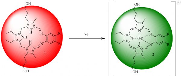
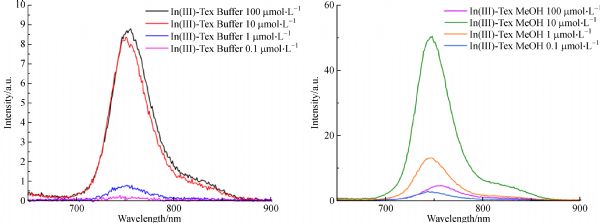
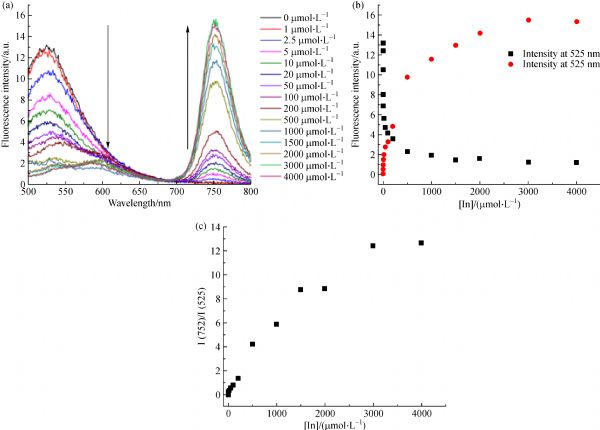
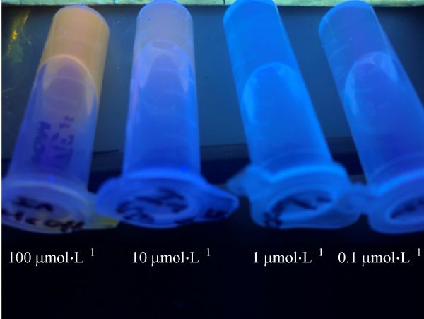
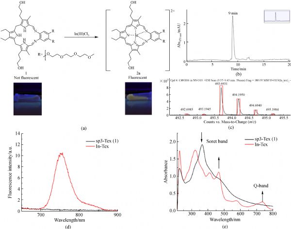
Abstract We report here a water-soluble metal cation sensor system based on the as-prepared or reduced form of an expanded porphyrin, texaphyrin. Upon metal complexation, a change in the redox state of the ligand occurs that is accompanied by a color change from red to green. Although long employed for synthesis in organic media, we have now found that this complexation-driven redox behavior may be used to achieve the naked eye detectable colorimetric sensing of several number of less-common metal ions in aqueous media. Exposure to In(III), Hg(II), Cd(II), Mn(II), Bi(III), Co(II), and Pb(II) cations leads to a colorimetric response within 10 min. This process is selective for Hg(II) under conditions of competitive analysis. Furthermore, among the subset of response-producing cations, In(III) proved unique in giving rise to a ratiometric change in the ligand-based fluorescence features, including an overall increase in intensity. The cation selectivity observed in aqueous media stands in contrast to what is seen in organic solvents, where a wide range of texaphyrin metal complexes may be prepared. The formation of metal cation complexes under the present aqueous conditions was confirmed by reversed phase high-performance liquid chromatography, ultra-violet-visible absorption and fluorescence spectroscopies, and high-resolution mass spectrometry.
|
| Keywords
texaphyrin
fluorescent sensor
ion-sensing
indium
mercury
|
|
Corresponding Author(s):
Jonathan L. Sessler
|
|
Online First Date: 07 January 2020
Issue Date: 20 January 2020
|
|
| 1 |
D Wu, A C Sedgwick, T Gunnlaugsson, E U Akkaya, J Yoon, T D James. Fluorescent chemosensors: The past, present, and future. Chemical Society Review , 2017, 46(23): 7105–7123
https://doi.org/10.1039/C7CS00240H
|
| 2 |
Z Li, J R Askim, K S Suslick. The optoelectronic nose: Colorimetric and fluorometric sensor arrays. Chemical Reviews, 2019, 119(1): 231–292
https://doi.org/10.1021/acs.chemrev.8b00226
|
| 3 |
B Kaur, N Kaur, S Kumar. Colorimetric metal ion sensors—a comprehensive review of the years 2011‒2016. Coordination Chemistry Reviews, 2018, 358: 13–69
https://doi.org/10.1016/j.ccr.2017.12.002
|
| 4 |
A Piriya, P Joseph, K Daniel, S Lakshmanan, T Kinoshita, S Muthusamy. Colorimetric sensors for rapid detection of various analytes. Materials Science and Engineering C, 2017, 78: 1231–1245
https://doi.org/10.1016/j.msec.2017.05.018
|
| 5 |
F Long, A Zhu, H Shi, H Wang, J Liu. Rapid on-site/in-situ detection of heavy metal ions in environmental water using a structure-switching DNA optical biosensor. Scientific Reports, 2013, 3(1): 1–7
https://doi.org/10.1038/srep02308
|
| 6 |
W Zhou, R Saran, J Liu. Metal sensing by DNA. Chemical Reviews, 2017, 117(12): 8272–8325
https://doi.org/10.1021/acs.chemrev.7b00063
|
| 7 |
E M Nolan, S J Lippard. Turn-on and ratiometric mercury sensing in water with a red-emitting probe. Journal of the American Chemical Society, 2007, 129(18): 5910–5918
https://doi.org/10.1021/ja068879r
|
| 8 |
N A Azmi, S H Ahmad, S C Low. Detection of mercury ions in water using a membrane-based colorimetric sensor. RSC Advances, 2018, 8(1): 251–261
https://doi.org/10.1039/C7RA11450H
|
| 9 |
J Chang, G Zhou, X Gao, S Mao, S Cui, L E Ocola, C Yuan, J Chen. Real-time detection of mercury ions in water using a reduced graphene oxide/DNA field-effect transistor with assistance of a passivation layer. Sensing and Bio-Sensing Research, 2015, 5: 97–104
https://doi.org/10.1016/j.sbsr.2015.07.009
|
| 10 |
K Karthikeyan, L Sujatha. Fluorometric sensor for mercury ion detection in a fluidic MEMS device. IEEE Sensors Journal, 2018, 18(13): 5225–5231
https://doi.org/10.1109/JSEN.2018.2840331
|
| 11 |
S Maher, B Bastani, B Smith, Z Jjunju, S Taylor, I S Young. Portable fluorescent sensing array for monitoring heavy metals in water. IEEE Sensors, 2016: 1–3
|
| 12 |
W He, L Luo, Q Liu, Z Chen. Colorimetric sensor array for discrimination of heavy metal ions in aqueous solution based on three kinds of thiols as receptors. Analytical Chemistry, 2018, 90(7): 4770–4775
https://doi.org/10.1021/acs.analchem.8b00076
|
| 13 |
L Niu, H Li, L Feng, Y Guan, Y Chen, C Duan, L Wu, Y Guan, C Tung, Q Yang. BODIPY-based fluorometric sensor array for the highly sensitive identification of heavy-metal ions. Analytica Chimica Acta, 2013, 775: 93–99
https://doi.org/10.1016/j.aca.2013.03.013
|
| 14 |
R K Singh, S Mishra, S Jena, B Panigrahi, B Das, R Jayabalan, P K Parhi, D Mandal. Rapid colorimetric sensing of gadolinium by EGCG-derived AgNPs: The development of a nanohybrid bioimaging probe. Chemical Communications, 2018, 54(32): 3981–3984
https://doi.org/10.1039/C8CC01777H
|
| 15 |
M Denis, J Pancholi, K Jobe, M Watkinson, S M Goldup. Chelating rotaxane ligands as fluorescent sensors for metal ions. Angewandte Chemie International Edition, 2018, 57(19): 5310–5314
https://doi.org/10.1002/anie.201712931
|
| 16 |
W Hong, W Li, X Hu, B Zhao, F Zhang, D Zhang. Highly sensitive colorimetric sensing for heavy metal ions by strong polyelectrolyte photonic hydrogels. Journal of Materials Chemistry, 2011, 21(43): 17193–17201
https://doi.org/10.1039/c1jm12785c
|
| 17 |
M R Moghaddam, S Carrara, C F Hogan. Multi-colour bipolar electrochemiluminescence for heavy metal ion detection. Chemical Communications, 2018, 55(8): 3–6
|
| 18 |
D W Boening. Ecological effects, transport, and fate of Mercury: A general review. Chemosphere, 2000, 40(12): 1335–1351
https://doi.org/10.1016/S0045-6535(99)00283-0
|
| 19 |
W Zheng, M Aschner, J Ghersi-egea. Brain barrier systems: A new frontier in metal neurotoxicological research. Toxicology and Applied Pharmacology, 2003, 192(1): 1–11
https://doi.org/10.1016/S0041-008X(03)00251-5
|
| 20 |
P D Selid, H Xu, E M Collins, M S Face-Collins, J X Zhao. Sensing mercury for biomedical and environmental monitoring. Sensors (Basel), 2009, 9(7): 5446–5459
https://doi.org/10.3390/s90705446
|
| 21 |
J Hu, T Wu, G Zhang, S Liu. Highly selective fluorescence sensing of mercury ions over a broad concentration range based on mixed polymeric micelles. Macromolecules, 2012, 45(9): 3939–3947
https://doi.org/10.1021/ma3006558
|
| 22 |
E M Nolan, S J Lippard. Tools and tactics for the optical detection of mercuric ion. Chemical Reviews, 2008, 108(9): 3443–3480
https://doi.org/10.1021/cr068000q
|
| 23 |
K Zhang, Y Wu, W Wang, B Li, Y Zhang, T Zuo. Resources, conservation and recycling indium from waste LCDs: A review. Resources, Conservation and Recycling, 2015, 104: 276–290
https://doi.org/10.1016/j.resconrec.2015.07.015
|
| 24 |
M L Thakur, M J Welch, J H Joist, R E Coleman. Indium-III labeled platelets: Studies on preparation and evaluation of in vitro and in vivo functions. Thrombosis Research, 1976, 9(4): 345–357
https://doi.org/10.1016/0049-3848(76)90135-3
|
| 25 |
M Thakur, J P Lavender, R Arnot, D J Silvester, A W Segal. Indium-III-labeled autologous leukocytes in man. Journal of Nuclear Medicine, 1977, 18(10): 1014–1021
|
| 26 |
H Zolata, F Abbasi, H Afarideh. Synthesis, characterization and theranostic evaluation of indium-III labeled multifunctional superparamagnetic iron oxide nanoparticles. Nuclear Medicine and Biology, 2015, 42(2): 164–170
https://doi.org/10.1016/j.nucmedbio.2014.09.007
|
| 27 |
A M Alfantazi, R R Moskalyk. Processing of indium: A review. Materials & Design, 2003, 16(8): 687–694
|
| 28 |
C H Lim, J Han, H Cho, M Kang. Studies on the toxicity and distribution of indium compounds according to particle size in sprague-dawley rats. Toxicological Research, 2014, 30(1): 55–63
https://doi.org/10.5487/TR.2014.30.1.055
|
| 29 |
A Tanaka, M Hirata, Y Kiyohara, M Nakano, K Omae, M Shiratani, K Koga. Review of pulmonary toxicity of indium compounds to animals and humans. Thin Solid Films, 2010, 518(11): 2934–2936
https://doi.org/10.1016/j.tsf.2009.10.123
|
| 30 |
P K Mehta, G W Hwang, J Park, K Lee. Highly sensitive ratiometric fluorescent detection of indium(III) using fluorescent probe based on phosphoserine as a receptor. Analytical Chemistry, 2018, 90(19): 11256–11264
https://doi.org/10.1021/acs.analchem.8b01440
|
| 31 |
Y C Wu, H Li, H Yang. A sensitive and highly selective fluorescent sensor for In3+. Organic & Biomolecular Chemistry, 2010, 8(15): 3394–3397
https://doi.org/10.1039/c0ob00002g
|
| 32 |
S K Kim, S H Kim, H J Kim, S H Lee, S W Lee, J Ko, R A Bartsch, J S Kim. Indium (III)-induced fluorescent excimer formation and extinction in calix[4]arene—fluoroionophores. Inorganic Chemistry, 2005, 44(22): 7866–7875
https://doi.org/10.1021/ic050702v
|
| 33 |
Y Ding, W Zhu, Y Xie. Development of ion chemosensors based on porphyrin analogues. Chemical Reviews, 2017, 117(4): 2203–2256
https://doi.org/10.1021/acs.chemrev.6b00021
|
| 34 |
J L Sessler, T D Mody, G W Hemmi, V Lynch. Synthesis and structural characterization of lanthanide(III) texaphyrins. Inorganic Chemistry, 1993, 32(14): 3175–3187
https://doi.org/10.1021/ic00066a032
|
| 35 |
C Preihs, J F Arambula, V M Lynch, H Siddik, J L Sessler. Bismuth- and lead-texaphyrin complexes: Towards potential α-core emitters for radiotherapy. Chemical Communications, 2010, 46(42): 7900–7902
https://doi.org/10.1039/c0cc03528a
|
| 36 |
G Thiabaud, V Radchenko, J J Wilson, K D John, E R Birnbaum, J L Sessler. Rapid insertion of bismuth radioactive isotopes into texaphyrin in aqueous media. Journal of Porphyrins and Phthalocyanines, 2017, 21(12): 882–886
https://doi.org/10.1142/S1088424617500870
|
| 37 |
B G Maiya, A Harriman, J L Sessler, G Hemmi, T Murai, T E Mallouk. Ground- and excited-state spectral and redox properties of cadmium(II) texaphyrin. Journal of Physical Chemistry, 1989, 93(24): 8111–8115
https://doi.org/10.1021/j100361a027
|
| 38 |
J L Sessler, T Murai, V Lynch, M Cyr. An “expanded porphyrin”: The synthesis and structure of a new aromatic pentadentate ligand of chemistry. Journal of the American Chemical Society, 1988, 110(16): 5586–5588
https://doi.org/10.1021/ja00224a062
|
| 39 |
J L Sessler, W C Dow, D O Connor, A Harriman, G Hemmi, T D Mody, R A Miller, F Qing, S Springs, K Woodburn, et al. Biomedical applications of lanthanide(III) texaphyrins lutetium(III) texaphyrins as potential photodynamic therapy photosensitizers. Journal of Alloys and Compounds, 1997, 249(1-2): 146–152
https://doi.org/10.1016/S0925-8388(96)02517-0
|
| 40 |
D Magda, R A Miller. Motexafin gadolinium: A novel redox active drug for cancer therapy. Seminars in Cancer Biology, 2006, 16(6): 466–476
https://doi.org/10.1016/j.semcancer.2006.09.002
|
| 41 |
S Hannah, V Lynch, D M Guldi, N Gerasimchuk, C L B Macdonald, D Magda, J L Sessler. Late first-row transition-metal complexes of texaphyrin. Journal of the American Chemical Society, 2002, 124(28): 8416–8427
https://doi.org/10.1021/ja012747a
|
| 42 |
G Thiabaud, J F Arambula, Z H Siddik, J L Sessler. Photoinduced reduction of Pt IV within an anti-proliferative Pt IV-texaphyrin conjugate. Chemistry (Weinheim an der Bergstrasse, Germany), 2014, 20(29): 8942–8947
https://doi.org/10.1002/chem.201490117
|
| 43 |
G Thiabaud, R Mccall, G He, J F Arambula, Z H Siddik, J L Sessler. Activation of platinum(IV) prodrugs by motexafin gadolinium as a redox mediator. Angewandte Chemie International Edition, 2016, 55(41): 12626–12631
https://doi.org/10.1002/anie.201604236
|
| 44 |
J F Arambula, J L Sessler, Z H Siddik. Overcoming biochemical pharmacologic mechanisms of platinum resistance with a texaphyrin-platinum conjugate. Bioorganic & Medicinal Chemistry Letters, 2011, 21(6): 1701–1705
https://doi.org/10.1016/j.bmcl.2011.01.092
|
| 45 |
J F Arambula, J L Sessler, Z H Siddik. A texaphyrin-oxaliplatin conjugate that overcomes both pharmacologic and molecular mechanisms of cisplatin resistance in cancer cells. MedChemComm, 2012, 3(10): 1275–1281
https://doi.org/10.1039/c2md20206a
|
| 46 |
M H Lee, E J Kim, S Y Park, K S Hong, S Kim, J L Sessler. Acid-triggered release of doxorubicin from a hydrazone-linked Gd3+-texaphyrin conjugate. Chemical Communications, 2016, 52(69): 10551–10554
https://doi.org/10.1039/C6CC05673C
|
| 47 |
M Blesic, E Melo, Z Petrovski, N V Plechkova, N C Lopes, K R Seddon, P N Rebelo. On the self-aggregation and fluorescence quenching aptitude of surfactant ionic liquids. Journal of Physical Chemistry B, 2008, 112(29): 8645–8650
https://doi.org/10.1021/jp802179j
|
| 48 |
J Mei, N L C Leung, R T K Kwok, J W Y Lam, B Z Tang. Aggregation-induced emission: Together we shine, united we soar! Chemical Reviews, 2015, 115(21): 11718–11940
https://doi.org/10.1021/acs.chemrev.5b00263
|
| 49 |
S D Quinn, S W Magennis. Optical detection of gadolinium(III) ions via quantum dot aggregation. RSC Advances, 2017, 7(40): 24730–24735
https://doi.org/10.1039/C7RA03969G
|
|
Viewed |
|
|
|
Full text
|
|
|
|
|
Abstract
|
|
|
|
|
Cited |
|
|
|
|
| |
Shared |
|
|
|
|
| |
Discussed |
|
|
|
|