|
|
|
Xiao Ke Qing improves glycometabolism and ameliorates insulin resistance by regulating the PI3K/Akt pathway in KKAy mice |
Xiaoqing Li, Xinxin Li( ), Genbei Wang, Yan Xu, Yuanyuan Wang, Ruijia Hao, Xiaohui Ma ), Genbei Wang, Yan Xu, Yuanyuan Wang, Ruijia Hao, Xiaohui Ma |
| Department of Pharmacology and Toxicology, Tasly Pharmaceutical Co., Ltd., Tianjin 300410, China |
|
|
|
|
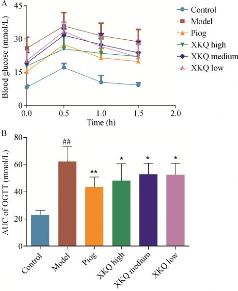
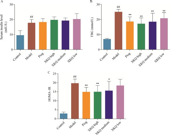
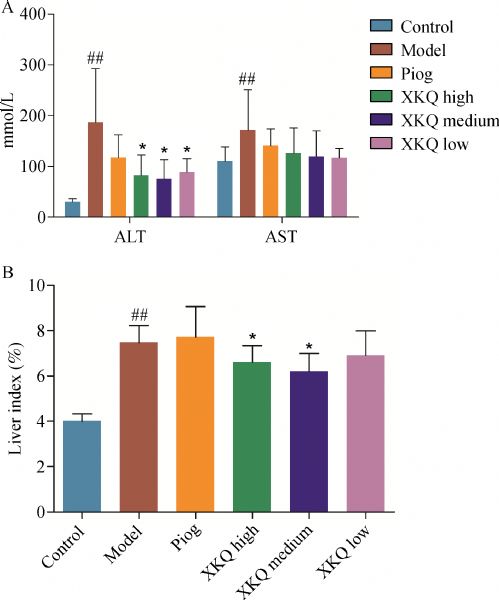
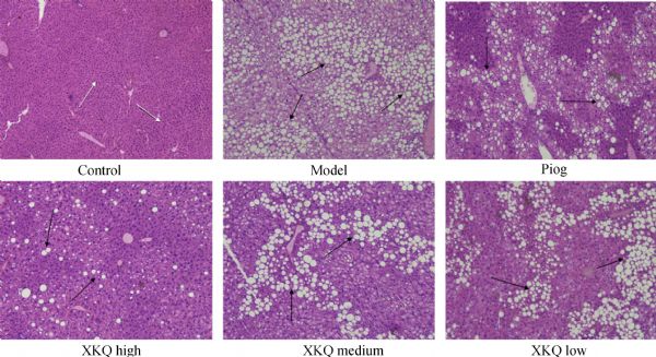
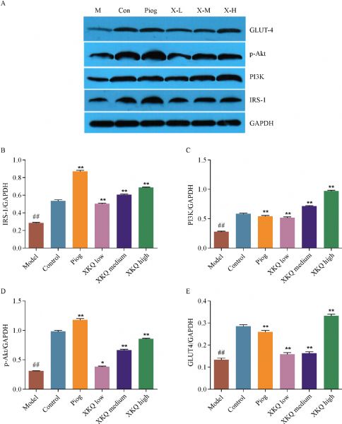
Abstract Xiao Ke Qing (XKQ) granule has been clinically used to treat type 2 diabetes mellitus (T2DM) for 10 years in Chinese traditional medication. However, its mechanisms against hyperglycemia remain poorly understood. This study aims to investigate XKQ mechanisms on diabetes and diabetic liver disease by using the KKAy mice model. Our results indicate that XKQ can significantly reduce food and water intake. XKQ treatment also remarkably decreases both the fasting blood glucose and blood glucose in the oral glucose tolerance test. Additionally, XKQ can significantly decrease the serum alanine aminotransferase level and liver index and can alleviate the fat degeneration in liver tissues. Moreover, XKQ can ameliorate insulin resistance and upregulate the expression of IRS-1, PI3K (p85), p-Akt, and GLUT4 in the skeletal muscle of KKAy mice. XKQ is an effective drug for T2DM by ameliorating insulin resistance and regulating the PI3K/Akt signaling pathway in the skeletal muscle.
|
| Keywords
XKQ
type 2 diabetes mellitus
KKAy mice
PI3K/Akt pathway
diabetic liver disease
|
|
Corresponding Author(s):
Xinxin Li
|
|
Just Accepted Date: 24 October 2018
Online First Date: 13 November 2018
Issue Date: 03 December 2018
|
|
| 1 |
Zimmet P, Alberti KG, Shaw J. Global and societal implications of the diabetes epidemic. Nature 2001; 414(6865): 782–787
https://doi.org/10.1038/414782a
pmid: 11742409
|
| 2 |
American Diabetes Association. Diagnosis and classification of diabetes mellitus. Diabetes Care 2010; 33(Suppl 1): S62–S69
https://doi.org/10.2337/dc10-S062
pmid: 20042775
|
| 3 |
Yabe D, Seino Y, Fukushima M, Seino S. β cell dysfunction versus insulin resistance in the pathogenesis of type 2 diabetes in East Asians. Curr Diab Rep 2015; 15(6): 602
https://doi.org/10.1007/s11892-015-0602-9
pmid: 25944304
|
| 4 |
Pessin JE, Saltiel AR. Signaling pathways in insulin action: molecular targets of insulin resistance. J Clin Invest 2000; 106(2): 165–169
https://doi.org/10.1172/JCI10582
pmid: 10903329
|
| 5 |
Brunetti A, Chiefari E, Foti D. Recent advances in the molecular genetics of type 2 diabetes mellitus. World J Diabetes 2014; 5(2): 128–140
https://doi.org/10.4239/wjd.v5.i2.128
pmid: 24748926
|
| 6 |
DeFronzo RA, Tripathy D. Skeletal muscle insulin resistance is the primary defect in type 2 diabetes. Diabetes Care 2009; 32(Suppl 2): S157–S163
https://doi.org/10.2337/dc09-S302
pmid: 19875544
|
| 7 |
Cusi K, Maezono K, Osman A, Pendergrass M, Patti ME, Pratipanawatr T, DeFronzo RA, Kahn CR, Mandarino LJ. Insulin resistance differentially affects the PI 3-kinase- and MAP kinase-mediated signaling in human muscle. J Clin Invest 2000; 105(3): 311–320
https://doi.org/10.1172/JCI7535
pmid: 10675357
|
| 8 |
Brachmann SM, Ueki K, Engelman JA, Kahn RC, Cantley LC. Phosphoinositide 3-kinase catalytic subunit deletion and regulatory subunit deletion have opposite effects on insulin sensitivity in mice. Mol Cell Biol 2005; 25(5): 1596–1607
https://doi.org/10.1128/MCB.25.5.1596-1607.2005
pmid: 15713620
|
| 9 |
Kanai F, Ito K, Todaka M, Hayashi H, Kamohara S, Ishii K, Okada T, Hazeki O, Ui M, Ebina Y. Insulin-stimulated GLUT4 translocation is relevant to the phosphorylation of IRS-1 and the activity of PI3-kinase. Biochem Biophys Res Commun 1993; 195(2): 762–768
https://doi.org/10.1006/bbrc.1993.2111
pmid: 8396927
|
| 10 |
Meex RCR, Watt MJ. Hepatokines: linking nonalcoholic fatty liver disease and insulin resistance. Nat Rev Endocrinol 2017; 13(9): 509–520
https://doi.org/10.1038/nrendo.2017.56
pmid: 28621339
|
| 11 |
Lin P, Zhou H. Relationship between insulin resistance, dyslipidemia and fatty liver in non-insulin-dependent diabetes mellitus. Guangzhou Med J (Guangzhou Yi Yao) 2001; 32(l): 41–42 (in Chinese)
|
| 12 |
Yun X, Yao D, Han T. Clinical observation of Xiao ke Qing in treating 42 cases of type 2 diabetic patients. Gansu J Tradit Chin Med (Gansu Zhong Yi) 2002; 15(4): 37–38 (in Chinese)
|
| 13 |
Li Y. Clinical observation of Xiaokeqing treating type 2 diabetes mellitus with relieving blood stasis. Dissertation. Liaoning: Liaoning University of Traditional Chinese Medicine, 2017 (in Chinese)
|
| 14 |
Chen XM, Li NI, Jin HL, Sun WL. Studies of hypoglycemic effects of xiaokeqing. Chin Hosp Pharm J (Zhongguo Yi Yuan Yao Xue Za Zhi) 2005; 25(2): 126–128 (in Chinese)
|
| 15 |
Qiu ZJ, Shi RS, Zhu XX, Chen Z. Experimental study on treatment of diabetes with Xiaokeqing soft extract. J Nanjing Univ Tradit Chin Med (Nanjing Zhong Yi Yao Da Xue Xue Bao) 2001; 17(3): 170–172 (in Chinese)
|
| 16 |
Wang LQ, Wang X, Tong L, Li XW, Liu WY, Zhou SP, Sun H. Establishment of UPLC-PDA-ELSD fingerprints of Xiaokeqing Granules and determination of its five main constituents. Chin Tradit Herbal Drugs (Zhong Cao Yao) 2013; 44(24): 3482–3488 (in Chinese)
|
| 17 |
Sellamuthu PS, Arulselvan P, Fakurazi S, Kandasamy M. Beneficial effects of mangiferin isolated from Salacia chinensis on biochemical and hematological parameters in rats with streptozotocin-induced diabetes. Pak J Pharm Sci 2014; 27(1): 161–167
pmid: 24374436
|
| 18 |
Lim J, Liu Z, Apontes P, Feng D, Pessin JE, Sauve AA, Angeletti RH, Chi Y. Dual mode action of mangiferin in mouse liver under high fat diet. PLoS One 2014; 9(3): e90137
https://doi.org/10.1371/journal.pone.0090137
pmid: 24598864
|
| 19 |
Na L, Zhang Q, Jiang S, Du S, Zhang W, Li Y, Sun C, Niu Y. Mangiferin supplementation improves serum lipid profiles in overweight patients with hyperlipidemia: a double-blind randomized controlled trial. Sci Rep 2015; 5(1): 10344
https://doi.org/10.1038/srep10344
pmid: 25989216
|
| 20 |
Li CM, Gao YL, Li M, Han B, Liu ZF. Effects of timosaponins on blood glucose level in mice. Pharm Clin Chin Materia Medica (Zhongguo Yao Li Yu Lin Chuang) 2005;21 (4):22–23 (in Chinese)
|
| 21 |
Yin J, Xing H, Ye J. Efficacy of berberine in patients with type 2 diabetes mellitus. Metabolism 2008; 57(5): 712–717
https://doi.org/10.1016/j.metabol.2008.01.013
pmid: 18442638
|
| 22 |
Lee YS, Kim WS, Kim KH, Yoon MJ, Cho HJ, Shen Y, Ye JM, Lee CH, Oh WK, Kim CT, Hohnen-Behrens C, Gosby A, Kraegen EW, James DE, Kim JB. Berberine, a natural plant product, activates AMP-activated protein kinase with beneficial metabolic effects in diabetic and insulin-resistant states. Diabetes 2006; 55(8): 2256–2264
https://doi.org/10.2337/db06-0006
pmid: 16873688
|
| 23 |
Yin J, Gao Z, Liu D, Liu Z, Ye J. Berberine improves glucose metabolism through induction of glycolysis. Am J Physiol Endocrinol Metab 2008; 294(1): E148–E156
https://doi.org/10.1152/ajpendo.00211.2007
pmid: 17971514
|
| 24 |
Yi P, Lu FE, Xu LJ, Chen G, Dong H, Wang KF. Berberine reverses free-fatty-acid-induced insulin resistance in 3T3-L1 adipocytes through targeting IKKβ. World J Gastroenterol 2008; 14(6): 876–883
https://doi.org/10.3748/wjg.14.876
pmid: 18240344
|
| 25 |
Chen Y, Li Y, Wang Y, Wen Y, Sun C. Berberine improves free-fatty-acid-induced insulin resistance in L6 myotubes through inhibiting peroxisome proliferator-activated receptor γ and fatty acid transferase expressions. Metabolism 2009; 58(12): 1694–1702
https://doi.org/10.1016/j.metabol.2009.06.009
pmid: 19767038
|
| 26 |
Leng SH, Lu FE, Xu LJ. Therapeutic effects of berberine in impaired glucose tolerance rats and its influence on insulin secretion. Acta Pharmacol Sin 2004; 25(4): 496–502
pmid: 15066220
|
| 27 |
Kong WJ, Zhang H, Song DQ, Xue R, Zhao W, Wei J, Wang YM, Shan N, Zhou ZX, Yang P, You XF, Li ZR, Si SY, Zhao LX, Pan HN, Jiang JD. Berberine reduces insulin resistance through protein kinase C-dependent up-regulation of insulin receptor expression. Metabolism 2009; 58(1): 109–119
https://doi.org/10.1016/j.metabol.2008.08.013
pmid: 19059538
|
| 28 |
Chen G, Lu FE, Wang ZS, Yi P, Wang KF, Zou X. Correlation between the amelioration of insulin resistance and protein expression of PI3K and GLUT4 in type 2 diabetic rats treated with berberine. Chin Pharmacol Bull (Zhongguo Yao Li Xue Tong Bao) 2008; 24(8): 1007–1010 (in Chinese)
|
| 29 |
Chen W, Li S, Jing X, Jia H, Wan Y, Che R. Research progress in animal models of type 2 diabetes KKAy mice. J Clin Med (Lin Chuang Yi Yao Wen Xian Za Zhi ) 2017; 4(54): 10681–10682 (in Chinese)
|
| 30 |
Cantley LC. The phosphoinositide 3-kinase pathway. Science 2002; 296(5573): 1655–1657
https://doi.org/10.1126/science.296.5573.1655
pmid: 12040186
|
| 31 |
Kohn AD, Summers SA, Birnbaum MJ, Roth RA. Expression of a constitutively active Akt Ser/Thr kinase in 3T3-L1 adipocytes stimulates glucose uptake and glucose transporter 4 translocation. J Biol Chem 1996; 271(49): 31372–31378
https://doi.org/10.1074/jbc.271.49.31372
pmid: 8940145
|
| 32 |
Gandhi GR, Stalin A, Balakrishna K, Ignacimuthu S, Paulraj MG, Vishal R. Insulin sensitization via partial agonism of PPARg and glucose uptake through translocation and activation of GLUT4 in PI3K/p-Akt signaling pathway by embelin in type 2 diabetic rats. Biochim Biophys Acta 2013; 1830(1): 2243–2255
https://doi.org/10.1016/j.bbagen.2012.10.016
pmid: 23104384
|
| 33 |
Pessin JE, Saltiel AR. Signaling pathways in insulin action: molecular targets of insulin resistance. J Clin Invest 2000; 106(2): 165–169
https://doi.org/10.1172/JCI10582
pmid: 10903329
|
| 34 |
Chi YJ, Jing LI, Guan YF, Yang JC. PI3K/Akt signaling axis in regulation of glucose homeostasis. Chin J Biochem Mol Biol (Zhongguo Sheng Wu Hua Xue Yu Fen Zi Sheng Wu Xue Bao) 2010; 26(10): 879–885 (in Chinese)
|
| 35 |
Tolman KG, Fonseca V, Tan MH, Dalpiaz A. Narrative review: hepatobiliary disease in type 2 diabetes mellitus. Ann Intern Med 2004; 141(12): 946–956
https://doi.org/10.7326/0003-4819-141-12-200412210-00011
pmid: 15611492
|
| 36 |
Targher G, Bertolini L, Rodella S, Tessari R, Zenari L, Lippi G, Arcaro G. Nonalcoholic fatty liver disease is independently associated with an increased incidence of cardiovascular events in type 2 diabetic patients. Diabetes Care 2007; 30(8): 2119–2121
https://doi.org/10.2337/dc07-0349
pmid: 17519430
|
| 37 |
Zhu X, Bian H, Gao X. The potential mechanisms of berberine in the treatment of nonalcoholic fatty liver disease. Molecules 2016; 21(10): 1336
https://doi.org/10.3390/molecules21101336
pmid: 27754444
|
| 38 |
Galbo T, Shulman GI. Lipid-induced hepatic insulin resistance. Aging (Albany NY) 2013; 5(8): 582–583
https://doi.org/10.18632/aging.100585
pmid: 23929893
|
|
Viewed |
|
|
|
Full text
|
|
|
|
|
Abstract
|
|
|
|
|
Cited |
|
|
|
|
| |
Shared |
|
|
|
|
| |
Discussed |
|
|
|
|