|
|
|
A comparison of the biological activities of ethyl acetate fractions from the stems and leaves of Penthorum chinense Pursh |
Zhaolei WANG1, Kai JIANG1, Qinchao DING1, Shujun LIU2, Xiaobing DOU1, Bin DING1( ) ) |
1. College of Life Science, Zhejiang Chinese Medical University, Hangzhou 310053, China
2. Beijing Stomatological Hospital/School of Stomatology, Capital Medical University, Beijing 100050, China |
|
|
|
|
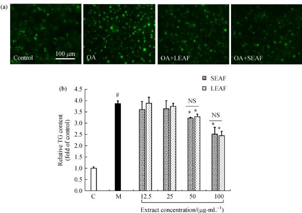
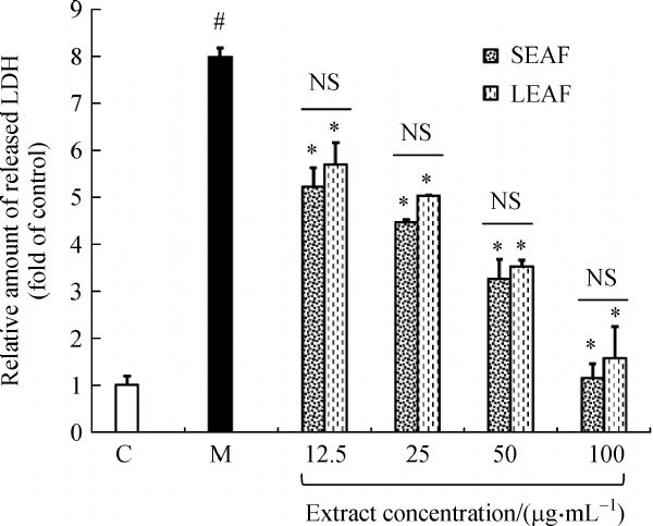
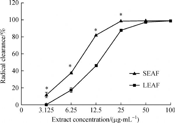
Abstract Penthorum chinense Pursh (PCP) is a popular traditional medicinal plant in China, widely used for the treatment of a variety of liver diseases. Although it has been long recognized that the main active elements of PCP are contained in ethyl acetate fraction (EAF), little is known so far in terms of the relative effectiveness of EAF derived from the stems versus leaves of this plant. In the current study, we prepared EAF by reflux extraction and sequential extraction from the stems (SEAF) and leaves (LEAF) of PCP and tested their hepatoprotective efficacies. The extract rates and flavonoid contents of LEAF were higher than those of SEAF. EAFs (>50 μg·mL−1) prevented lipid accumulation in cells and protected against lipotoxicity injury when the concentration exceeded 25 μg·mL−1. More than 95% free radicals released by 2,2-diphenyl-1-picrylhydrazyl (DPPH) were eliminated by 25 μg·mL−1 SEAF and 50 μg·mL−1 LEAF, respectively. Further, EAFs (25 μg·mL−1) also showed protective antioxidant effects, with the activity of LEAF being significantly higher than that of SEAF. EAFs (10 mg·mL−1) also showed similar unspecific bacteriostatic activity. In comparison with SEAF, LEAF contained more flavonoids and had a higher anti-oxidation capability and for these reasons we suggest it should be better for clinical use.
|
| Keywords
antibacterial
anti-oxidation
lipid accumulation
lipotoxicity
Penthorum chinense Pursh
|
|
Corresponding Author(s):
Bin DING
|
|
Just Accepted Date: 31 May 2019
Online First Date: 28 June 2019
Issue Date: 06 November 2020
|
|
| 1 |
J M Schattenberg, I Bergheim. Nutritional intake and the risk for non-alcoholic fatty liver disease (NAFLD). Nutrients, 2019, 11(3): 588–592
https://doi.org/10.3390/nu11030588
pmid: 30862016
|
| 2 |
B A Neuschwander-Tetri. Hepatic lipotoxicity and the pathogenesis of nonalcoholic steatohepatitis: the central role of nontriglyceride fatty acid metabolites. Hepatology, 2010, 52(2): 774–788
https://doi.org/10.1002/hep.23719
pmid: 20683968
|
| 3 |
H Malhi, G J Gores. Molecular mechanisms of lipotoxicity in nonalcoholic fatty liver disease. Seminars in Liver Disease, 2008, 28(4): 360–369
https://doi.org/10.1055/s-0028-1091980
pmid: 18956292
|
| 4 |
E D’Adamo, N Santoro, S Caprio. Metabolic syndrome in pediatrics: old concepts revised, new concepts discussed. Pediatric Clinics of North America, 2011, 58(5): 1241–1255
https://doi.org/10.1016/j.pcl.2011.07.005
pmid: 21981958
|
| 5 |
H Cichoż-Lach, A Michalak. Oxidative stress as a crucial factor in liver diseases. World Journal of Gastroenterology, 2014, 20(25): 8082–8091
https://doi.org/10.3748/wjg.v20.i25.8082
pmid: 25009380
|
| 6 |
R N Jadeja, R V Devkar, S Nammi. Oxidative stress in liver diseases: pathogenesis, prevention, and therapeutics. Oxidative Medicine and Cellular Longevity, 2017, 2017: 8341286
https://doi.org/10.1155/2017/8341286
pmid: 28529677
|
| 7 |
I Milosevic, A Vujovic, A Barac, M Djelic, M Korac, A Radovanovic Spurnic, I Gmizic, O Stevanovic, V Djordjevic, N Lekic, E Russo, A Amedei. Gut-liver axis, gut microbiota, and its modulation in the management of liver diseases: a review of the literature. International Journal of Molecular Sciences, 2019, 20(2): 395
https://doi.org/10.3390/ijms20020395
pmid: 30658519
|
| 8 |
Y Fitriakusumah, C R A Lesmana, W P Bastian, C O M Jasirwan, I Hasan, M Simadibrata, J Kurniawan, A S Sulaiman, R A Gani. The role of small intestinal bacterial overgrowth (SIBO) in non-alcoholic fatty liver disease (NAFLD) patients evaluated using controlled attenuation parameter (CAP) transient elastography (TE): a tertiary referral center experience. BMC Gastroenterology, 2019, 19(1): 43
https://doi.org/10.1186/s12876-019-0960-x
pmid: 30894137
|
| 9 |
F M Zhang, X Lu, Y T Zhang, D Y Zhang, X Ran, F J Zeng. Research on edible proportion of Penthorum chinense Pursh through population survey in Gulin. Food Industries, 2016, 37(11): 232–236 (in Chinese)
|
| 10 |
A Wang, L Lin, Y Wang. Traditional Chinese herbal medicine Penthorum chinense Pursh. A phytochemical and pharmacological review. American Journal of Chinese Medicine, 2015, 43(4): 601–620
https://doi.org/10.1142/S0192415X15500378
pmid: 26119956
|
| 11 |
Y J Chen. A study on effect of “Penthorum chinense Pursh” on liver fibrosis due to chronic hepatitis B and HSC. Guangzhou University of Chinese Medicine, 2008 (in Chinese)
|
| 12 |
L Yu, Y Tang, R Zhang, B Wu. Systematic evaluation on Gansu granules for treating chronic hepatitis B. China Pharmaceuticals, 2012, 21: 20–22 (in Chinese)
|
| 13 |
W J Yu, S Q Wang, C E Gen, X F Yin, H J Shi. 86 clinic cases of hepatitis B report: Gansu granule combined with liver disease therapeutic instrument treatment. Liaoning Journal of Traditional Chinese Medicine, 2007, 34(2): 173–174 (in Chinese)
|
| 14 |
J B Wang, Z Li, Y H Zhao. Clinical observation of Ganhuangcao compound treating patients with alcoholic fatty liver disease. Chinese Journal of Experimental Traditional Medical Formulae, 2016, 22(13): 156–160 (in Chinese)
|
| 15 |
C F Zhang, Z H Li, X F Pan, B Y Xue, X J Jin. Clinical observation of Gansu granule in the treatment of non-alcoholic steatohepatitis. Chinese Journal of Pharmacoepidemiology, 2007, 16(1): 5–7 (in Chinese)
|
| 16 |
B Y Zhu, G M Tang. Clinical observation on adefovir dipivoxil combined with Gansu capsule for active liver cirrhosis. Chinese Journal of Clinical Rational Drug Use, 2009, 2(11): 17 (in Chinese)
|
| 17 |
D Huang, Y Jiang, W Chen, F Yao, G Huang, L Sun. Evaluation of hypoglycemic effects of polyphenols and extracts from Penthorum chinense. Journal of Ethnopharmacology, 2015, 163: 256–263
https://doi.org/10.1016/j.jep.2015.01.014
pmid: 25620384
|
| 18 |
F X Yu, M X Chen, Q Cheng, T Chen, L Fu, C B Ding. Classified extraction and activity of total flavonoids from Penthorum chinense Purse. Natural Product Research and Development, 2017, 6(29): 976–982 (in Chinese)
|
| 19 |
W W Guo, X Wang, X Q Chen, Y Y Ba, N Zhang, R R Xu, W W Zhao, X Wu. Flavonones from Penthorum chinense ameliorate hepatic steatosis by activating the SIRT1/AMPK Pathway in HepG2 Cells. International Journal of Molecular Sciences, 2018, 19(9): 2555
https://doi.org/10.3390/ijms19092555
pmid: 30154382
|
| 20 |
J Lei, M Xiao, R Zhu, J R Xia, Z Q Yang. Preliminary antimicrobial activity of different solvent extracts from Phethorum chinense Pursh. Asia-Pacific Traditional Medicine, 2012, 8(8): 29–30 (in Chinese)
|
| 21 |
G Shu, H Cao, J C Lin, C Lv, W Zhang. Study on the in vitro antibacterial effect of Penthorum chinense extracts combined with antibiotics against Staphylococcus aureus. Journal of Anhui Agricultural Sciences, 2012, 40(2):837–863, 926 (in Chinese)
|
| 22 |
X H He, L Xu, M L Tan, F L Du, J G Zeng. DPPH radical scavenging effect of Penthorum chinense Pursh extract. Lishizhen Medicine and Materia Medical Research, 2009, 20(8): 1924–1926 (in Chinese)
|
| 23 |
L He, S Zhang, C Luo, Y Sun, Q Lu, L Huang, F Chen, L Tang. Functional teas from the stems of Penthorum chinense Pursh: phenolic constituents, antioxidant and hepatoprotective activity. Plant Foods for Human Nutrition, 2019, 74(1): 83–90
https://doi.org/10.1007/s11130-018-0701-2
pmid: 30552560
|
| 24 |
Y T Zhou, C W Zhao. Popularity investigation and expanding strategy of genuine medicinal materials Penthorum chinense Pursh in Luzhou. Journal of Anhui Agricultural Sciences, 2016, 44(14): 172–174 (in Chinese)
|
| 25 |
P Sun, W Tong, X Yang, L L Huang, S Q Hu. Study on variation of plant weight and active component contents in herba penthori at different growth stages. Southwest China Journal of Agricultural Sciences, 2013, 26(6): 2666–2668 (in Chinese)
|
| 26 |
L P Xiao, Y Y Song, Y X Zhou, J L Liu, S He, D Y Zhang, X F Xie, C Peng. Experiment research about resistant effects of Penthorum chinese on nonalcoholic fatty fiver. Chinese Journal of Experimental Traditional Medical Formulae, 2014, 20(10): 125–129 (in Chinese)
|
| 27 |
W Han, H Zhang, Z J Zhang, H Li. Comparison of HPLC characteristic fingerprints of stems and leaves of Penthorum chinense. Journal of Chinese Medicinal Materials, 2013, 36(3): 387–391 (in Chinese)
pmid: 24010318
|
| 28 |
Y L Tuo, L Jin, X Zhang. Comparison of HPLC fingerprint analysis of Flos, Caulis and Folium from Penthorum chinense. Chinese Journal of Experimental Traditional Medical Formulae, 2015, 21(15): 61–64 (in Chinese)
|
| 29 |
T Gabaldón. Peroxisome diversity and evolution. Philosophical Transactions of the Royal Society of London. Series B: Biological Sciences, 2010, 365(1541): 765–773
https://doi.org/10.1098/rstb.2009.0240
pmid: 20124343
|
| 30 |
X Chen, H Xue, W Fang, K Chen, S Chen, W Yang, T Shen, X Chen, P Zhang, W Ling. Adropin protects against liver injury in nonalcoholic steatohepatitis via the Nrf2 mediated antioxidant capacity. Redox Biology, 2019, 21: 101068
https://doi.org/10.1016/j.redox.2018.101068
pmid: 30684890
|
| 31 |
A Nagappan, D Y Jung, J H Kim, H Lee, M H Jung. Gomisin N alleviates ethanol-induced liver injury through ameliorating lipid metabolism and oxidative stress. International Journal of Molecular Sciences, 2018, 19(9): 2601
https://doi.org/10.3390/ijms19092601
pmid: 30200508
|
| 32 |
S Kumar, A K Pandey. Chemistry and biological activities of flavonoids: an overview. The Scientific World Journal, 2013, 2013: 162750
https://doi.org/10.1155/2013/162750
pmid: 24470791
|
| 33 |
X H He, X S Wang, J G Zen. Determination of quercetin, quercetol and pinocembrin-7-O-glucoside in Penthorum chinense Pursh by HPLC. Chinese Traditional and Herbal Drugs, 2009, 40(6): 981–983 (in Chinese)
|
| 34 |
W Guo, Y Jiang, X Chen, P Yu, M Wang, X Wu, D Zhang. Identification and quantitation of major phenolic compounds from Penthorum chinense Pursh by HPLC with tandem mass spectrometry and HPLC with diode array detection. Journal of Separation Science, 2015, 38(16): 2789–2796
https://doi.org/10.1002/jssc.201500303
pmid: 26037645
|
| 35 |
Z L Sun, Y Z Zhang, F Zhang, J W Zhang, G C Zheng, L Tan, C Z Wang, L D Zhou, Q H Zhang, C S Yuan. Quality assessment of Penthorum chinense Pursh through multicomponent qualification and fingerprint, chemometric, and antihepatocarcinoma analyses. Food & Function, 2018, 9(7): 3807–3814
https://doi.org/10.1039/C8FO00754C
pmid: 29932194
|
| 36 |
S Wang, J Yao, B Zhou, J Yang, M T Chaudry, M Wang, F Xiao, Y Li, W Yin. Bacteriostatic effect of quercetin as an antibiotic alternative in vivo and its antibacterial mechanism in vitro. Journal of Food Protection, 2018, 81(1): 68–78
https://doi.org/10.4315/0362-028X.JFP-17-214
pmid: 29271686
|
| 37 |
F L Hu. Advances on the chemical composition, quality control and biology activity of propolis. Journal of Economic Animal, 2017, 21(4): 187–196, 200 (in Chinese)
|
|
Viewed |
|
|
|
Full text
|
|
|
|
|
Abstract
|
|
|
|
|
Cited |
|
|
|
|
| |
Shared |
|
|
|
|
| |
Discussed |
|
|
|
|