|
|
|
CHARACTERISTICS OF HERBIVORY/WOUND-ELICITED ELECTRICAL SIGNAL TRANSDUCTION IN TOMATO |
Chaoyi HU1, Siqi DUAN1, Jie ZHOU1, Jingquan YU1,2( ) ) |
1. Department of Horticulture, Zijingang Campus, Zhejiang University, Hangzhou 310058, China.
2. Key Laboratory of Horticultural Plant Growth and Development, Ministry of Agriculture and Rural Affairs of China, Hangzhou 310058, China. |
|
|
|
|
Abstract • Herbivory and mechanical wounding elicited electrical signals. • Petiole wounding elicited stronger electrical signals than did leaflet wounding. • Leaflet wounding elicited electrical signals and JA signaling within a compound leaf. • GLR3.3 and GLR3.5 mediated leaflet-to-leaflet electrical signal transduction. • JA synthesis and Helicoverpa armigera resistance were reduced in glr3.3/3.5 plants.
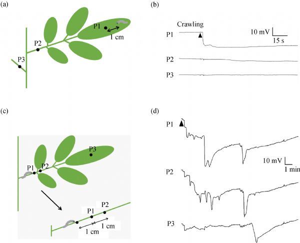
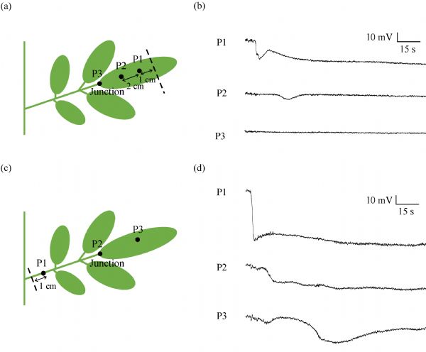
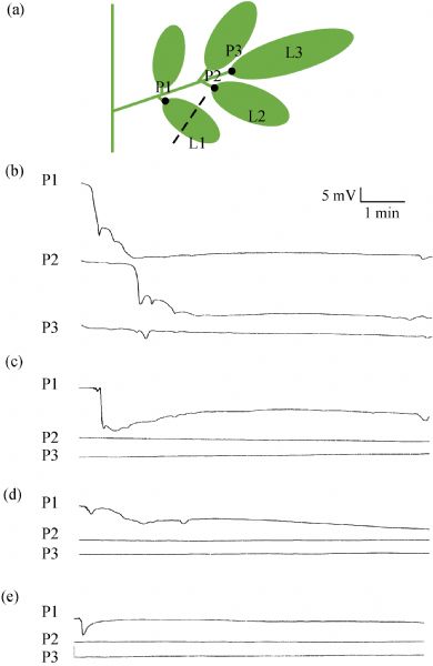
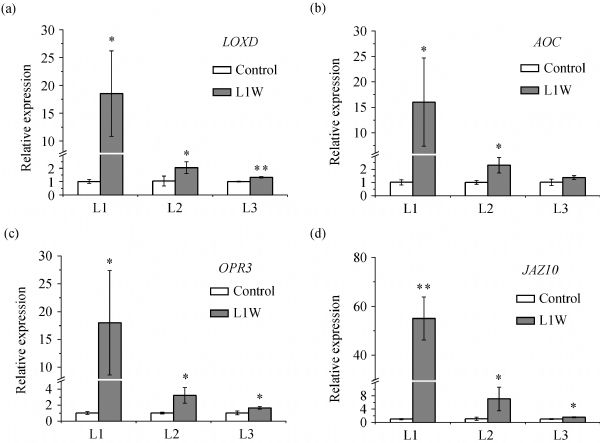
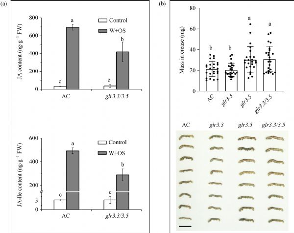
Electrical signals commonly occur in plants in response to various environmental changes and have a dominant function in plant acclimation. The transduction of wound-elicited electrical signals in the model plant species Arabidopsis has been characterized but the characteristics of electrical signal transduction in response to herbivory or wounding in crop species remain unknown. Here, the features of electrical signals elicited by insect herbivory and wounding in tomato were investigated. Unlike those in Arabidopsis, wounding tomato leaves did not cause leaf-to-leaf electrical signal transduction. In contrast, electrical signals elicited in response to petiole wounding were stronger and more strongly transduced. Leaflet wounding also activated electrical signal transduction and jasmonic acid (JA) signaling within the whole compound leaf. It was also demonstrated that tomato glutamate receptor-like 3.3 (GLR3.3) and GLR3.5 mediated leaflet-to-leaflet electrical signal transduction. Herbivory-induced JA accumulation and Helicoverpa armigera resistance were reduced in glr3.3/3.5 plants. This work reveals the nature of electrical signal transduction in tomato and emphasizes the key roles of GLR3.3 and GLR3.5 in electrical signal transduction and JA signaling activation.
|
| Keywords
electrical signal
glutamate receptor-like
herbivory
jasmonic acid
tomato
|
|
Corresponding Author(s):
Jingquan YU
|
|
Just Accepted Date: 30 April 2021
Online First Date: 24 May 2021
Issue Date: 13 July 2021
|
|
| 1 |
W G Choi, G Miller, I Wallace, J Harper, R Mittler, S Gilroy. Orchestrating rapid long-distance signaling in plants with Ca2+, ROS and electrical signals. Plant Journal, 2017, 90(4): 698–707
https://doi.org/10.1111/tpj.13492
pmid: 28112437
|
| 2 |
G Miller, K Schlauch, R Tam, D Cortes, M A Torres, V Shulaev, J L Dangl, R Mittler. The plant NADPH oxidase RBOHD mediates rapid systemic signaling in response to diverse stimuli. Science Signaling, 2009, 2(84): ra45
https://doi.org/10.1126/scisignal.2000448
pmid: 19690331
|
| 3 |
S Gilroy, N Suzuki, G Miller, W G Choi, M Toyota, A R Devireddy, R Mittler. A tidal wave of signals: calcium and ROS at the forefront of rapid systemic signaling. Trends in Plant Science, 2014, 19(10): 623–630
https://doi.org/10.1016/j.tplants.2014.06.013
pmid: 25088679
|
| 4 |
V Kiep, J Vadassery, J Lattke, J P Maaß, W Boland, E Peiter, A Mithöfer. Systemic cytosolic Ca2+ elevation is activated upon wounding and herbivory in Arabidopsis. New Phytologist, 2015, 207(4): 996–1004
https://doi.org/10.1111/nph.13493
pmid: 25996806
|
| 5 |
M Toyota, D Spencer, S Sawai-Toyota, W Jiaqi, T Zhang, A J Koo, G A Howe, S Gilroy. Glutamate triggers long-distance, calcium-based plant defense signaling. Science, 2018, 361(6407): 1112–1115
https://doi.org/10.1126/science.aat7744
pmid: 30213912
|
| 6 |
S A R Mousavi, A Chauvin, F Pascaud, S Kellenberger, E E Farmer. GLUTAMATE RECEPTOR-LIKE genes mediate leaf-to-leaf wound signalling. Nature, 2013, 500(7463): 422–426
https://doi.org/10.1038/nature12478
pmid: 23969459
|
| 7 |
R Hedrich, V Salvador-Recatalà, I Dreyer. Electrical wiring and long-distance plant communication. Trends in Plant Science, 2016, 21(5): 376–387
https://doi.org/10.1016/j.tplants.2016.01.016
pmid: 26880317
|
| 8 |
J Burdon-Sanderson. Note on the electrical phenomena which accompany irritation of the leaf of Dionaea muscipula. Proceedings of the Royal Society of London, 1872, 21: 495–496
|
| 9 |
C R Darwin. Insectivorous plants. London: John Murray, 1875
|
| 10 |
D C Wildon, J F Thain, P E H Minchin, I R Gubb, A J Reilly, Y D Skipper, H M Doherty, P J Odonnell, D J Bowles. Electrical signaling and systemic proteinase-inhibitor induction in the wounded plant. Nature, 1992, 360(6399): 62–65
https://doi.org/10.1038/360062a0
|
| 11 |
M Białasek, M Górecka, R Mittler, S Karpiński. Evidence for the involvement of electrical, calcium and ROS signaling in the systemic regulation of non- photochemical quenching and photosynthesis. Plant & Cell Physiology, 2017, 58(2): 207–215
https://doi.org/10.1093/pcp/pcw232
pmid: 28184891
|
| 12 |
G Wang, C Hu, J Zhou, Y Liu, J Cai, C Pan, Y Wang, X Wu, K Shi, X Xia, Y Zhou, C H Foyer, J Yu. Systemic root-shoot signaling drives jasmonate-based root defense against nematodes. Current Biology, 2019, 29(20): 3430–3438.e4
https://doi.org/10.1016/j.cub.2019.08.049
pmid: 31588001
|
| 13 |
E Michard, P T Lima, F Borges, A C Silva, M T Portes, J E Carvalho, M Gilliham, L H Liu, G Obermeyer, J A Feijó. Glutamate receptor-like genes form Ca2+ channels in pollen tubes and are regulated by pistil D-serine. Science, 2011, 332(6028): 434–437
https://doi.org/10.1126/science.1201101
pmid: 21415319
|
| 14 |
E D Vincill, A E Clarin, J N Molenda, E P Spalding. Interacting glutamate receptor-like proteins in Phloem regulate lateral root initiation in Arabidopsis. Plant Cell, 2013, 25(4): 1304–1313
https://doi.org/10.1105/tpc.113.110668
pmid: 23590882
|
| 15 |
S K Singh, C T Chien, I F Chang. The Arabidopsis glutamate receptor-like gene GLR3.6 controls root development by repressing the Kip-related protein gene KRP4. Journal of Experimental Botany, 2016, 67(6): 1853–1869
https://doi.org/10.1093/jxb/erv576
pmid: 26773810
|
| 16 |
Y Cheng, X Zhang, T Sun, Q Tian, W H Zhang. Glutamate receptor homolog3.4 is involved in regulation o seed germination under salt stress in Arabidopsis. Plant & Cell Physiology, 2018, 59(5): 978–988
https://doi.org/10.1093/pcp/pcy034
pmid: 29432559
|
| 17 |
P H Wang, C E Lee, Y S Lin, M H Lee, P Y Chen, H C Chang, I F Chang. The glutamate receptor-like protein GLR3.7 interacts with 14–3-3 omega and participates in salt stress response in Arabidopsis thaliana. Frontiers in Plant Science, 2019, 10: 1169
https://doi.org/10.3389/fpls.2019.01169
pmid: 31632419
|
| 18 |
Y Zheng, L Luo, J Wei, Q Chen, Y Yang, X Hu, X Kong. The glutamate receptors AtGLR1.2 and AtGLR1.3 increase cold tolerance by regulating jasmonate signaling in Arabidopsis thaliana. Biochemical and Biophysical Research Communications, 2018, 506(4): 895–900
https://doi.org/10.1016/j.bbrc.2018.10.153
pmid: 30392908
|
| 19 |
H Li, X Jiang, X Lv, G J Ahammed, Z Guo, Z Qi, J Yu, Y Zhou. Tomato GLR3.3 and GLR3.5 mediate cold acclimation-induced chilling tolerance by regulating apoplastic H2O2 production and redox homeostasis. Plant, Cell & Environment, 2019, 42(12): 3326–3339
https://doi.org/10.1111/pce.13623
pmid: 31329293
|
| 20 |
H Manzoor, J Kelloniemi, A Chiltz, D Wendehenne, A Pugin, B Poinssot, A Garcia-Brugger. Involvement of the glutamate receptor AtGLR3.3 in plant defense signaling and resistance to Hyaloperonospora arabidopsidis. Plant Journal, 2013, 76(3): 466–480
https://doi.org/10.1111/tpj.12311
pmid: 23952652
|
| 21 |
F Li, J Wang, C Ma, Y Zhao, Y Wang, A Hasi, Z Qi. Glutamate receptor-like channel3.3 is involved in mediating glutathione-triggered cytosolic calcium transients, transcriptional changes, and innate immunity responses in Arabidopsis. Plant Physiology, 2013, 162(3): 1497–1509
https://doi.org/10.1104/pp.113.217208
pmid: 23656893
|
| 22 |
C T Nguyen, A Kurenda, S Stolz, A Chételat, E E Farmer. Identification of cell populations necessary for leaf-to-leaf electrical signaling in a wounded plant. Proceedings of the National Academy of Sciences of the United States of America, 2018, 115(40): 10178–10183
https://doi.org/10.1073/pnas.1807049115
pmid: 30228123
|
| 23 |
Y Lei, L Lu, H Y Liu, S Li, F Xing, L L Chen. CRISPR-P: a web tool for synthetic single-guide RNA design of CRISPR-system in plants. Molecular Plant, 2014, 7(9): 1494–1496
|
| 24 |
C Pan, L Ye, L Qin, X Liu, Y He, J Wang, L Chen, G Lu. CRISPR/Cas9-mediated efficient and heritable targeted mutagenesis in tomato plants in the first and later generations. Scientific Reports, 2016, 6(1): 24765
https://doi.org/10.1038/srep24765
pmid: 27097775
|
| 25 |
J Fillatti, J Kiser, R Rose, L Comai. Efficient transfer of a glyphosate tolerance gene into tomato using a binary Agrobacterium tumefaciens vector. Bio/Technology, 1987, 5: 726–730
|
| 26 |
K J Livak, T D Schmittgen. Analysis of relative gene expression data using real-time quantitative PCR and the 2− ΔΔCT Method. Methods, 2001, 25(4): 402–408
https://doi.org/10.1006/meth.2001.1262
pmid: 11846609
|
| 27 |
H Zhang, Z Hu, C Lei, C Zheng, J Wang, S Shao, X Li, X Xia, X Cai, J Zhou, Y Zhou, J Yu, C H Foyer, K Shi. A plant phytosulfokine peptide initiates auxin-dependent immunity through cytosolic Ca2+ signaling in tomato. Plant Cell, 2018, 30(3): 652–667
https://doi.org/10.1105/tpc.17.00537
pmid: 29511053
|
| 28 |
J Browse. Jasmonate passes muster: a receptor and targets for the defense hormone. Annual Review of Plant Biology, 2009, 60(1): 183–205
https://doi.org/10.1146/annurev.arplant.043008.092007
pmid: 19025383
|
| 29 |
C L Ballaré. Jasmonate-induced defenses: a tale of intelligence, collaborators and rascals. Trends in Plant Science, 2011, 16(5): 249–257
https://doi.org/10.1016/j.tplants.2010.12.001
pmid: 21216178
|
| 30 |
C Wasternack, B Hause. Jasmonates: biosynthesis, perception, signal transduction and action in plant stress response, growth and development. An update to the 2007 review in Annals of Botany. Annals of Botany, 2013, 111(6): 1021–1058
https://doi.org/10.1093/aob/mct067
pmid: 23558912
|
| 31 |
G Glauser, E Grata, L Dubugnon, S Rudaz, E E Farmer, J L Wolfender. Spatial and temporal dynamics of jasmonate synthesis and accumulation in Arabidopsis in response to wounding. Journal of Biological Chemistry, 2008, 283(24): 16400–16407
https://doi.org/10.1074/jbc.M801760200
pmid: 18400744
|
| 32 |
G Glauser, L Dubugnon, S A R Mousavi, S Rudaz, J L Wolfender, E E Farmer. Velocity estimates for signal propagation leading to systemic jasmonic acid accumulation in wounded Arabidopsis. Journal of Biological Chemistry, 2009, 284(50): 34506–34513
https://doi.org/10.1074/jbc.M109.061432
pmid: 19846562
|
| 33 |
A J K Koo, X Gao, A Daniel Jones, G A Howe. A rapid wound signal activates the systemic synthesis of bioactive jasmonates in Arabidopsis. Plant Journal, 2009, 59(6): 974–986
https://doi.org/10.1111/j.1365-313X.2009.03924.x
pmid: 19473329
|
| 34 |
L Li, C Li, G I Lee, G A Howe. Distinct roles for jasmonate synthesis and action in the systemic wound response of tomato. Proceedings of the National Academy of Sciences of the United States of America, 2002, 99(9): 6416–6421
https://doi.org/10.1073/pnas.072072599
pmid: 11959903
|
| 35 |
C Koziolek, T E E Grams, U Schreiber, R Matyssek, J Fromm. Transient knockout of photosynthesis mediated by electrical signals. New Phytologist, 2004, 161(3): 715–722
https://doi.org/10.1111/j.1469-8137.2004.00985.x
pmid: 33873726
|
|
Viewed |
|
|
|
Full text
|
|
|
|
|
Abstract
|
|
|
|
|
Cited |
|
|
|
|
| |
Shared |
|
|
|
|
| |
Discussed |
|
|
|
|