|
|
|
Bacteriophages in water pollution control: Advantages and limitations |
Mengzhi Ji1, Zichen Liu1, Kaili Sun1, Zhongfang Li2( ), Xiangyu Fan1(), Qiang Li1 ), Xiangyu Fan1(), Qiang Li1 |
1. School of Biological Science and Technology, University of Jinan, Jinan 250022, China
2. College of Food and Bioengineering, Hezhou University, Hezhou 542899, China |
|
|
|
|
Abstract •Phages can be better indicators of enteric viruses than fecal indicator bacteria. •Multiple phages should be added to the microbial source tracking toolbox. •Engineered phage or phage cocktail can effectively target resistant bacteria. •In phage use, phage-mediated horizontal gene transfer cannot be ignored. •More schemes are needed to prevent phage concentration from decreasing.
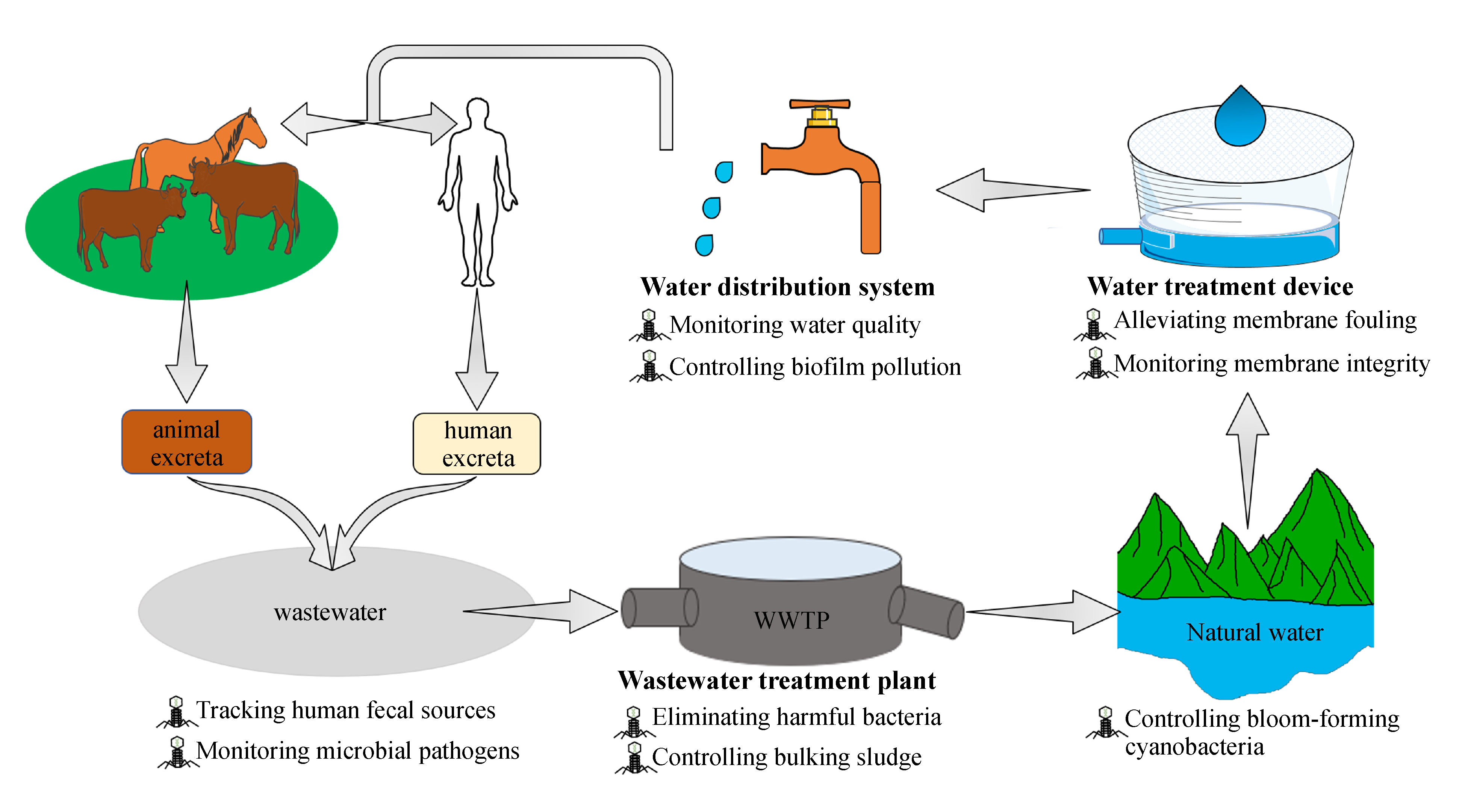
Wastewater is a breeding ground for many pathogens, which may pose a threat to human health through various water transmission pathways. Therefore, a simple and effective method is urgently required to monitor and treat wastewater. As bacterial viruses, bacteriophages (phages) are the most widely distributed and abundant organisms in the biosphere. Owing to their capacity to specifically infect bacterial hosts, they have recently been used as novel tools in water pollution control. The purpose of this review is to summarize and evaluate the roles of phages in monitoring pathogens, tracking pollution sources, treating pathogenic bacteria, infecting bloom-forming cyanobacteria, and controlling bulking sludge and biofilm pollution in wastewater treatment systems. We also discuss the limitations of phage usage in water pollution control, including phage-mediated horizontal gene transfer, the evolution of bacterial resistance, and phage concentration decrease. This review provides an integrated outlook on the use of phages in water pollution control.
|
| Keywords
Phage
Water pollution monitoring
Harmful bacteria biocontrol
Horizontal gene transfer
Bacterial resistance
|
|
Corresponding Author(s):
Zhongfang Li,Xiangyu Fan
|
|
Issue Date: 07 December 2020
|
|
| 1 |
W Ahmed, A Lobos, J Senkbeil, J Peraud, J Gallard, V J Harwood (2018). Evaluation of the novel crAssphage marker for sewage pollution tracking in storm drain outfalls in Tampa, Florida. Water Research, 131: 142–150
https://doi.org/10.1016/j.watres.2017.12.011
|
| 2 |
M Amarasiri, M Kitajima, T H Nguyen, S Okabe, D Sano (2017). Bacteriophage removal efficiency as a validation and operational monitoring tool for virus reduction in wastewater reclamation. Water Research, 121: 258–269
https://doi.org/10.1016/j.watres.2017.05.035
|
| 3 |
S Aracic, S Manna, S Petrovski, J L Wiltshire, G Mann, A E Franks (2015). Innovative biological approaches for monitoring and improving water quality. Frontiers in Microbiology, 6: 826
https://doi.org/10.3389/fmicb.2015.00826
|
| 4 |
S Ayyaru, J Choi, Y H Ahn (2018). Biofouling reduction in a MBR by the application of a lytic phage on a modified nanocomposite membrane. Environmental Science. Water Research & Technology, 4(10): 1624–1638
https://doi.org/10.1039/C8EW00316E
|
| 5 |
K Bányai, M K Estes, V Martella, U D Parashar (2018). Viral gastroenteritis. Lancet, 392(10142): 175–186
https://doi.org/10.1016/S0140-6736(18)31128-0
|
| 6 |
S Batinovic, F Wassef, S A Knowler, D T Rice, C R Stanton, J Rose, J Tucci, T Nittami, A Vinh, G R Drummond, C G Sobey, H T Chan, R J Seviour, S Petrovski, A E Franks (2019). Bacteriophages in Natural and Artificial Environments. Pathogens (Basel, Switzerland), 8(3): 100
https://doi.org/10.3390/pathogens8030100
|
| 7 |
A S Bhattacharjee, J Choi, A M Motlagh, S T Mukherji, R Goel (2015). Bacteriophage therapy for membrane biofouling in membrane bioreactors and antibiotic‐resistant bacterial biofilms. Biotechnology and Bioengineering, 112(8): 1644–1654
https://doi.org/10.1002/bit.25574
|
| 8 |
A Bivins, K Crank, J Greaves, D North, Z Wu, K Bibby (2020). Cross-assembly phage and pepper mild mottle virus as viral water quality monitoring tools–potential, research gaps, and way forward. Current Opinion in Environmental Science & Health, 16: 54-61
https://doi.org/10.1016/j.coesh.2020.02.001
|
| 9 |
M Brown-Jaque, W Calero-Cáceres, P Espinal, J Rodríguez-Navarro, E Miró, J J González-López, T Cornejo, J C, Hurtado F Navarro, M Muniesa (2018). Antibiotic resistance genes in phage particles isolated from human faeces and induced from clinical bacterial isolates. International Journal of Antimicrobial Agents, 51(3): 434–442
https://doi.org/10.1016/j.ijantimicag.2017.11.014
|
| 10 |
W Calero-Cáceres, J Méndez, J Martín-Díaz, M Muniesa (2017). The occurrence of antibiotic resistance genes in a Mediterranean river and their persistence in the riverbed sediment. Environmental Pollution, 223: 384–394
https://doi.org/10.1016/j.envpol.2017.01.035
|
| 11 |
W Calero-Cáceres, M Muniesa (2016). Persistence of naturally occurring antibiotic resistance genes in the bacteria and bacteriophage fractions of wastewater. Water Research, 95: 11–18
https://doi.org/10.1016/j.watres.2016.03.006
|
| 12 |
W Calero-Cáceres, M Ye, J L Balcázar (2019). Bacteriophages as environmental reservoirs of antibiotic resistance. Trends in microbiology, 27(7): 570–577
https://doi.org/10.1016/j.tim.2019.02.008
|
| 13 |
W N Chaudhry, I U Haq, S Andleeb, I Qadri (2014). Characterization of a virulent bacteriophage LK1 specific for Citrobacter freundii isolated from sewage water. Journal of Basic Microbiology, 54(6): 531–541
https://doi.org/10.1002/jobm.201200710
|
| 14 |
J Chen, N Carpena, N Quiles-Puchalt, G Ram, R P Novick, J R Penadés (2015). Intra-and inter-generic transfer of pathogenicity island-encoded virulence genes by cos phages. ISME Journal, 9(5): 1260–1263
https://doi.org/10.1038/ismej.2014.187
|
| 15 |
X Cheng, H M Delanka-Pedige, S P Munasinghe-Arachchige, I S Abeysiriwardana-Arachchige, G B Smith, N Nirmalakhandan, Y Zhang (2020). Removal of antibiotic resistance genes in an algal-based wastewater treatment system employing Galdieria sulphuraria: A comparative study. Science of the Total Environment, 711: 134435
https://doi.org/10.1016/j.scitotenv.2019.134435
|
| 16 |
N Chyerochana, A Kongprajug, P Somnark, P Leelapanang Kamphaengthong, S Mongkolsuk, K Sirikanchana (2020). Distributions of enterococci and human-specific bacteriophages of enterococci in a tropical watershed. International Journal of Hygiene and Environmental Health, 226: 113482
https://doi.org/10.1016/j.ijheh.2020.113482
|
| 17 |
O Cinek, K Mazankova, L Kramna, R Odeh, A Alassaf, M U Ibekwe, G Ahmadov, H Mekki, M A Abdullah, B M Elmahi, H Hyöty, P Rainetova (2018). Quantitative CrAssphage real-time PCR assay derived from data of multiple geographically distant populations. Journal of Medical Virology, 90(4): 767–771
https://doi.org/10.1002/jmv.25012
|
| 18 |
K Crank, X Li, D North, G B Ferraro, M Iaconelli, P Mancini, G La Rosa, K Bibby (2020). CrAssphage abundance and correlation with molecular viral markers in Italian wastewater. Water Research, 184: 116161
https://doi.org/10.1016/j.watres.2020.116161
|
| 19 |
A de Brauwere, N K Ouattara, P Servais (2014). Modeling fecal indicator bacteria concentrations in natural surface waters: A review. Critical Reviews in Environmental Science and Technology, 44(21): 2380–2453
https://doi.org/10.1080/10643389.2013.829978
|
| 20 |
Y Deng, B Li, T Zhang (2018). Bacteria that make a meal of sulfonamide antibiotics: Blind spots and emerging opportunities. Environmental Science & Technology, 52(7): 3854–3868
https://doi.org/10.1021/acs.est.7b06026
|
| 21 |
E Dias, J Ebdon, H Taylor (2018). The application of bacteriophages as novel indicators of viral pathogens in wastewater treatment systems. Water Research, 129: 172–179
https://doi.org/10.1016/j.watres.2017.11.022
|
| 22 |
D Diston, M Sinreich, S Zimmermann, A Baumgartner, R Felleisen (2015). Evaluation of molecular-and culture-dependent MST markers to detect fecal contamination and indicate viral presence in good quality groundwater. Environmental Science & Technology, 49(12): 7142–7151
https://doi.org/10.1021/acs.est.5b00515
|
| 23 |
D Diston, M Wicki (2015). Occurrence of bacteriophages infecting Bacteroides host strains (ARABA 84 and GB-124) in fecal samples of human and animal origin. Journal of Water and Health, 13(3): 654–661
https://doi.org/10.2166/wh.2014.199
|
| 24 |
J Doss, K Culbertson, D Hahn, J Camacho, N Barekzi (2017). A review of phage therapy against bacterial pathogens of aquatic and terrestrial organisms. Viruses, 9(3): 50
https://doi.org/10.3390/v9030050
|
| 25 |
P Dow, K Kotz, S Gruszka, J Holder, J Fiering (2018). Acoustic separation in plastic microfluidics for rapid detection of bacteria in blood using engineered bacteriophage. Lab on a Chip, 18(6): 923–932
https://doi.org/10.1039/C7LC01180F
|
| 26 |
B E Dutilh, N Cassman, K Mcnair, S E Sanchez, G G Silva, L Boling, J J Barr, D R Speth, V Seguritan, R K Aziz, B Felts, E A Dinsdale, J L Mokili, R A Edwards (2014). A highly abundant bacteriophage discovered in the unknown sequences of human faecal metagenomes. Nature Communications, 5(1): 4498
https://doi.org/10.1038/ncomms5498
|
| 27 |
R A Edwards, A A Vega, H M Norman, M Ohaeri, K Levi, E A Dinsdale, O Cinek, R K Aziz, K Mcnair, J J Barr, K Bibby, S J J Brouns, A Cazares, P A de Jonge, C Desnues, S L Díaz Muñoz, P C Fineran, A Kurilshikov, R Lavigne, K Mazankova, D T McCarthy, F L Nobrega, A Reyes Muñoz, G Tapia, N Trefault, A V Tyakht, P Vinuesa, J Wagemans, A Zhernakova, F M Aarestrup, G Ahmadov, A Alassaf, J Anton, A Asangba, E K Billings, V A Cantu, J M Carlton, D Cazares, G S Cho, T Condeff, P Cortés, M Cranfield, D A Cuevas, R De la Iglesia, P Decewicz, M P Doane, N J Dominy, L Dziewit, B M Elwasila, A M Eren, C Franz, J Fu, C Garcia-Aljaro, E Ghedin, K M Gulino, J M Haggerty, S R Head, R S Hendriksen, C Hill, H Hyöty, E N Ilina, M T Irwin, T C Jeffries, J Jofre, R E Junge, S T Kelley, M Khan Mirzaei, M Kowalewski, D Kumaresan, S R Leigh, D Lipson, E S Lisitsyna, M Llagostera, J M Maritz, L C Marr, A McCann, S Molshanski-Mor, S Monteiro, B Moreira-Grez, M Morris, L Mugisha, M Muniesa, H Neve, N Nguyen, O D Nigro, A S Nilsson, T O’Connell, R Odeh, A Oliver, M Piuri, A J Prussin II, U Qimron, Z X Quan, P Rainetova, A Ramírez-Rojas, R Raya, K Reasor, G A O Rice, A Rossi, R Santos, J Shimashita, E N Stachler, L C Stene, R Strain, R Stumpf, P J Torres, A Twaddle, M A Ugochi Ibekwe, N Villagra, S Wandro, B White, A Whiteley, K L Whiteson, C Wijmenga, M M Zambrano, H Zschach, B E Dutilh (2019). Global phylogeography and ancient evolution of the widespread human gut virus crAssphage. Nature Microbiology, 4(10): 1727–1736
https://doi.org/10.1038/s41564-019-0494-6
|
| 28 |
G Ertürk, R Lood (2018). Bacteriophages as biorecognition elements in capacitive biosensors: Phage and host bacteria detection. Sensors and Actuators. B, Chemical, 258: 535–543
https://doi.org/10.1016/j.snb.2017.11.117
|
| 29 |
K Farkas, D I Walker, E M Adriaenssens, J E Mcdonald, L S Hillary, S K Malham, D L Jones (2020). Viral indicators for tracking domestic wastewater contamination in the aquatic environment. Water Research, 181: 115926
https://doi.org/10.1016/j.watres.2020.115926
|
| 30 |
N Gabiatti, P Yu, J Mathieu, G W Lu, X Wang, H Zhang, H M Soares, P J Alvarez (2018). Bacterial endospores as phage genome carriers and protective shells. Applied and Environmental Microbiology, 84(18): e01186-18
https://doi.org/10.1128/AEM.01186-18
|
| 31 |
E B Gao, J F Gui, Q Y Zhang (2012). A novel cyanophage with a cyanobacterial nonbleaching protein A gene in the genome. Journal of Virology, 86(1): 236–245
https://doi.org/10.1128/JVI.06282-11
|
| 32 |
C García‐Aljaro, E Ballesté M Muniesa, J Jofre (2017). Determination of crAssphage in water samples and applicability for tracking human faecal pollution. Microbial Biotechnology, 10(6): 1775–1780 doi:10.1111/1751-7915.12841
|
| 33 |
J Gu, B Han, J Wang (2020). COVID-19: gastrointestinal manifestations and potential fecal-oral transmission. Gastroenterology, 158(6): 1518–1519
https://doi.org/10.1053/j.gastro.2020.02.054
|
| 34 |
C Guzmán, L Mocé-Llivina, F Lucena, J Jofre (2008). Evaluation of Escherichia coli host strain CB390 for simultaneous detection of somatic and F-specific coliphages. Applied and Environmental Microbiology, 74(2): 531–534
https://doi.org/10.1128/AEM.01710-07
|
| 35 |
C Hagedorn, A R Blanch, V J Harwood (2011). Microbial Source Tracking: Methods, Applications, and Case Studies. Dordrecht: Springer Science & Business Media
|
| 36 |
H G Hampton, B N Watson, P C Fineran (2020). The arms race between bacteria and their phage foes. Nature, 577(7790): 327–336
https://doi.org/10.1038/s41586-019-1894-8
|
| 37 |
I A Hamza, K Bibby (2019). Critical issues in application of molecular methods to environmental virology. Journal of Virological Methods, 266: 11–24
https://doi.org/10.1016/j.jviromet.2019.01.008
|
| 38 |
E Haramoto, S Fujino, M Otagiri (2015). Distinct behaviors of infectious F-specific RNA coliphage genogroups at a wastewater treatment plant. Science of the Total Environment, 520: 32–38
https://doi.org/10.1016/j.scitotenv.2015.03.034
|
| 39 |
K R Hodgson, V A Torok, A R Turnbull (2017). Bacteriophages as enteric viral indicators in bivalve mollusc management. Food Microbiology, 65: 284–293
https://doi.org/10.1016/j.fm.2017.03.003
|
| 40 |
S A Jassim, R G Limoges (2013). Impact of external forces on cyanophage–host interactions in aquatic ecosystems. World Journal of Microbiology & Biotechnology, 29(10): 1751–1762
https://doi.org/10.1007/s11274-013-1358-5
|
| 41 |
S A Jassim, R G Limoges, H El-Cheikh (2016). Bacteriophages biocontrol in wastewater treatment. World Journal of Microbiology & Biotechnology, 32(4): 70
https://doi.org/10.1007/s11274-0.16-2028-1
|
| 42 |
S A Jassim, R G Limoges (2017). Bacteriophages: Practical Applications for Nature’s Biocontrol. Dordrecht: Springer
|
| 43 |
J Jofre, A R Blanch, F Lucena, M Muniesa (2014). Bacteriophages infecting Bacteroides as a marker for microbial source tracking. Water Research, 55: 1–11
https://doi.org/10.1016/j.watres.2014.02.006
|
| 44 |
J Jofre, F Lucena, A R Blanch, M Muniesa (2016). Coliphages as model organisms in the characterization and management of water resources. Water (Basel), 8(5): 199
https://doi.org/10.3390/w8050199
|
| 45 |
J W Jun, S S Giri, H J Kim, S K Yun, C Chi, J Y Chai, B C Lee, S C Park (2016). Bacteriophage application to control the contaminated water with Shigella. Scientific Reports, 6: 22636
https://doi.org/10.1038/srep22636
|
| 46 |
N Karumidze, I Kusradze, S Rigvava, M Goderdzishvili, K Rajakumar, Z Alavidze (2013). Isolation and characterisation of lytic bacteriophages of Klebsiella pneumoniae and Klebsiella oxytoca. Current Microbiology, 66(3): 251–258
https://doi.org/10.1007/s00284-012-0264-7
|
| 47 |
F M Khan, R Gupta (2020). Escherichia coli (E. coli) as an Indicator of Fecal Contamination in Groundwater: A Review. New York: Springer, 225–235
|
| 48 |
S M Kotay, T Datta, J Choi, R Goel (2011). Biocontrol of biomass bulking caused by Haliscomenobacter hydrossis using a newly isolated lytic bacteriophage. Water Research, 45(2): 694–704
https://doi.org/10.1016/j.watres.2010.08.038
|
| 49 |
M Kwiatek, S Parasion, P Rutyna, L Mizak, R Gryko, M Niemcewicz, A Olender, M Łobocka (2017). Isolation of bacteriophages and their application to control Pseudomonas aeruginosa in planktonic and biofilm models. Research in Microbiology, 168(3): 194–207
https://doi.org/10.1016/j.resmic.2016.10.009
|
| 50 |
O Larrañaga, M Brown-Jaque, P Quirós, C Gómez-Gómez, A R Blanch, L Rodríguez-Rubio, M Muniesa (2018). Phage particles harboring antibiotic resistance genes in fresh-cut vegetables and agricultural soil. Environment International, 115: 133–141
https://doi.org/10.1016/j.envint.2018.03.019
|
| 51 |
I Lekunberri, J Subirats, C M Borrego, J L Balcázar (2017a). Exploring the contribution of bacteriophages to antibiotic resistance. Environmental Pollution, 220: 981–984
https://doi.org/10.1016/j.envpol.2016.11.059
|
| 52 |
I Lekunberri, M Villagrasa, J L Balcázar, C M Borrego (2017b). Contribution of bacteriophage and plasmid DNA to the mobilization of antibiotic resistance genes in a river receiving treated wastewater discharges. Science of the Total Environment, 601-602: 206–209
https://doi.org/10.1016/j.scitotenv.2017.05.174
|
| 53 |
S S Leung, S Morales, W Britton, E Kutter, H K Chan (2018). Microfluidic-assisted bacteriophage encapsulation into liposomes. International Journal of Pharmaceutics, 545(1–2): 176–182
https://doi.org/10.1016/j.ijpharm.2018.04.063
|
| 54 |
B Li, F Ju, L Cai, T Zhang (2015). Profile and fate of bacterial pathogens in sewage treatment plants revealed by high-throughput metagenomic approach. Environmental Science & Technology, 49(17): 10492–10502
https://doi.org/10.1021/acs.est.5b02345
|
| 55 |
L L Li, P Yu, X Wang, S S Yu, J Mathieu, H Q Yu, P J Alvarez (2017). Enhanced biofilm penetration for microbial control by polyvalent phages conjugated with magnetic colloidal nanoparticle clusters (CNCs). Environmental Science. Nano, 4(9): 1817–1826
https://doi.org/10.1039/C7EN00414A
|
| 56 |
Y Liang, X Jin, Y Huang, S Chen (2018). Development and application of a real-time polymerase chain reaction assay for detection of a novel gut bacteriophage (CrAssphage). Journal of Medical Virology, 90(3): 464–468
https://doi.org/10.1002/jmv.24974
|
| 57 |
N T Lin, P Y Chiou, K C Chang, L K Chen, M J Lai (2010). Isolation and characterization of AB2: A novel bacteriophage of Acinetobacter baumannii. Research in Microbiology, 161(4): 308–314
https://doi.org/10.1016/j.resmic.2010.03.007
|
| 58 |
M Liu, J J Gill, R Young, E J Summer (2015). Bacteriophages of wastewater foaming-associated filamentous Gordonia reduce host levels in raw activated sludge. Scientific Reports, 5(1): 13754
https://doi.org/10.1038/srep13754
|
| 59 |
T K Lu, J J Collins (2007). Dispersing biofilms with engineered enzymatic bacteriophage. Proceedings of the National Academy of Sciences of the United States of America, 104(27): 11197–11202
https://doi.org/10.1073/pnas.0704624104
|
| 60 |
K B Maal, A S Delfan, S Salmanizadeh (2015). Isolation and identification of two novel Escherichia Coli bacteriophages and their application in wastewater treatment and coliform’s phage therapy. Jundishapur Journal of Microbiology, 8(3): e14945
https://doi.org/10.5812/jjm.14945
|
| 61 |
J Mathieu, P Yu, P Zuo, M L Da Silva, P J Alvarez (2019). Going viral: Emerging opportunities for phage-based bacterial control in water treatment and reuse. Accounts of Chemical Research, 52(4): 849–857
https://doi.org/10.1021/acs.accounts.8b00576
|
| 62 |
B R McMinn, N J Ashbolt, A Korajkic (2017). Bacteriophages as indicators of faecal pollution and enteric virus removal. Letters in Applied Microbiology, 65(1): 11–26
https://doi.org/10.1111/lam.12736
|
| 63 |
M Moazeni, M Nikaeen, M Hadi, S Moghim, L Mouhebat, M Hatamzadeh, A Hassanzadeh (2017). Estimation of health risks caused by exposure to enteroviruses from agricultural application of wastewater effluents. Water Research, 125: 104–113
https://doi.org/10.1016/j.watres.2017.08.028
|
| 64 |
R Monteiro, D P Pires, A R Costa, J Azeredo (2019). Phage therapy: going temperate? Trends in Microbiology, 27(4): 368–378
https://doi.org/10.1016/j.tim.2018.10.008
|
| 65 |
A M Motlagh, A S Bhattacharjee, R Goel (2016). Biofilm control with natural and genetically-modified phages. World Journal of Microbiology & Biotechnology, 32(4): 67
https://doi.org/10.1007/s11274-016-2009-4
|
| 66 |
M Muniesa, E Ballesté, L Imamovic, M Pascual-Benito, D Toribio-Avedillo, F Lucena, A Blanch, J Jofre (2018). Bluephage: A rapid method for the detection of somatic coliphages used as indicators of fecal pollution in water. Water Research, 128: 10–19
https://doi.org/10.1016/j.watres.2017.10.030
|
| 67 |
S Petrovski, R J Seviour, D Tillett (2011a). Characterization of the genome of the polyvalent lytic bacteriophage GTE2, which has potential for biocontrol of Gordonia-, Rhodococcus-, and Nocardia-stabilized foams in activated sludge plants. Applied and Environmental Microbiology, 77(12): 3923–3929
https://doi.org/10.1128/AEM.00025-11
|
| 68 |
S Petrovski, R J Seviour, D Tillett (2011b). Prevention of Gordonia and Nocardia stabilized foam formation by using bacteriophage GTE7. Applied and Environmental Microbiology, 77(21): 7864–7867
https://doi.org/10.1128/AEM.05692-11
|
| 69 |
D P Pires, L D R Melo, D Vilas Boas, S Sillankorva, J Azeredo (2017). Phage therapy as an alternative or complementary strategy to prevent and control biofilm-related infections. Current Opinion in Microbiology, 39: 48–56
https://doi.org/10.1016/j.mib.2017.09.004
|
| 70 |
S Purnell, J Ebdon, H Wilkins, H Taylor (2018). Human-specific phages infecting Enterococcus host strain MW47: are they reliable microbial source tracking markers? Journal of Applied Microbiology, 124(5): 1274–1282
https://doi.org/10.1111/jam.13700
|
| 71 |
S E Purnell, J E Ebdon, H D Taylor (2011). Bacteriophage lysis of Enterococcus host strains: A tool for microbial source tracking? Environmental Science & Technology, 45(24): 10699–10705
https://doi.org/10.1021/es202141x
|
| 72 |
B Rémy, S Mion, L Plener, M Elias, E Chabrière, D Daudé (2018). Interference in bacterial quorum sensing: A biopharmaceutical perspective. Frontiers in Pharmacology, 9: 203
https://doi.org/10.3389/fphar.2018.00203
|
| 73 |
Ł Richter, M Janczuk-Richter, J Niedziółka-Jönsson, J Paczesny, R Hołyst (2018). Recent advances in bacteriophage-based methods for bacteria detection. Drug Discovery Today, 23(2): 448–455
https://doi.org/10.1016/j.drudis.2017.11.007
|
| 74 |
S Rodriguez-Mozaz, S Chamorro, E Marti, B Huerta, M Gros, A Sànchez-Melsió, C M Borrego, D Barceló, J L Balcázar (2015). Occurrence of antibiotics and antibiotic resistance genes in hospital and urban wastewaters and their impact on the receiving river. Water Research, 69: 234–242
https://doi.org/10.1016/j.watres.2014.11.021
|
| 75 |
Ş Şener, E Şener, A Davraz (2017). Evaluation of water quality using water quality index (WQI) method and GIS in Aksu River (SW-Turkey). Science of the Total Environment, 584-585: 131–144
https://doi.org/10.1016/j.scitotenv.2017.01.102
|
| 76 |
A N Shkoporov, E V Khokhlova, C B Fitzgerald, S R Stockdale, L A Draper, R P Ross, C Hill (2018). CrAss001 represents the most abundant bacteriophage family in the human gut and infects Bacteroides intestinalis. Nature Communications, 9(1): 4781
https://doi.org/10.1038/s41467-018-07225-7
|
| 77 |
J A Soller, M E Schoen, T Bartrand, J E Ravenscroft, N J Ashbolt (2010). Estimated human health risks from exposure to recreational waters impacted by human and non-human sources of faecal contamination. Water Research, 44(16): 4674–4691
https://doi.org/10.1016/j.watres.2010.06.049
|
| 78 |
E Stachler, C Kelty, M Sivaganesan, X Li, K Bibby, O C Shanks (2017). Quantitative CrAssphage PCR Assays for Human Fecal Pollution Measurement. Environmental Science & Technology, 51(16): 9146–9154
https://doi.org/10.1021/acs.est.7b02703
|
| 79 |
J Q Su, X L An, B Li, Q L Chen, M R Gillings, H Chen, T Zhang, Y G Zhu (2017). Metagenomics of urban sewage identifies an extensively shared antibiotic resistome in China. Microbiome, 5(1): 84
https://doi.org/10.1186/s40168-017-0298-y
|
| 80 |
D Toribio-Avedillo, J Martín-Díaz, J Jofre, A R Blanch, M Muniesa (2019). New approach for the simultaneous detection of somatic coliphages and F-specific RNA coliphages as indicators of fecal pollution. Science of the Total Environment, 655: 263–272
https://doi.org/10.1016/j.scitotenv.2018.11.198
|
| 81 |
M Touchon, J A Moura de Sousa, E P Rocha (2017). Embracing the enemy: the diversification of microbial gene repertoires by phage-mediated horizontal gene transfer. Current Opinion in Microbiology, 38: 66–73
https://doi.org/10.1016/j.mib.2017.04.010
|
| 82 |
Y Turki, H Ouzari, I Mehri, A B Ammar, A Hassen (2012). Evaluation of a cocktail of three bacteriophages for the biocontrol of Salmonella of wastewater. Food Research International, 45(2): 1099–1105
https://doi.org/10.1016/j.foodres.2011.05.041
|
| 83 |
K Vijayavel, M N Byappanahalli, J Ebdon, H Taylor, R Whitman, D Kashian (2014). Enterococcus phages as potential tool for identifying sewage inputs in the Great Lakes region. Journal of Great Lakes Research, 40(4): 989–993
https://doi.org/10.1016/j.jglr.2014.09.011
|
| 84 |
K Vijayavel, R Fujioka, J Ebdon, H Taylor (2010). Isolation and characterization of Bacteroides host strain HB-73 used to detect sewage specific phages in Hawaii. Water Research, 44(12): 3714–3724
https://doi.org/10.1016/j.watres.2010.04.012
|
| 85 |
M Wang, P Liu, Q Zhou, W Tao, Y Sun, Z Zeng (2018). Estimating the contribution of bacteriophage to the dissemination of antibiotic resistance genes in pig feces. Environmental Pollution, 238: 291–298
https://doi.org/10.1016/j.envpol.2018.03.024
|
| 86 |
B Wangkahad, S Mongkolsuk, K Sirikanchana (2017). Integrated multivariate analysis with nondetects for the development of human sewage source-tracking tools using bacteriophages of Enterococcus faecalis. Environmental Science & Technology, 51(4): 2235–2245
https://doi.org/10.1021/acs.est.6b04714
|
| 87 |
Y Wei, A Kirby, B R Levin (2011). The population and evolutionary dynamics of Vibrio cholerae and its bacteriophage: Conditions for maintaining phage-limited communities. American Naturalist, 178(6): 715–725
https://doi.org/10.1086/662677
|
| 88 |
B Wu, R Wang, A G Fane (2017). The roles of bacteriophages in membrane-based water and wastewater treatment processes: A review. Water Research, 110: 120–132
https://doi.org/10.1016/j.watres.2016.12.004
|
| 89 |
Z Wu, J Greaves, L Arp, D Stone, K Bibby (2020). Comparative fate of CrAssphage with culturable and molecular fecal pollution indicators during activated sludge wastewater treatment. Environment International, 136: 105452
https://doi.org/10.1016/j.envint.2019.105452
|
| 90 |
Y Yang, W Shi, S Y Lu, J Liu, H Liang, Y Yang, G Duan, Y Li, H Wang, A Zhang (2018). Prevalence of antibiotic resistance genes in bacteriophage DNA fraction from Funan River water in Sichuan, China. Science of the Total Environment, 626: 835–841
https://doi.org/10.1016/j.scitotenv.2018.01.148
|
| 91 |
Y Yang, X Xie, M Tang, J Liu, H Tuo, J Gu, Y Tang, C Lei, H Wang, A Zhang (2020). Exploring the profile of antimicrobial resistance genes harboring by bacteriophage in chicken feces. Science of the Total Environment, 700: 134446
https://doi.org/10.1016/j.scitotenv.2019.134446
|
| 92 |
M Yen, L S Cairns, A Camilli (2017). A cocktail of three virulent bacteriophages prevents Vibrio cholerae infection in animal models. Nature Communications, 8(1): 14187
https://doi.org/10.1038/ncomms14187
|
| 93 |
Y Yoshida-Takashima, M Yoshida, H Ogata, K Nagasaki, S Hiroishi, T Yoshida (2012). Cyanophage infection in the bloom-forming cyanobacteria Microcystis aeruginosa in surface freshwater. Microbes and Environments, 27(4): 350–355
https://doi.org/10.1264/jsme2.ME12037
|
| 94 |
P Yu, J Mathieu, M Li, Z Dai, P J Alvarez (2016). Isolation of polyvalent bacteriophages by sequential multiple-host approaches. Applied and Environmental Microbiology, 82(3): 808–815
https://doi.org/10.1128/AEM.02382-15
|
| 95 |
P Yu, J Mathieu, G W Lu, N Gabiatti, P J Alvarez (2017). Control of antibiotic-resistant bacteria in activated sludge using polyvalent phages in conjunction with a production host. Environmental Science & Technology Letters, 4(4): 137–142
https://doi.org/10.1021/acs.estlett.7b00045
|
| 96 |
H Yue, Y He, E Fan, L Wang, S Lu, Z Fu (2017). Label-free electrochemiluminescent biosensor for rapid and sensitive detection of pseudomonas aeruginosa using phage as highly specific recognition agent. Biosensors & Bioelectronics, 94: 429–432
https://doi.org/10.1016/j.bios.2017.03.033
|
| 97 |
A Zhang, D R Call, T E Besser, J Liu, L Jones, H Wang, M A Davis (2019).β-lactam resistance genes in bacteriophage and bacterial DNA from wastewater, river water, and irrigation water in Washington State. Water Research, 161: 335–340
https://doi.org/10.1016/j.watres.2019.06.026
|
| 98 |
Y Zhou, A Marar, P Kner, R P Ramasamy (2017). Charge-directed immobilization of bacteriophage on nanostructured electrode for whole-cell electrochemical biosensors. Analytical Chemistry, 89(11): 5734–5741
https://doi.org/10.1021/acs.analchem.6b03751
|
|
Viewed |
|
|
|
Full text
|
|
|
|
|
Abstract
|
|
|
|
|
Cited |
|
|
|
|
| |
Shared |
|
|
|
|
| |
Discussed |
|
|
|
|