|
|
|
Acetylated HOXB9 at lysine 27 is of differential diagnostic value in patients with pancreatic ductal adenocarcinoma |
Xiaoran Sun1,2, Jiagui Song1, Jing Zhang1, Jun Zhan1( ), Weigang Fang1,2(), Hongquan Zhang1() ), Weigang Fang1,2(), Hongquan Zhang1() |
1. Department of Human Anatomy, Histology and Embryology, Key Laboratory of Carcinogenesis and Translational Research, Ministry of Education, and State Key Laboratory of Natural and Biomimetic Drugs, Peking University Health Science Center, Beijing 100191, China
2. Department of Pathology, Peking University Health Science Center, Beijing 100191, China |
|
|
|
|
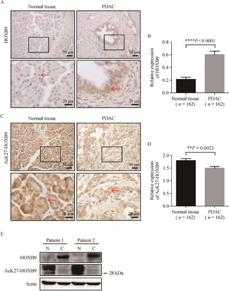
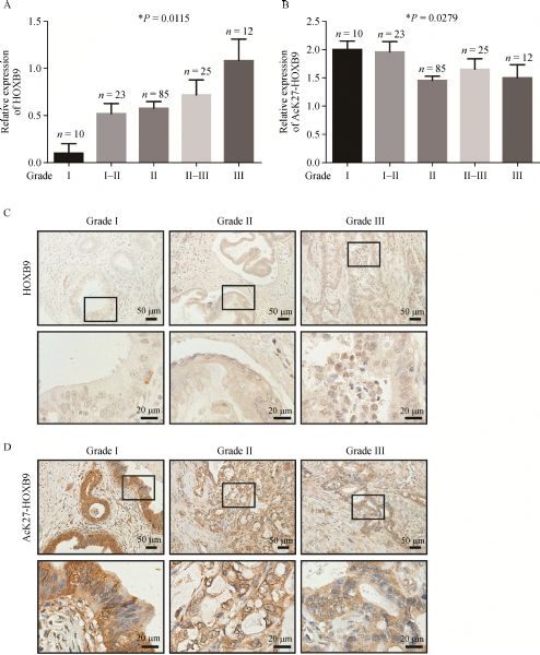
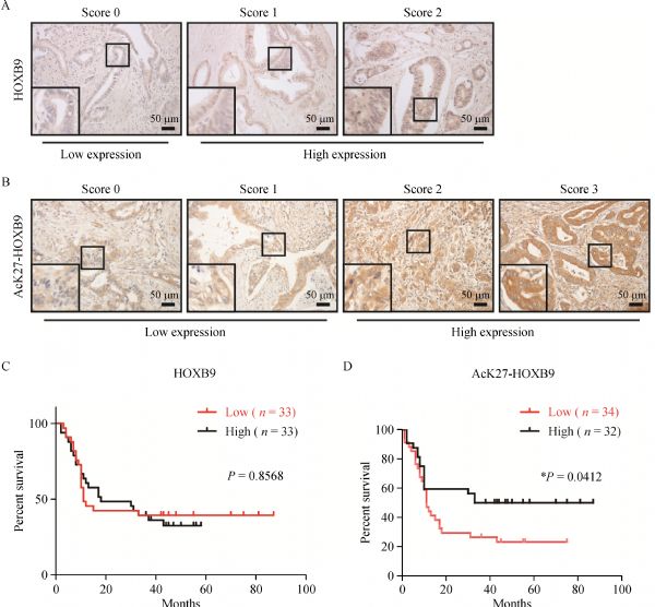
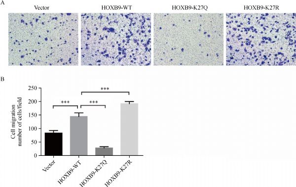
Abstract Pancreatic ductal adenocarcinoma (PDAC) is the ninth most common human malignancy and the sixth leading cause of cancer-related death in China. AcK27-HOXB9 is a newly identified HOXB9 post-transcriptional modification that can predict the outcome in lung adenocarcinoma and colon cancer well. However, the role of AcK27-HOXB9 in PDAC is unclear. The present study aims to investigate the differential diagnostic role of patients with AcK27-HOXB9 PDAC. Tissue microarrays consisting of 162 pancreatic tumor tissue samples from patients with PDAC and paired normal subjects were used to examine HOXB9 and AcK27-HOXB9 levels and localizations by immunohistochemical analysis and Western blot assay, respectively. HOXB9 was upregulated (P<0.0001), and AcK27-HOXB9 (P=0.0023) was downregulated in patients with PDAC. HOXB9 promoted (P=0.0115), while AcK27-HOXB9 (P=0.0279) inhibited PDAC progression. AcK27-HOXB9 predicted favorable outcome in patients with PDAC (P=0.0412). AcK27-HOXB9 also suppressed PDAC cell migration in a cell migration assay. The results of this study showed that HOXB9 promoted and AcK27-HOXB9 suppressed PDAC progression. The determination of ratio between HOXB9 and AcK27-HOXB9 exhibited potential diagnostic value in patients with PDAC.
|
| Keywords
HOXB9
AcK27-HOXB9
PDAC
|
|
Corresponding Author(s):
Jun Zhan,Weigang Fang,Hongquan Zhang
|
|
Just Accepted Date: 06 June 2019
Online First Date: 05 August 2019
Issue Date: 02 March 2020
|
|
| 1 |
W Chen, R Zheng, PD Baade, S Zhang, H Zeng, F Bray, A Jemal, XQ Yu, J He. Cancer statistics in China, 2015. CA Cancer J Clin 2016; 66(2): 115–132
https://doi.org/10.3322/caac.21338
pmid: 26808342
|
| 2 |
L Rahib, BD Smith, R Aizenberg, AB Rosenzweig, JM Fleshman, LM Matrisian. Projecting cancer incidence and deaths to 2030: the unexpected burden of thyroid, liver, and pancreas cancers in the United States. Cancer Res 2014; 74(11): 2913–2921
https://doi.org/10.1158/0008-5472.CAN-14-0155
pmid: 24840647
|
| 3 |
H Oettle, P Neuhaus, A Hochhaus, JT Hartmann, K Gellert, K Ridwelski, M Niedergethmann, C Zülke, J Fahlke, MB Arning, M Sinn, A Hinke, H Riess. Adjuvant chemotherapy with gemcitabine and long-term outcomes among patients with resected pancreatic cancer: the CONKO-001 randomized trial. JAMA 2013; 310(14): 1473–1481
https://doi.org/10.1001/jama.2013.279201
pmid: 24104372
|
| 4 |
WF Regine, KA Winter, RA Abrams, H Safran, JP Hoffman, A Konski, AB Benson, JS Macdonald, MR Kudrimoti, ML Fromm, MG Haddock, P Schaefer, CG Willett, TA Rich. Fluorouracil vs gemcitabine chemotherapy before and after fluorouracil-based chemoradiation following resection of pancreatic adenocarcinoma: a randomized controlled trial. JAMA 2008; 299(9): 1019–1026
https://doi.org/10.1001/jama.299.9.1019
pmid: 18319412
|
| 5 |
J Garcia-Fernàndez. The genesis and evolution of homeobox gene clusters. Nat Rev Genet 2005; 6(12): 881–892
https://doi.org/10.1038/nrg1723
pmid: 16341069
|
| 6 |
C Abate-Shen. Deregulated homeobox gene expression in cancer: cause or consequence? Nat Rev Cancer 2002; 2(10): 777–785
https://doi.org/10.1038/nrc907
pmid: 12360280
|
| 7 |
Q Chang, L Zhang, C He, B Zhang, J Zhang, B Liu, N Zeng, Z Zhu. HOXB9 induction of mesenchymal-to-epithelial transition in gastric carcinoma is negatively regulated by its hexapeptide motif. Oncotarget 2015; 6(40): 42838–42853
https://doi.org/10.18632/oncotarget.5814
pmid: 26536658
|
| 8 |
SY Wu, R Rupaimoole, F Shen, S Pradeep, CV Pecot, C Ivan, AS Nagaraja, KM Gharpure, E Pham, H Hatakeyama, MH McGuire, M Haemmerle, V Vidal-Anaya, C Olsen, C Rodriguez-Aguayo, J Filant, EA Ehsanipour, SM Herbrich, SN Maiti, L Huang, JH Kim, X Zhang, HD Han, GN Armaiz-Pena, EG Seviour, S Tucker, M Zhang, D Yang, LJ Cooper, R Ali-Fehmi, M Bar-Eli, JS Lee, PT Ram, KA Baggerly, G Lopez-Berestein, MC Hung, AK Sood. A miR-192-EGR1-HOXB9 regulatory network controls the angiogenic switch in cancer. Nat Commun 2016; 7(1): 11169
https://doi.org/10.1038/ncomms11169
pmid: 27041221
|
| 9 |
T Hayashida, F Takahashi, N Chiba, E Brachtel, M Takahashi, N Godin-Heymann, KW Gross, M Vivanco, V Wijendran, T Shioda, D Sgroi, PK Donahoe, S Maheswaran. HOXB9, a gene overexpressed in breast cancer, promotes tumorigenicity and lung metastasis. Proc Natl Acad Sci USA 2010; 107(3): 1100–1105
https://doi.org/10.1073/pnas.0912710107
pmid: 20080567
|
| 10 |
Z Kelly, C Moller-Levet, S McGrath, S Butler-Manuel, T Kavitha Madhuri, AM Kierzek, H Pandha, R Morgan, A Michael. The prognostic significance of specific HOX gene expression patterns in ovarian cancer. Int J Cancer 2016; 139(7): 1608–1617
https://doi.org/10.1002/ijc.30204
pmid: 27225067
|
| 11 |
J Zhan, P Wang, M Niu, Y Wang, X Zhu, Y Guo, H Zhang. High expression of transcriptional factor HoxB9 predicts poor prognosis in patients with lung adenocarcinoma. Histopathology 2015; 66(7): 955–965
https://doi.org/10.1111/his.12585
pmid: 25324169
|
| 12 |
J Wan, H Liu, Q Feng, J Liu, L Ming. HOXB9 promotes endometrial cancer progression by targeting E2F3. Cell Death Dis 2018; 9(5): 509
https://doi.org/10.1038/s41419-018-0556-3
pmid: 29724991
|
| 13 |
J Wan, W Xu, J Zhan, J Ma, X Li, Y Xie, J Wang, WG Zhu, J Luo, H Zhang. PCAF-mediated acetylation of transcriptional factor HOXB9 suppresses lung adenocarcinoma progression by targeting oncogenic protein JMJD6. Nucleic Acids Res 2016; 44(22): 10662–10675
https://doi.org/10.1093/nar/gkw808
pmid: 27613418
|
| 14 |
J Song, T Wang, W Xu, P Wang, J Wan, Y Wang, J Zhan, H Zhang. HOXB9 acetylation at K27 is responsible for its suppression of colon cancer progression. Cancer Lett 2018; 426: 63–72
https://doi.org/10.1016/j.canlet.2018.04.002
pmid: 29654889
|
| 15 |
C Chen, X Sun, Q Ran, KD Wilkinson, TJ Murphy, JW Simons, JT Dong. Ubiquitin-proteasome degradation of KLF5 transcription factor in cancer and untransformed epithelial cells. Oncogene 2005; 24(20): 3319–3327
https://doi.org/10.1038/sj.onc.1208497
pmid: 15735697
|
| 16 |
DG Grier, A Thompson, A Kwasniewska, GJ McGonigle, HL Halliday, TR Lappin. The pathophysiology of HOX genes and their role in cancer. J Pathol 2005; 205(2): 154–171
https://doi.org/10.1002/path.1710
pmid: 15643670
|
| 17 |
H Chen, S Sukumar. HOX genes: emerging stars in cancer. Cancer Biol Ther 2003; 2(5): 524–525
https://doi.org/10.4161/cbt.2.5.525
pmid: 14614319
|
| 18 |
K Rhoads, G Arderiu, A Charboneau, SL Hansen, W Hoffman, N Boudreau. A role for HOx A5 in regulating angiogenesis and vascular patterning. Lymphat Res Biol 2005; 3(4): 240–252
https://doi.org/10.1089/lrb.2005.3.240
pmid: 16379594
|
| 19 |
C Fromental-Ramain, X Warot, S Lakkaraju, B Favier, H Haack, C Birling, A Dierich, P Doll e, P Chambon. Specific and redundant functions of the paralogous Hoxa-9 and Hoxd-9 genes in forelimb and axial skeleton patterning. Development 1996; 122(2): 461–472
pmid: 8625797
|
| 20 |
F Chen, MR Capecchi. Paralogous mouse Hox genes, Hoxa9, Hoxb9, and Hoxd9, function together to control development of the mammary gland in response to pregnancy. Proc Natl Acad Sci USA 1999; 96(2): 541–546
https://doi.org/10.1073/pnas.96.2.541
pmid: 9892669
|
| 21 |
S Sha, Y Gu, B Xu, H Hu, Y Yang, X Kong, K Wu. Decreased expression of HOXB9 is related to poor overall survival in patients with gastric carcinoma. Dig Liver Dis 2013; 45: 422–429
https://doi.org/DOI:10.1016/j.dld.2012.12.004
pmid: PMID:23332081
|
|
Viewed |
|
|
|
Full text
|
|
|
|
|
Abstract
|
|
|
|
|
Cited |
|
|
|
|
| |
Shared |
|
|
|
|
| |
Discussed |
|
|
|
|