|
|
|
Keratin 5-Cre-driven deletion of Ncstn in an acne inversa-like mouse model leads to a markedly increased IL-36a and Sprr2 expression |
Jun Yang1, Lianqing Wang1,2, Yingzhi Huang1, Keqiang Liu1, Chaoxia Lu1, Nuo Si1, Rongrong Wang1, Yaping Liu1( ), Xue Zhang1() ), Xue Zhang1() |
1. McKusick-Zhang Center for Genetic Medicine, State Key Laboratory of Medical Molecular Biology, Institute of Basic Medical Sciences Chinese Academy of Medical Sciences, School of Basic Medicine Peking Union Medical College, Beijing 100005, China
2. Center of Translational Medicine, Central Hospital of Zibo, Shandong University, Zibo 255036, China |
|
|
|
|
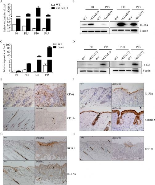
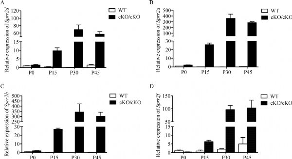
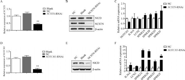
Abstract Familial acne inversa (AI) is an autoinflammatory disorder that affects hair follicles and is caused by loss-of-function mutations in g-secretase component genes. We and other researchers showed that nicastrin (NCSTN) is the most frequently mutated gene in familial AI. In this study, we generated a keratin 5-Cre-driven epidermis-specific Ncstn conditional knockout mutant in mice. We determined that this mutant recapitulated the major phenotypes of AI, including hyperkeratosis of hair follicles and inflammation. In Ncstnflox/flox;K5-Cre mice, the IL-36a expression level markedly increased starting from postnatal day 0 (P0), and this increase occurred much earlier than those of TNF-α, IL-23A, IL-1b, and TLR4. RNA-Seq analysis indicated that Sprr2d, a member of the small proline-rich protein 2 family, in the skin tissues of the Ncstnflox/flox;K5-Cre mice was also upregulated on P0. Quantitative reverse-transcription polymerase chain reaction showed that other Sprr2 genes had a similar expression pattern. Our findings suggested that IL-36a might be a key inflammatory cytokine in the pathophysiology of AI and implicate malfunction of the skin barrier in the pathogenesis of AI.
|
| Keywords
acne inversa mouse model
interleukin 1 family, member 6
small proline rich protein 2D
key inflammatory cytokine
|
|
Corresponding Author(s):
Yaping Liu,Xue Zhang
|
|
Just Accepted Date: 21 November 2019
Online First Date: 25 December 2019
Issue Date: 08 June 2020
|
|
| 1 |
J Revuz. Hidradenitis suppurativa. J Eur Acad Dermatol Venereol 2009; 23(9): 985–998
https://doi.org/10.1111/j.1468-3083.2009.03356.x
pmid: 19682181
|
| 2 |
A Garg, JS Kirby, J Lavian, G Lin, A Strunk. Sex- and age-adjusted population analysis of prevalence estimates for hidradenitis suppurativa in the United States. JAMA Dermatol 2017; 153(8): 760–764
https://doi.org/10.1001/jamadermatol.2017.0201
pmid: 28492923
|
| 3 |
JE Revuz, F Canoui-Poitrine, P Wolkenstein, C Viallette, G Gabison, F Pouget, F Poli, O Faye, JC Roujeau, G Bonnelye, JJ Grob, S Bastuji-Garin. Prevalence and factors associated with hidradenitis suppurativa: results from two case-control studies. J Am Acad Dermatol 2008; 59(4): 596–601
https://doi.org/10.1016/j.jaad.2008.06.020
pmid: 18674845
|
| 4 |
C Coughlan, W Ledger, Q Wang, F Liu, A Demirol, T Gurgan, R Cutting, K Ong, H Sallam, TC Li. Recurrent implantation failure: definition and management. Reprod Biomed Online 2014; 28(1): 14–38
https://doi.org/10.1016/j.rbmo.2013.08.011
pmid: 24269084
|
| 5 |
GB Jemec, M Heidenheim, NH Nielsen. The prevalence of hidradenitis suppurativa and its potential precursor lesions. J Am Acad Dermatol 1996; 35(2 Pt 1): 191–194
https://doi.org/10.1016/S0190-9622(96)90321-7
pmid: 8708018
|
| 6 |
JR Ingram. The genetics of hidradenitis suppurativa. Dermatol Clin 2016; 34(1): 23–28
https://doi.org/10.1016/j.det.2015.07.002
pmid: 26617354
|
| 7 |
JV Schmitt, G Bombonatto, M Martin, HA Miot. Risk factors for hidradenitis suppurativa: a pilot study. An Bras Dermatol 2012; 87(6): 936–938
https://doi.org/10.1590/S0365-05962012000600024
pmid: 23197222
|
| 8 |
I Brook, EH Frazier. Aerobic and anaerobic microbiology of axillary hidradenitis suppurativa. J Med Microbiol 1999; 48(1): 103–105
https://doi.org/10.1099/00222615-48-1-103
pmid: 9920133
|
| 9 |
I Jansen, P Altmeyer, G Piewig. Acne inversa (alias hidradenitis suppurativa). J Eur Acad Dermatol Venereol 2001; 15(6): 532–540
https://doi.org/10.1046/j.1468-3083.2001.00303.x
pmid: 11843212
|
| 10 |
M Gao, PG Wang, Y Cui, S Yang, YH Zhang, D Lin, KY Zhang, YH Liang, LD Sun, KL Yan, FL Xiao, W Huang, XJ Zhang. Inversa acne (hidradenitis suppurativa): a case report and identification of the locus at chromosome 1p21.1-1q25.3. J Invest Dermatol 2006; 126(6): 1302–1306
https://doi.org/10.1038/sj.jid.5700272
pmid: 16543891
|
| 11 |
JM Von Der Werth, HC Williams, JA Raeburn. The clinical genetics of hidradenitis suppurativa revisited. Br J Dermatol 2015; 142: 947–953
https://doi.org/DOI: 10.1046/j.1365-2133.2000.03476.x
|
| 12 |
Y Nomura, T Nomura, S Suzuki, M Takeda, O Mizuno, Y Ohguchi, R Abe, Y Murata, H Shimizu. A novel NCSTN mutation alone may be insufficient for the development of familial hidradenitis suppurativa. J Dermatol Sci 2014; 74(2): 180–182
https://doi.org/10.1016/j.jdermsci.2014.01.013
pmid: 24581508
|
| 13 |
B Wang, W Yang, W Wen, J Sun, B Su, B Liu, D Ma, D Lv, Y Wen, T Qu, M Chen, M Sun, Y Shen, X Zhang. γ-Secretase gene mutations in familial acne inversa. Science 2010; 330(6007): 1065
https://doi.org/10.1126/science.1196284
pmid: 20929727
|
| 14 |
RJ Kelleher 3rd, J Shen. Genetics. γ-Secretase and human disease. Science 2010; 330(6007): 1055–1056
https://doi.org/10.1126/science.1198668
pmid: 21097925
|
| 15 |
E Prens, I Deckers. Pathophysiology of hidradenitis suppurativa: an update. J Am Acad Dermatol 2015; 73(5 Suppl 1): S8–S11
https://doi.org/10.1016/j.jaad.2015.07.045
pmid: 26470623
|
| 16 |
A Alikhan, PJ Lynch, DB Eisen. Hidradenitis suppurativa: a comprehensive review. J Am Acad Dermatol 2009; 60(4): 539–561, quiz 562–563
https://doi.org/10.1016/j.jaad.2008.11.911
pmid: 19293006
|
| 17 |
RK Andersen, GB Jemec. Treatments for hidradenitis suppurativa. Clin Dermatol 2017; 35(2): 218–224
https://doi.org/10.1016/j.clindermatol.2016.10.018
pmid: 28274363
|
| 18 |
EJ Giamarellos-Bourboulis, A Antonopoulou, C Petropoulou, M Mouktaroudi, E Spyridaki, F Baziaka, A Pelekanou, H Giamarellou, NG Stavrianeas. Altered innate and adaptive immune responses in patients with hidradenitis suppurativa. Br J Dermatol 2007; 156(1): 51–56
https://doi.org/10.1111/j.1365-2133.2006.07556.x
pmid: 17199566
|
| 19 |
Q Chen, H Bao, H Wu, S Zhao, S Huang, F Zhao. Diagnosis of cobalamin C deficiency with renal abnormality from onset in a Chinese child by next generation sequencing: a case report. Exp Ther Med 2017; 14(4): 3637–3643
https://doi.org/10.3892/etm.2017.4970
pmid: 29042959
|
| 20 |
TP Sullivan, E Welsh, FA Kerdel, AE Burdick, RS Kirsner. Infliximab for hidradenitis suppurativa. Br J Dermatol 2003; 149(5): 1046–1049
https://doi.org/10.1111/j.1365-2133.2003.05663.x
pmid: 14632813
|
| 21 |
A Kurayev, H Ashkar, A Saraiya, AB Gottlieb. Hidradenitis suppurativa: review of the pathogenesis and treatment. J Drugs Dermatol 2016; 15(8): 1017–1022
pmid: 27538005
|
| 22 |
G Kelly, R Hughes, T McGarry, M van den Born, K Adamzik, R Fitzgerald, C Lawlor, AM Tobin, CM Sweeney, B Kirby. Dysregulated cytokine expression in lesional and nonlesional skin in hidradenitis suppurativa. Br J Dermatol 2015; 173(6): 1431–1439
https://doi.org/10.1111/bjd.14075
pmid: 26282467
|
| 23 |
L Tong, RM Corrales, Z Chen, AL Villarreal, CS De Paiva, R Beuerman, DQ Li, SC Pflugfelder. Expression and regulation of cornified envelope proteins in human corneal epithelium. Invest Ophthalmol Vis Sci 2006; 47(5): 1938–1946
https://doi.org/10.1167/iovs.05-1129
pmid: 16639001
|
| 24 |
Affymetrix. GeneChip® Expression Analysis. Data Analysis Fundamentals. 2004
|
| 25 |
S Anders, W Huber. Differential expression of RNA-Seq data at the gene level — the DESeq package. DESeq version 1.38.0. European Molecular Biology Laboratory (EMBL). 2013
|
| 26 |
L Wang, Z Feng, X Wang, X Wang, X Zhang. DEGseq: an R package for identifying differentially expressed genes from RNA-seq data. Bioinformatics 2010; 26(1): 136–138
https://doi.org/10.1093/bioinformatics/btp612
pmid: 19855105
|
| 27 |
Y Nomura, T Nomura, S Suzuki, M Takeda, O Mizuno, Y Ohguchi, R Abe, Y Murata, H Shimizu. A novel NCSTN mutation alone may be insufficient for the development of familial hidradenitis suppurativa. J Dermatol Sci 2014; 74(2): 180–182
https://doi.org/10.1016/j.jdermsci.2014.01.013
pmid: 24581508
|
| 28 |
X Zhang, SS Sisodia. Acne inversa caused by missense mutations in NCSTN is not fully compatible with impairments in Notch signaling. J Invest Dermatol 2015; 135(2): 618–620
https://doi.org/10.1038/jid.2014.399
pmid: 25211177
|
| 29 |
X Xiao, Y He, C Li, X Zhang, H Xu, B Wang. Nicastrin mutations in familial acne inversa impact keratinocyte proliferation and differentiation through the Notch and phosphoinositide 3-kinase/AKT signalling pathways. Br J Dermatol 2016; 174(3): 522–532
https://doi.org/10.1111/bjd.14223
pmid: 26473517
|
| 30 |
L Yang, C Mao, Y Teng, W Li, J Zhang, X Cheng, X Li, X Han, Z Xia, H Deng, X Yang. Targeted disruption of Smad4 in mouse epidermis results in failure of hair follicle cycling and formation of skin tumors. Cancer Res 2005; 65(19): 8671–8678
https://doi.org/10.1158/0008-5472.CAN-05-0800
pmid: 16204035
|
| 31 |
A Ramirez, A Page, A Gandarillas, J Zanet, S Pibre, M Vidal, L Tusell, A Genesca, DA Whitaker, DW Melton, JL Jorcano. A keratin K5Cre transgenic line appropriate for tissue-specific or generalized Cre-mediated recombination. Genesis 2004; 39(1): 52–57
https://doi.org/10.1002/gene.20025
pmid: 15124227
|
| 32 |
B Wang, W Yang, W Wen, J Sun, B Su, B Liu, D Ma, D Lv, Y Wen, T Qu, M Chen, M Sun, Y Shen, X Zhang. γ-Secretase gene mutations in familial acne inversa. Science 2010; 330(6007): 1065
https://doi.org/10.1126/science.1196284
pmid: 20929727
|
| 33 |
AE Pink, MA Simpson, N Desai, RC Trembath, JNW Barker. γ-Secretase mutations in hidradenitis suppurativa: new insights into disease pathogenesis. J Invest Dermatol 2013; 133(3): 601–607
https://doi.org/10.1038/jid.2012.372
pmid: 23096707
|
| 34 |
MA Boutet, G Bart, M Penhoat, J Amiaud, B Brulin, C Charrier, F Morel, JC Lecron, M Rolli-Derkinderen, A Bourreille, S Vigne, C Gabay, G Palmer, B Le Goff, F Blanchard. Distinct expression of interleukin (IL)-36a, b and g, their antagonist IL-36Ra and IL-38 in psoriasis, rheumatoid arthritis and Crohn’s disease. Clin Exp Immunol 2016; 184(2): 159–173
https://doi.org/10.1111/cei.12761
pmid: 26701127
|
| 35 |
AE Pink, MA Simpson, GW Brice, CH Smith, N Desai, PS Mortimer, JN Barker, RC Trembath. PSENEN and NCSTN mutations in familial hidradenitis suppurativa (acne inversa). J Invest Dermatol 2011; 131(7): 1568–1570
https://doi.org/10.1038/jid.2011.42
pmid: 21412258
|
| 36 |
H Xu, X Xiao, Y Hui, X Zhang, Y He, C Li, B Wang. Phenotype of 53 Chinese individuals with nicastrin gene mutations in association with familial hidradenitis suppurativa (acne inversa). Br J Dermatol 2016; 174(4): 927–929
https://doi.org/10.1111/bjd.14268
pmid: 26522179
|
| 37 |
HH van der Zee, JD Laman, J Boer, EP Prens. Hidradenitis suppurativa: viewpoint on clinical phenotyping, pathogenesis and novel treatments. Exp Dermatol 2012; 21(10): 735–739
https://doi.org/10.1111/j.1600-0625.2012.01552.x
pmid: 22882284
|
| 38 |
JW van der Meer, A Simon. The challenge of autoinflammatory syndromes: with an emphasis on hyper-IgD syndrome. Rheumatology (Oxford) 2016; 55(suppl 2): ii23–ii29
https://doi.org/10.1093/rheumatology/kew351
pmid: 27856657
|
| 39 |
S Hessam, M Sand, T Gambichler, M Skrygan, I Rüddel, FG Bechara. Interleukin-36 in hidradenitis suppurativa: evidence for a distinctive proinflammatory role and a key factor in the development of an inflammatory loop. Br J Dermatol 2018; 178(3): 761–767
https://doi.org/10.1111/bjd.16019
pmid: 28975626
|
| 40 |
E Witte-Händel, K Wolk, A Tsaousi, ML Irmer, R Mößner, O Shomroni, T Lingner, K Witte, D Kunkel, G Salinas, S Jodl, N Schmidt, W Sterry, HD Volk, EJ Giamarellos-Bourboulis, A Pokrywka, WD Döcke, S Schneider-Burrus, R Sabat. The IL-1 pathway is hyperactive in hidradenitis suppurativa and contributes to skin infiltration and destruction. J Invest Dermatol 2019; 139(6): 1294–1305
https://doi.org/10.1016/j.jid.2018.11.018
pmid: 30528824
|
| 41 |
R Thomi, M Kakeda, N Yawalkar C Schlapbach, RE Hunger. Increased expression of the interleukin-36 cytokines in lesions of hidradenitis suppurativa. J Eur Acad Dermatol Venereol 2017; 31(12): 2091–2096
https://doi.org/10.1111/jdv.14389
|
| 42 |
R Di Caprio, A Balato, G Caiazzo, S Lembo, A Raimondo, G Fabbrocini, G Monfrecola. IL-36 cytokines are increased in acne and hidradenitis suppurativa. Arch Dermatol Res 2017; 309(8): 673–678
https://doi.org/10.1007/s00403-017-1769-5
pmid: 28852851
|
| 43 |
DE Smith, BR Renshaw, RR Ketchem, M Kubin, KE Garka, JE Sims. Four new members expand the interleukin-1 superfamily. J Biol Chem 2000; 275(2): 1169–1175
https://doi.org/10.1074/jbc.275.2.1169
pmid: 10625660
|
| 44 |
Y Carrier, HL Ma, HE Ramon, L Napierata, C Small, M O’Toole, DA Young, LA Fouser, C Nickerson-Nutter, M Collins, K Dunussi-Joannopoulos, QG Medley. Inter-regulation of Th17 cytokines and the IL-36 cytokines in vitro and in vivo: implications in psoriasis pathogenesis. J Invest Dermatol 2011; 131(12): 2428–2437
https://doi.org/10.1038/jid.2011.234
pmid: 21881584
|
| 45 |
H Blumberg, H Dinh, ES Trueblood, J Pretorius, D Kugler, N Weng, ST Kanaly, JE Towne, CR Willis, MK Kuechle, JE Sims, JJ Peschon. Opposing activities of two novel members of the IL-1 ligand family regulate skin inflammation. J Exp Med 2007; 204(11): 2603–2614
https://doi.org/10.1084/jem.20070157
pmid: 17908936
|
| 46 |
JE Towne, KE Garka, BR Renshaw, GD Virca, JE Sims. Interleukin (IL)-1F6, IL-1F8, and IL-1F9 signal through IL-1Rrp2 and IL-1RAcP to activate the pathway leading to NF-κB and MAPKs. J Biol Chem 2004; 279(14): 13677–13688
https://doi.org/10.1074/jbc.M400117200
pmid: 14734551
|
| 47 |
S Vigne, G Palmer, P Martin, C Lamacchia, D Strebel, E Rodriguez, ML Olleros, D Vesin, I Garcia, F Ronchi, F Sallusto, JE Sims, C Gabay. IL-36 signaling amplifies Th1 responses by enhancing proliferation and Th1 polarization of naive CD4+ T cells. Blood 2012; 120(17): 3478–3487
https://doi.org/10.1182/blood-2012-06-439026
pmid: 22968459
|
| 48 |
BC Melnik, G Plewig. Impaired Notch-MKP-1 signalling in hidradenitis suppurativa: an approach to pathogenesis by evidence from translational biology. Exp Dermatol 2013; 22(3): 172–177
https://doi.org/10.1111/exd.12098
pmid: 23489419
|
| 49 |
K Wolk, J Wenzel, A Tsaousi, E Witte-Händel, N Babel, C Zelenak, HD Volk, W Sterry, S Schneider-Burrus, R Sabat. Lipocalin-2 is expressed by activated granulocytes and keratinocytes in affected skin and reflects disease activity in acne inversa/hidradenitis suppurativa. Br J Dermatol 2017; 177(5): 1385–1393
https://doi.org/10.1111/bjd.15424
pmid: 28256718
|
| 50 |
PM Steinert, E Candi, T Kartasova, L Marekov. Small proline-rich proteins are cross-bridging proteins in the cornified cell envelopes of stratified squamous epithelia. J Struct Biol 1998; 122(1-2): 76–85
https://doi.org/10.1006/jsbi.1998.3957
pmid: 9724607
|
| 51 |
N Wakabayashi, S Shin, SL Slocum, ES Agoston, J Wakabayashi, MK Kwak, V Misra, S Biswal, M Yamamoto, TW Kensler. Regulation of notch1 signaling by nrf2: implications for tissue regeneration. Sci Signal 2010; 3(130): ra52
https://doi.org/10.1126/scisignal.2000762
pmid: 20628156
|
| 52 |
M Schfer, H Farwanah, AH Willrodt, AJ Huebner, K Sandhoff, D Roop, D Hohl, W Bloch, S Werner. Nrf2 links epidermal barrier function with antioxidant defense. EMBO Mol Med 2012(4): 364–379
https://doi.org/10.1002/emmm.201200219
|
| 53 |
N Wakabayashi, DV Chartoumpekis, TW Kensler. Crosstalk between Nrf2 and Notch signaling. Free Radic Biol Med 2015; 88(Pt B): 158–167
https://doi.org/10.1016/j.freeradbiomed.2015.05.017
pmid: 26003520
|
| 54 |
N Wakabayashi, JJ Skoko, DV Chartoumpekis, S Kimura, SL Slocum, K Noda, DL Palliyaguru, M Fujimuro, PA Boley, Y Tanaka, N Shigemura, S Biswal, M Yamamoto, TW Kensler. Notch-Nrf2 axis: regulation of Nrf2 gene expression and cytoprotection by Notch signaling. Mol Cell Biol 2014; 34(4): 653–663
https://doi.org/10.1128/MCB.01408-13
pmid: 24298019
|
|
Viewed |
|
|
|
Full text
|
|
|
|
|
Abstract
|
|
|
|
|
Cited |
|
|
|
|
| |
Shared |
|
|
|
|
| |
Discussed |
|
|
|
|