|
|
|
Altered white matter microarchitecture in Parkinson’s disease: a voxel-based meta-analysis of diffusion tensor imaging studies |
Xueling Suo1, Du Lei1,2( ), Wenbin Li1,2, Lei Li1, Jing Dai3, Song Wang1, Nannan Li4, Lan Cheng4, Rong Peng4, Graham J Kemp5, Qiyong Gong1,6() ), Wenbin Li1,2, Lei Li1, Jing Dai3, Song Wang1, Nannan Li4, Lan Cheng4, Rong Peng4, Graham J Kemp5, Qiyong Gong1,6() |
1. Huaxi MR Research Center (HMRRC), Department of Radiology, West China Hospital of Sichuan University, Chengdu 610041, China
2. Department of Psychiatry and Behavioral Neuroscience, University of Cincinnati, Cincinnati, Ohio, USA
3. Department of Psychoradiology, Chengdu Mental Health Center, Chengdu 610041, China
4. Department of Neurology, West China Hospital of Sichuan University, Chengdu 610041, China
5. Liverpool Magnetic Resonance Imaging Centre (LiMRIC) and Institute of Ageing and Chronic Disease, University of Liverpool, Liverpool L69 3GE, United Kingdom
6. Psychoradiology Research Unit of Chinese Academy of Medical Sciences, West China Hospital of Sichuan University, Chengdu 610041, China |
|
|
|
|
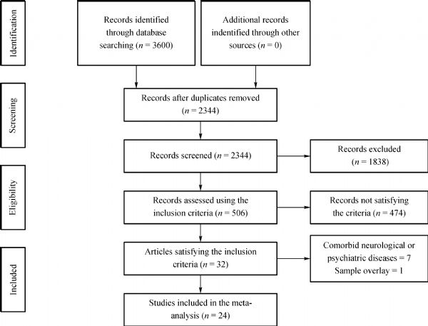
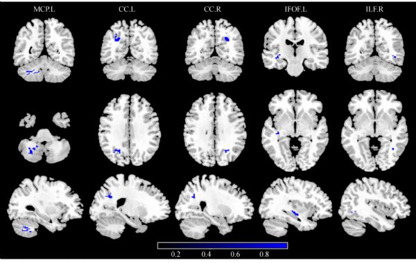
Abstract This study aimed to define the most consistent white matter microarchitecture pattern in Parkinson’s disease (PD) reflected by fractional anisotropy (FA), addressing clinical profiles and methodology-related heterogeneity. Web-based publication databases were searched to conduct a meta-analysis of whole-brain diffusion tensor imaging studies comparing PD with healthy controls (HC) using the anisotropic effect size–signed differential mapping. A total of 808 patients with PD and 760 HC coming from 27 databases were finally included. Subgroup analyses were conducted considering heterogeneity with respect to medication status, disease stage, analysis methods, and the number of diffusion directions in acquisition. Compared with HC, patients with PD had decreased FA in the left middle cerebellar peduncle, corpus callosum (CC), left inferior fronto-occipital fasciculus, and right inferior longitudinal fasciculus. Most of the main results remained unchanged in subgroup meta-analyses of medicated patients, early stage patients, voxel-based analysis, and acquisition with ˂30 diffusion directions. The subgroup meta-analysis of medication-free patients showed FA decrease in the right olfactory cortex. The cerebellum and CC, associated with typical motor impairment, showed the most consistent FA decreases in PD. Medication status, analysis approaches, and the number of diffusion directions have an important impact on the findings, needing careful evaluation in future meta-analyses.
|
| Keywords
Parkinson’s disease
diffusion tensor imaging
fractional anisotropy
meta-analysis
anisotropic effect size–signed differential mapping
|
|
Corresponding Author(s):
Du Lei,Qiyong Gong
|
|
Online First Date: 27 May 2020
Issue Date: 11 February 2021
|
|
| 1 |
LM de Lau, MM Breteler. Epidemiology of Parkinson’s disease. Lancet Neurol 2006; 5(6): 525–535
https://doi.org/10.1016/S1474-4422(06)70471-9
pmid: 16713924
|
| 2 |
H Reichmann, MD Brandt, L Klingelhoefer. The nonmotor features of Parkinson’s disease: pathophysiology and management advances. Curr Opin Neurol 2016; 29(4): 467–473
https://doi.org/10.1097/WCO.0000000000000348
pmid: 27262147
|
| 3 |
M Kubicki, CF Westin, SE Maier, H Mamata, M Frumin, H Ersner-Hershfield, R Kikinis, FA Jolesz, R McCarley, ME Shenton. Diffusion tensor imaging and its application to neuropsychiatric disorders. Harv Rev Psychiatry 2002; 10(6): 324–336
https://doi.org/10.1080/10673220216231
pmid: 12485979
|
| 4 |
WD Taylor, E Hsu, KR Krishnan, JR MacFall. Diffusion tensor imaging: background, potential, and utility in psychiatric research. Biol Psychiatry 2004; 55(3): 201–207
https://doi.org/10.1016/j.biopsych.2003.07.001
pmid: 14744459
|
| 5 |
D Le Bihan, JF Mangin, C Poupon, CA Clark, S Pappata, N Molko, H Chabriat. Diffusion tensor imaging: concepts and applications. J Magn Reson Imaging 2001; 13(4): 534–546
https://doi.org/10.1002/jmri.1076
pmid: 11276097
|
| 6 |
B Chen, G Fan, W Sun, X Shang, S Shi, S Wang, G Lv, C. Wu Usefulness of diffusion-tensor MRI in the diagnosis of Parkinson variant of multiple system atrophy and Parkinson’s disease: a valuable tool to differentiate between them? Clin Radiol 2017; 72(7): 610.e9–610.e15
https://doi.org/10.1016/j.crad.2017.02.005
pmid: 28318507
|
| 7 |
L Ji, Y Wang, D Zhu, W Liu, J Shi. White matter differences between multiple system atrophy (parkinsonian type) and Parkinson’s disease: a diffusion tensor image study. Neuroscience 2015; 305: 109–116
https://doi.org/10.1016/j.neuroscience.2015.07.060
pmid: 26215920
|
| 8 |
S Vercruysse, I Leunissen, G Vervoort, W Vandenberghe, S Swinnen, A Nieuwboer. Microstructural changes in white matter associated with freezing of gait in Parkinson’s disease. Mov Disord 2015; 30(4): 567–576
https://doi.org/10.1002/mds.26130
pmid: 25640958
|
| 9 |
AT Karagulle Kendi, S Lehericy, M Luciana, K Ugurbil, P Tuite. Altered diffusion in the frontal lobe in Parkinson disease. AJNR Am J Neuroradiol 2008; 29(3): 501–505
https://doi.org/10.3174/ajnr.A0850
pmid: 18202242
|
| 10 |
PL Chiang, HL Chen, CH Lu, PC Chen, MH Chen, IH Yang, NW Tsai, WC Lin. White matter damage and systemic inflammation in Parkinson’s disease. BMC Neurosci 2017; 18(1): 48
https://doi.org/10.1186/s12868-017-0367-y
pmid: 28595572
|
| 11 |
MH Chen, PC Chen, CH Lu, HL Chen, YP Chao, SH Li, YW Chen, WC Lin. Plasma DNA mediate autonomic dysfunctions and white matter injuries in patients with Parkinson’s disease. Oxid Med Cell Longev 2017; 2017: 7371403
https://doi.org/10.1155/2017/7371403
pmid: 28232858
|
| 12 |
K Zhang, C Yu, Y Zhang, X Wu, C Zhu, P Chan, K Li. Voxel-based analysis of diffusion tensor indices in the brain in patients with Parkinson’s disease. Eur J Radiol 2011; 77(2): 269–273
https://doi.org/10.1016/j.ejrad.2009.07.032
pmid: 19692193
|
| 13 |
C Luo, W Song, Q Chen, J Yang, Q Gong, HF Shang. White matter microstructure damage in tremor-dominant Parkinson’s disease patients. Neuroradiology 2017; 59(7): 691–698
https://doi.org/10.1007/s00234-017-1846-7
pmid: 28540401
|
| 14 |
A Worker, C Blain, J Jarosz, KR Chaudhuri, GJ Barker, SC Williams, RG Brown, PN Leigh, F Dell’Acqua, A Simmons. Diffusion tensor imaging of Parkinson’s disease, multiple system atrophy and progressive supranuclear palsy: a tract-based spatial statistics study. PLoS One 2014; 9(11): e112638
https://doi.org/10.1371/journal.pone.0112638
pmid: 25405990
|
| 15 |
F Agosta, E Canu, T Stojković, M Pievani, A Tomić, L Sarro, N Dragašević, M Copetti, G Comi, VS Kostić, M Filippi. The topography of brain damage at different stages of Parkinson’s disease. Hum Brain Mapp 2013; 34(11): 2798–2807
https://doi.org/10.1002/hbm.22101
pmid: 22528144
|
| 16 |
ST Schwarz, M Abaei, V Gontu, PS Morgan, N Bajaj, DP Auer. Diffusion tensor imaging of nigral degeneration in Parkinson’s disease: a region-of-interest and voxel-based study at 3 T and systematic review with meta-analysis. Neuroimage Clin 2013; 3: 481–488
https://doi.org/10.1016/j.nicl.2013.10.006
pmid: 24273730
|
| 17 |
C Atkinson-Clement, S Pinto, A Eusebio, O Coulon. Diffusion tensor imaging in Parkinson’s disease: review and meta-analysis. Neuroimage Clin 2017; 16: 98–110
https://doi.org/10.1016/j.nicl.2017.07.011
pmid: 28765809
|
| 18 |
CJ Cochrane, KP Ebmeier. Diffusion tensor imaging in parkinsonian syndromes: a systematic review and meta-analysis. Neurology 2013; 80(9): 857–864
https://doi.org/10.1212/WNL.0b013e318284070c
pmid: 23439701
|
| 19 |
F Albrecht, T Ballarini, J Neumann, ML Schroeter. FDG-PET hypometabolism is more sensitive than MRI atrophy in Parkinson’s disease: a whole-brain multimodal imaging meta-analysis. Neuroimage Clin 2019; 21: 101594
https://doi.org/10.1016/j.nicl.2018.11.004
pmid: 30514656
|
| 20 |
D Moher, A Liberati, J Tetzlaff, DG Altman; PRISMA Group. Preferred reporting items for systematic reviews and meta-analyses: the PRISMA statement. PLoS Med 2009; 6(7): e1000097
https://doi.org/10.1371/journal.pmed.1000097
pmid: 19621072
|
| 21 |
P Pan, H Zhan, M Xia, Y Zhang, D Guan, Y Xu. Aberrant regional homogeneity in Parkinson’s disease: a voxel-wise meta-analysis of resting-state functional magnetic resonance imaging studies. Neurosci Biobehav Rev 2017; 72: 223–231
https://doi.org/10.1016/j.neubiorev.2016.11.018
pmid: 27916710
|
| 22 |
AM Shepherd, SL Matheson, KR Laurens, VJ Carr, MJ Green. Systematic meta-analysis of insula volume in schizophrenia. Biol Psychiatry 2012; 72(9): 775–784
https://doi.org/10.1016/j.biopsych.2012.04.020
pmid: 22621997
|
| 23 |
J Radua, D Mataix-Cols. Voxel-wise meta-analysis of grey matter changes in obsessive-compulsive disorder. Br J Psychiatry 2009; 195(5): 393–402
https://doi.org/10.1192/bjp.bp.108.055046
pmid: 19880927
|
| 24 |
J Radua, D Mataix-Cols, ML Phillips, W El-Hage, DM Kronhaus, N Cardoner, S Surguladze. A new meta-analytic method for neuroimaging studies that combines reported peak coordinates and statistical parametric maps. Eur Psychiatry 2012; 27(8): 605–611
https://doi.org/10.1016/j.eurpsy.2011.04.001
pmid: 21658917
|
| 25 |
J Radua, K Rubia, EJ Canales-Rodríguez, E Pomarol-Clotet, P Fusar-Poli, D Mataix-Cols. Anisotropic kernels for coordinate-based meta-analyses of neuroimaging studies. Front Psychiatry 2014; 5: 13
https://doi.org/10.3389/fpsyt.2014.00013
pmid: 24575054
|
| 26 |
T Wise, J Radua, G Nortje, AJ Cleare, AH Young, D Arnone. Voxel-based meta-analytical evidence of structural disconnectivity in major depression and bipolar disorder. Biol Psychiatry 2016; 79(4): 293–302
https://doi.org/10.1016/j.biopsych.2015.03.004
pmid: 25891219
|
| 27 |
J Radua, M Grau, OA van den Heuvel, M Thiebaut de Schotten, DJ Stein, EJ Canales-Rodríguez, M Catani, D Mataix-Cols. Multimodal voxel-based meta-analysis of white matter abnormalities in obsessive-compulsive disorder. Neuropsychopharmacology 2014; 39(7): 1547–1557
https://doi.org/10.1038/npp.2014.5
pmid: 24407265
|
| 28 |
S Chen, P Chan, S Sun, H Chen, B Zhang, W Le, C Liu, G Peng, B Tang, L Wang, Y Cheng, M Shao, Z Liu, Z Wang, X Chen, M Wang, X Wan, H Shang, Y Liu, P Xu, J Wang, T Feng, X Chen, X Hu, A Xie, Q Xiao. The recommendations of Chinese Parkinson’s disease and movement disorder society consensus on therapeutic management of Parkinson’s disease. Transl Neurodegener 2016; 5(1): 12
https://doi.org/10.1186/s40035-016-0059-z
pmid: 27366321
|
| 29 |
DK Jones. The effect of gradient sampling schemes on measures derived from diffusion tensor MRI: a Monte Carlo study. Magn Reson Med 2004; 51(4): 807–815
https://doi.org/10.1002/mrm.20033
pmid: 15065255
|
| 30 |
X Guan, P Huang, Q Zeng, C Liu, H Wei, M Xuan, Q Gu, X Xu, N Wang, X Yu, X Luo, M Zhang. Quantitative susceptibility mapping as a biomarker for evaluating white matter alterations in Parkinson’s disease. Brain Imaging Behav 2019; 13(1): 220–231
https://doi.org/10.1007/s11682-018-9842-z
pmid: 29417492
|
| 31 |
MC Wen, HSE Heng, Z Lu, Z Xu, LL Chan, EK Tan, LCS Tan. Differential white matter regional alterations in motor subtypes of early drug-naive Parkinson’s disease patients. Neurorehabil Neural Repair 2018; 32(2): 129–141
https://doi.org/10.1177/1545968317753075
pmid: 29347868
|
| 32 |
P Péran, G Barbagallo, F Nemmi, M Sierra, M Galitzky, AP Traon, P Payoux, WG Meissner, O Rascol. MRI supervised and unsupervised classification of Parkinson’s disease and multiple system atrophy. Mov Disord 2018; 33(4): 600–608
https://doi.org/10.1002/mds.27307
pmid: 29473662
|
| 33 |
I Rektor, A Svátková, L Vojtíšek, I Zikmundová, J Vaníček, A Király, N Szabó. White matter alterations in Parkinson’s disease with normal cognition precede grey matter atrophy. PLoS One 2018; 13(1): e0187939
https://doi.org/10.1371/journal.pone.0187939
pmid: 29304183
|
| 34 |
J Acosta-Cabronero, A Cardenas-Blanco, MJ Betts, M Butryn, JP Valdes-Herrera, I Galazky, PJ Nestor. The whole-brain pattern of magnetic susceptibility perturbations in Parkinson’s disease. Brain 2017; 140(1): 118–131
https://doi.org/10.1093/brain/aww278
pmid: 27836833
|
| 35 |
S Zanigni, S Evangelisti, C Testa, DN Manners, G Calandra-Buonaura, M Guarino, A Gabellini, LL Gramegna, G Giannini, L Sambati, P Cortelli, R Lodi, C Tonon. White matter and cortical changes in atypical parkinsonisms: a multimodal quantitative MR study. Parkinsonism Relat Disord 2017; 39: 44–51
https://doi.org/10.1016/j.parkreldis.2017.03.001
pmid: 28291592
|
| 36 |
G Vervoort, I Leunissen, M Firbank, E Heremans, E Nackaerts, W Vandenberghe, A Nieuwboer. Structural brain alterations in motor subtypes of Parkinson’s disease: evidence from probabilistic tractography and shape analysis. PLoS One 2016; 11(6): e0157743
https://doi.org/10.1371/journal.pone.0157743
pmid: 27314952
|
| 37 |
F Agosta, E Canu, E Stefanova, L Sarro, A Tomić, V Špica, G Comi, VS Kostić, M Filippi. Mild cognitive impairment in Parkinson’s disease is associated with a distributed pattern of brain white matter damage. Hum Brain Mapp 2014; 35(5): 1921–1929
https://doi.org/10.1002/hbm.22302
pmid: 23843285
|
| 38 |
K Kamagata, Y Motoi, H Tomiyama, O Abe, K Ito, K Shimoji, M Suzuki, M Hori, A Nakanishi, T Sano, R Kuwatsuru, K Sasai, S Aoki, N Hattori. Relationship between cognitive impairment and white-matter alteration in Parkinson’s disease with dementia: tract-based spatial statistics and tract-specific analysis. Eur Radiol 2013; 23(7): 1946–1955
https://doi.org/10.1007/s00330-013-2775-4
pmid: 23404139
|
| 39 |
HJ Kim, SJ Kim, HS Kim, CG Choi, N Kim, S Han, EH Jang, SJ Chung, CS Lee. Alterations of mean diffusivity in brain white matter and deep gray matter in Parkinson’s disease. Neurosci Lett 2013; 550: 64–68
https://doi.org/10.1016/j.neulet.2013.06.050
pmid: 23831353
|
| 40 |
TR Melzer, R Watts, MR MacAskill, TL Pitcher, L Livingston, RJ Keenan, JC Dalrymple-Alford, TJ Anderson. White matter microstructure deteriorates across cognitive stages in Parkinson disease. Neurology 2013; 80(20): 1841–1849
https://doi.org/10.1212/WNL.0b013e3182929f62
pmid: 23596076
|
| 41 |
T Hattori, S Orimo, S Aoki, K Ito, O Abe, A Amano, R Sato, K Sakai, H Mizusawa. Cognitive status correlates with white matter alteration in Parkinson’s disease. Hum Brain Mapp 2012; 33(3): 727–739
https://doi.org/10.1002/hbm.21245
pmid: 21495116
|
| 42 |
K Kamagata, A Zalesky, T Hatano, R Ueda, MA Di Biase, A Okuzumi, K Shimoji, M Hori, K Caeyenberghs, C Pantelis, N Hattori, S Aoki. Gray matter abnormalities in idiopathic Parkinson’s disease: evaluation by diffusional kurtosis imaging and neurite orientation dispersion and density imaging. Hum Brain Mapp 2017; 38(7): 3704–3722
https://doi.org/10.1002/hbm.23628
pmid: 28470878
|
| 43 |
J Rosskopf, HP Müller, HJ Huppertz, AC Ludolph, EH Pinkhardt, J Kassubek. Frontal corpus callosum alterations in progressive supranuclear palsy but not in Parkinson’s disease. Neurodegener Dis 2014; 14(4): 184–193
https://doi.org/10.1159/000367693
pmid: 25377379
|
| 44 |
E Ziegler, M Rouillard, E André, T Coolen, J Stender, E Balteau, C Phillips, G Garraux. Mapping track density changes in nigrostriatal and extranigral pathways in Parkinson’s disease. Neuroimage 2014; 99: 498–508
https://doi.org/10.1016/j.neuroimage.2014.06.033
pmid: 24956065
|
| 45 |
CJ Stoodley, JD Schmahmann. Evidence for topographic organization in the cerebellum of motor control versus cognitive and affective processing. Cortex 2010; 46(7): 831–844
https://doi.org/10.1016/j.cortex.2009.11.008
pmid: 20152963
|
| 46 |
T Wu, M Hallett. The cerebellum in Parkinson’s disease. Brain 2013; 136(3): 696–709
https://doi.org/10.1093/brain/aws360
pmid: 23404337
|
| 47 |
PM Schweder, PC Hansen, AL Green, G Quaghebeur, J Stein, TZ Aziz. Connectivity of the pedunculopontine nucleus in parkinsonian freezing of gait. Neuroreport 2010; 21(14): 914–916
https://doi.org/10.1097/WNR.0b013e32833ce5f1
pmid: 20729769
|
| 48 |
E Canu, F Agosta, E Sarasso, MA Volontè, S Basaia, T Stojkovic, E Stefanova, G Comi, A Falini, VS Kostic, R Gatti, M Filippi. Brain structural and functional connectivity in Parkinson’s disease with freezing of gait. Hum Brain Mapp 2015; 36(12): 5064–5078
https://doi.org/10.1002/hbm.22994
pmid: 26359798
|
| 49 |
C Tessa, C Lucetti, S Diciotti, L Paoli, P Cecchi, M Giannelli, F Baldacci, A Ginestroni, C Vignali, M Mascalchi, U Bonuccelli. Hypoactivation of the primary sensorimotor cortex in de novo Parkinson’s disease: a motor fMRI study under controlled conditions. Neuroradiology 2012; 54(3): 261–268
https://doi.org/10.1007/s00234-011-0955-y
pmid: 21927866
|
| 50 |
JM Hall, KA Ehgoetz Martens, CC Walton, C O’Callaghan, PE Keller, SJ Lewis, AA Moustafa. Diffusion alterations associated with Parkinson’s disease symptomatology: a review of the literature. Parkinsonism Relat Disord 2016; 33: 12–26
https://doi.org/10.1016/j.parkreldis.2016.09.026
pmid: 27765426
|
| 51 |
S Sobhani, F Rahmani, MH Aarabi, AV Sadr. Exploring white matter microstructure and olfaction dysfunction in early Parkinson disease: diffusion MRI reveals new insight. Brain Imaging Behav 2019; 13(1): 210–219
https://doi.org/10.1007/s11682-017-9781-0
pmid: 29134611
|
| 52 |
M Catani, RJ Howard, S Pajevic, DK Jones. Virtual in vivo interactive dissection of white matter fasciculi in the human brain. Neuroimage 2002; 17(1): 77–94
https://doi.org/10.1006/nimg.2002.1136
pmid: 12482069
|
| 53 |
A Cronin-Golomb. Parkinson’s disease as a disconnection syndrome. Neuropsychol Rev 2010; 20(2): 191–208
https://doi.org/10.1007/s11065-010-9128-8
pmid: 20383586
|
| 54 |
X Hu, J Zhang, X Jiang, C Zhou, L Wei, X Yin, Y Wu, J Li, Y Zhang, J Wang. Decreased interhemispheric functional connectivity in subtypes of Parkinson’s disease. J Neurol 2015; 262(3): 760–767
https://doi.org/10.1007/s00415-014-7627-x
pmid: 25577177
|
| 55 |
C Luo, X Guo, W Song, B Zhao, B Cao, J Yang, Q Gong, HF Shang. Decreased resting-state interhemispheric functional connectivity in Parkinson’s disease. BioMed Res Int 2015; 2015: 692684
https://doi.org/10.1155/2015/692684
pmid: 26180807
|
| 56 |
J Li, Y Yuan, M Wang, J Zhang, L Zhang, S Jiang, X Wang, J Ding, K Zhang. Decreased interhemispheric homotopic connectivity in Parkinson’s disease patients with freezing of gait: a resting state fMRI study. Parkinsonism Relat Disord 2018; 52: 30–36
https://doi.org/10.1016/j.parkreldis.2018.03.015
pmid: 29602542
|
| 57 |
Z Zheng, S Shemmassian, C Wijekoon, W Kim, SY Bookheimer, N Pouratian. DTI correlates of distinct cognitive impairments in Parkinson’s disease. Hum Brain Mapp 2014; 35(4): 1325–1333
https://doi.org/10.1002/hbm.22256
pmid: 23417856
|
| 58 |
M Gorges, HP Müller, I Liepelt-Scarfone, A Storch, R Dodel; Consortium LANDSCAPE, R Hilker-Roggendorf , D Berg, MS Kunz, E Kalbe, S Baudrexel, J Kassubek, J Kassubek. Structural brain signature of cognitive decline in Parkinson’s disease: DTI-based evidence from the LANDSCAPE study. Ther Adv Neurol Disorder 2019; 12: 1756286419843447
https://doi.org/10.1177/1756286419843447
pmid: 31205489
|
| 59 |
LF Vasconcellos, JS Pereira, M Adachi, D Greca, M Cruz, AL Malak, H Charchat-Fichman. Volumetric brain analysis as a predictor of a worse cognitive outcome in Parkinson’s disease. J Psychiatr Res 2018; 102: 254–260
https://doi.org/10.1016/j.jpsychires.2018.04.016
pmid: 29729620
|
| 60 |
LL Chan, KM Ng, H Rumpel, S Fook-Chong, HH Li, EK Tan. Transcallosal diffusion tensor abnormalities in predominant gait disorder parkinsonism. Parkinsonism Relat Disord 2014; 20(1): 53–59
https://doi.org/10.1016/j.parkreldis.2013.09.017
pmid: 24126023
|
| 61 |
S Galantucci, F Agosta, I Stankovic, I Petrovic, T Stojkovic, V Kostic, M Filippi. Corpus callosum damage and motor function in Parkinson’s disease (P2.006). Neurology 2014; 82(10 Supplement):P2.006
|
| 62 |
M Catani, DK Jones, R Donato, DH Ffytche. Occipito-temporal connections in the human brain. Brain 2003; 126(9): 2093–2107
https://doi.org/10.1093/brain/awg203
pmid: 12821517
|
| 63 |
CJ Fox, G Iaria, JJ Barton. Disconnection in prosopagnosia and face processing. Cortex 2008; 44(8): 996–1009
https://doi.org/10.1016/j.cortex.2008.04.003
pmid: 18597749
|
| 64 |
DH Ffytche. The hodology of hallucinations. Cortex 2008; 44(8): 1067–1083
https://doi.org/10.1016/j.cortex.2008.04.005
pmid: 18586234
|
| 65 |
ED Ross. Sensory-specific amnesia and hypoemotionality in humans and monkeys: gateway for developing a hodology of memory. Cortex 2008; 44(8): 1010–1022
https://doi.org/10.1016/j.cortex.2008.02.002
pmid: 18585698
|
| 66 |
M Catani. From hodology to function. Brain 2007; 130(3): 602–605
https://doi.org/10.1093/brain/awm008
pmid: 17322561
|
| 67 |
M Catani, M Mesulam. The arcuate fasciculus and the disconnection theme in language and aphasia: history and current state. Cortex 2008; 44(8): 953–961
https://doi.org/10.1016/j.cortex.2008.04.002
pmid: 18614162
|
| 68 |
D Rudrauf, S Mehta, TJ Grabowski. Disconnection’s renaissance takes shape: formal incorporation in group-level lesion studies. Cortex 2008; 44(8): 1084–1096
https://doi.org/10.1016/j.cortex.2008.05.005
pmid: 18625495
|
| 69 |
M Haghshomar, M Dolatshahi, F Ghazi Sherbaf, H Sanjari Moghaddam, M Shirin Shandiz, MH Aarabi. Disruption of inferior longitudinal fasciculus microstructure in Parkinson’s disease: a systematic review of diffusion tensor imaging studies. Front Neurol 2018; 9: 598
https://doi.org/10.3389/fneur.2018.00598
pmid: 30093877
|
| 70 |
E Lee, JE Lee, K Yoo, JY Hong, J Oh, MK Sunwoo, JS Kim, Y Jeong, PH Lee, YH Sohn, SY Kang. Neural correlates of progressive reduction of bradykinesia in de novo Parkinson’s disease. Parkinsonism Relat Disord 2014; 20(12): 1376–1381
https://doi.org/10.1016/j.parkreldis.2014.09.027
pmid: 25304859
|
| 71 |
M Wang, S Jiang, Y Yuan, L Zhang, J Ding, J Wang, J Zhang, K Zhang, J Wang. Alterations of functional and structural connectivity of freezing of gait in Parkinson’s disease. J Neurol 2016; 263(8): 1583–1592
https://doi.org/10.1007/s00415-016-8174-4
pmid: 27230857
|
| 72 |
SYZ Tan, NCH Keong, RMP Selvan, H Li, LQR Ooi, EK Tan, LL Chan. Periventricular white matter abnormalities on diffusion tensor imaging of postural instability gait disorder parkinsonism. AJNR Am J Neuroradiol 2019; 40(4): 609–613
https://doi.org/10.3174/ajnr.A5993
pmid: 30872421
|
| 73 |
JY Wu, Y Zhang, WB Wu, G Hu, Y Xu. Impaired long contact white matter fibers integrity is related to depression in Parkinson’s disease. CNS Neurosci Ther 2018; 24(2): 108–114
https://doi.org/10.1111/cns.12778
pmid: 29125694
|
| 74 |
GW Duncan, MJ Firbank, AJ Yarnall, TK Khoo, DJ Brooks, RA Barker, DJ Burn, JT O’Brien. Gray and white matter imaging: a biomarker for cognitive impairment in early Parkinson’s disease? Mov Disord 2016; 31(1): 103–110
https://doi.org/10.1002/mds.26312
pmid: 26202802
|
| 75 |
JS Reijnders, U Ehrt, WE Weber, D Aarsland, AF Leentjens. A systematic review of prevalence studies of depression in Parkinson’s disease. Mov Disord 2008; 23(2): 183–189, quiz 313
https://doi.org/10.1002/mds.21803
pmid: 17987654
|
| 76 |
J Yu, CLM Lam, TMC Lee. White matter microstructural abnormalities in amnestic mild cognitive impairment: a meta-analysis of whole-brain and ROI-based studies. Neurosci Biobehav Rev 2017; 83: 405–416
https://doi.org/10.1016/j.neubiorev.2017.10.026
pmid: 29092777
|
| 77 |
K Bromis, M Calem, AATS Reinders, SCR Williams, MJ Kempton. Meta-analysis of 89 structural MRI studies in posttraumatic stress disorder and comparison with major depressive disorder. Am J Psychiatry 2018; 175(10): 989–998
https://doi.org/10.1176/appi.ajp.2018.17111199
pmid: 30021460
|
| 78 |
CH Hawkes, K Del Tredici, H Braak. Parkinson’s disease: a dual-hit hypothesis. Neuropathol Appl Neurobiol 2007; 33(6): 599–614
https://doi.org/10.1111/j.1365-2990.2007.00874.x
pmid: 17961138
|
| 79 |
S Nigro, R Riccelli, L Passamonti, G Arabia, M Morelli, R Nisticò, F Novellino, M Salsone, G Barbagallo, A Quattrone. Characterizing structural neural networks in de novo Parkinson disease patients using diffusion tensor imaging. Hum Brain Mapp 2016; 37(12): 4500–4510
https://doi.org/10.1002/hbm.23324
pmid: 27466157
|
| 80 |
RJ Zatorre, M Jones-Gotman. Human olfactory discrimination after unilateral frontal or temporal lobectomy. Brain 1991; 114(Pt 1A): 71–84
pmid: 1998891
|
| 81 |
RL Doty. Olfactory dysfunction in Parkinson disease. Nat Rev Neurol 2012; 8(6): 329–339
https://doi.org/10.1038/nrneurol.2012.80
pmid: 22584158
|
| 82 |
BD Berman, J Smucny, KP Wylie, E Shelton, E Kronberg, M Leehey, JR Tregellas. Levodopa modulates small-world architecture of functional brain networks in Parkinson’s disease. Mov Disord 2016; 31(11): 1676–1684
https://doi.org/10.1002/mds.26713
pmid: 27461405
|
| 83 |
DC Dean 3rd, J Sojkova, S Hurley, S Kecskemeti, O Okonkwo, BB Bendlin, F Theisen, SC Johnson, AL Alexander, CL Gallagher. Alterations of myelin content in Parkinson’s disease: a cross-sectional neuroimaging study. PLoS One 2016; 11(10): e0163774
https://doi.org/10.1371/journal.pone.0163774
pmid: 27706215
|
| 84 |
B Degirmenci, M Yaman, A Haktanir, R Albayrak, M Acar, G Caliskan. The effects of levodopa use on diffusion coefficients in various brain regions in Parkinson’s disease. Neurosci Lett 2007; 416(3): 294–298
https://doi.org/10.1016/j.neulet.2007.02.022
pmid: 17317000
|
| 85 |
SM Smith, M Jenkinson, H Johansen-Berg, D Rueckert, TE Nichols, CE Mackay, KE Watkins, O Ciccarelli, MZ Cader, PM Matthews, TE Behrens. Tract-based spatial statistics: voxelwise analysis of multi-subject diffusion data. Neuroimage 2006; 31(4): 1487–1505
https://doi.org/10.1016/j.neuroimage.2006.02.024
pmid: 16624579
|
| 86 |
A Zalesky. Moderating registration misalignment in voxelwise comparisons of DTI data: a performance evaluation of skeleton projection. Magn Reson Imaging 2011; 29(1): 111–125
https://doi.org/10.1016/j.mri.2010.06.027
pmid: 20933352
|
| 87 |
G Nortje, DJ Stein, J Radua, D Mataix-Cols, N Horn. Systematic review and voxel-based meta-analysis of diffusion tensor imaging studies in bipolar disorder. J Affect Disord 2013; 150(2): 192–200
https://doi.org/10.1016/j.jad.2013.05.034
pmid: 23810479
|
|
Viewed |
|
|
|
Full text
|
|
|
|
|
Abstract
|
|
|
|
|
Cited |
|
|
|
|
| |
Shared |
|
|
|
|
| |
Discussed |
|
|
|
|