|
|
|
Clinical laboratory features of Meigs’ syndrome: a retrospective study from 2009 to 2018 |
Wenwen Shang, Lei Wu, Rui Xu, Xian Chen, Shasha Yao, Peijun Huang, Fang Wang( ) ) |
| Department of Laboratory Medicine, the First Affiliated Hospital of Nanjing Medical University, Nanjing 210029, China; National Key Clinical Department of Laboratory Medicine, Nanjing 210029, China |
|
|
|
|
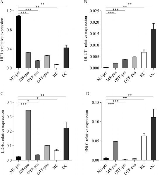
Abstract Meigs’ syndrome (MS), a rare complication of benign ovarian tumors, is easily misdiagnosed as ovarian cancer (OC). We retrospectively reviewed the clinical laboratory data of patients diagnosed with MS from 2009 to 2018. Serum carbohydrate antigen 125 and HE4 levels were higher in the MS group than in the ovarian thecoma-fibroma (OTF) and healthy control groups (all P <0.05). However, the serum HE4 levels were lower in the MS group than in the OC group (P <0.001). A routine blood test showed that the absolute counts and percentages of lymphocytes were significantly lower in the MS group than in the OTF and control groups (all P <0.05). However, these variables were higher in the MS group than in the OC group (both P <0.05). The neutrophil-to-lymphocyte ratio (NLR) was also significantly lower, whereas the lymphocyte-to-monocyte ratio was higher in the MS group than in the OC group (both P <0.05). The NLR, platelet-to-lymphocyte ratio, and systemic immune index were significantly higher in the MS group than in the OTF and control groups (all P <0.05). The hypoxia-inducible factor-1 mRNA levels were also significantly higher, whereas the glucose transporter 1, lactate dehydrogenase, and enolase 1 mRNA levels were lower in peripheral CD4+ T cells obtained preoperatively in a patient with MS than those in patients with OTF, patients with OC, and controls (all P <0.05). The expression of these four glucose metabolism genes was preferentially restored to normal levels after the tumor resection of MS (P <0.001). These clinical laboratory features can be useful in improving the preoperative diagnostic accuracy of MS.
|
| Keywords
Meigs’ syndrome
ovarian thecoma-fibroma
NLR (neutrophil to lymphocyte ratio)
CD4+ T cells
glucose metabolism
|
|
Corresponding Author(s):
Fang Wang
|
|
Just Accepted Date: 27 April 2020
Online First Date: 13 July 2020
Issue Date: 11 February 2021
|
|
| 1 |
A Chechia, L Attia, RB Temime, T Makhlouf, A Koubaa. Incidence, clinical analysis, and management of ovarian fibromas and fibrothecomas. Am J Obstet Gynecol 2008; 199(5): 473.e1–473.e4
https://doi.org/10.1016/j.ajog.2008.03.053
pmid: 18501324
|
| 2 |
E Sfar, K Ben Ammar, S Mahjoub, S Zine, N Kchir, H Chelli, M Khrouf, M Chelli. Anatomo-clinical characteristics of ovarian fibrothecal tumors. 19 cases over 12 years: 1981–1992. Rev Fr Gynecol Obstet 1994; 89(6): 315–321 (in French)
pmid: 8085103
|
| 3 |
V Sivanesaratnam, R Dutta, P Jayalakshmi. Ovarian fibroma--clinical and histopathological characteristics. Int J Gynaecol Obstet 1990; 33(3): 243–247
https://doi.org/10.1016/0020-7292(90)90009-A
pmid: 1977643
|
| 4 |
JV Meigs. Fibroma of the ovary with ascites and hydrothorax: Meigs’ syndrome. Am J Obstet Gynecol 1954; 67(5): 962–985
https://doi.org/10.1016/0002-9378(54)90258-6
pmid: 13148256
|
| 5 |
JJ Nicoll, PJ Cox. Leiomyoma of the ovary with ascites and hydrothorax. Am J Obstet Gynecol 1989; 161(1): 177–178
https://doi.org/10.1016/0002-9378(89)90260-3
pmid: 2750800
|
| 6 |
JV Meigs, JW Cass. Fibroma of the ovary with ascites and hydrothorax: with a report of seven cases. Am J Obstet Gynecol 1937; 33(2): 249–267
https://doi.org/10.1016/S0002-9378(37)80015-0
|
| 7 |
JV Meigs. Fibroma of the ovary with ascites and hydrothorax: a further report. Ann Surg 1939; 110(4): 731–754
https://doi.org/10.1097/00000658-193910000-00019
pmid: 17857484
|
| 8 |
K Okuda, S Noguchi, O Narumoto, M Ikemura, Y Yamauchi, G Tanaka, D Takai, M Fukayama, T Nagase. A case of Meigs’ syndrome with preceding pericardial effusion in advance of pleural effusion. BMC Pulm Med 2016; 16(1): 71
https://doi.org/10.1186/s12890-016-0241-1
pmid: 27160723
|
| 9 |
MC Renaud, M Plante, M Roy. Ovarian thecoma associated with a large quantity of ascites and elevated serum CA 125 and CA 15-3. J Obstet Gynaecol Can 2002; 24(12): 963–965
https://doi.org/10.1016/S1701-2163(16)30596-5
pmid: 12464996
|
| 10 |
D Timmerman, P Moerman, I Vergote. Meigs’ syndrome with elevated serum CA 125 levels: two case reports and review of the literature. Gynecol Oncol 1995; 59(3): 405–408
https://doi.org/10.1006/gyno.1995.9952
pmid: 8522265
|
| 11 |
A Morán-Mendoza, G Alvarado-Luna, G Calderillo-Ruiz, A Serrano-Olvera, CM López-Graniel, D Gallardo-Rincón. Elevated CA125 level associated with Meigs’ syndrome: case report and review of the literature. Int J Gynecol Cancer 2006; 16(Suppl 1): 315–318
pmid: 16515612
|
| 12 |
R Dong, C Jin, Q Zhang, X Yang, B Kong. Cellular leiomyoma with necrosis and mucinous degeneration presenting as pseudo-Meigs’ syndrome with elevated CA125. Oncol Rep 2015; 33(6): 3033–3037
https://doi.org/10.3892/or.2015.3912
pmid: 25891047
|
| 13 |
J Danilos, W Michał Kwaśniewski, D Mazurek, W Bednarek, J Kotarski. Meigs’ syndrome with elevated CA-125 and HE-4: a case of luteinized fibrothecoma. Przegl Menopauz 2015; 14(2): 152–154
https://doi.org/10.5114/pm.2015.52157
pmid: 26327905
|
| 14 |
CE Son, JS Choi, JH Lee, SW Jeon, JH Hong, JW Bae. Laparoscopic surgical management and clinical characteristics of ovarian fibromas. JSLS 2011; 15(1): 16–20
https://doi.org/10.4293/108680810X12924466009087
pmid: 21902936
|
| 15 |
AS Laganà, D Vergara, A Favilli, VL La Rosa, A Tinelli, S Gerli, M Noventa, A Vitagliano, O Triolo, AMC Rapisarda, SG Vitale. Epigenetic and genetic landscape of uterine leiomyomas: a current view over a common gynecological disease. Arch Gynecol Obstet 2017; 296(5): 855–867
https://doi.org/10.1007/s00404-017-4515-5
pmid: 28875276
|
| 16 |
AS Laganà, F Colonese, E Colonese, V Sofo, FM Salmeri, R Granese, B Chiofalo, L Ciancimino, O Triolo. Cytogenetic analysis of epithelial ovarian cancer’s stem cells: an overview on new diagnostic and therapeutic perspectives. Eur J Gynaecol Oncol 2015; 36(5): 495–505
pmid: 26513872
|
| 17 |
A Bellia, SG Vitale, AS Laganà, F Cannone, G Houvenaeghel, S Rua, A Ladaique, C Jauffret, G Ettore, E Lambaudie. Feasibility and surgical outcomes of conventional and robot-assisted laparoscopy for early-stage ovarian cancer: a retrospective, multicenter analysis. Arch Gynecol Obstet 2016; 294(3): 615–622
https://doi.org/10.1007/s00404-016-4087-9
pmid: 27040423
|
| 18 |
SI Grivennikov, FR Greten, M Karin. Immunity, inflammation, and cancer. Cell 2010; 140(6): 883–899
https://doi.org/10.1016/j.cell.2010.01.025
pmid: 20303878
|
| 19 |
A Mantovani, P Allavena, A Sica, F Balkwill. Cancer-related inflammation. Nature 2008; 454(7203): 436–444
https://doi.org/10.1038/nature07205
pmid: 18650914
|
| 20 |
J Margetts, LF Ogle, SL Chan, AWH Chan, KCA Chan, D Jamieson, CE Willoughby, DA Mann, CL Wilson, DM Manas, W Yeo, HL Reeves. Neutrophils: driving progression and poor prognosis in hepatocellular carcinoma? Br J Cancer 2018; 118(2): 248–257
https://doi.org/10.1038/bjc.2017.386
pmid: 29123264
|
| 21 |
P Sanchez-Salcedo, JP de-Torres, D Martinez-Urbistondo, J Gonzalez-Gutierrez, J Berto, A Campo, AB Alcaide, JJ Zulueta. The neutrophil to lymphocyte and platelet to lymphocyte ratios as biomarkers for lung cancer development. Lung Cancer 2016; 97: 28–34
https://doi.org/10.1016/j.lungcan.2016.04.010
pmid: 27237024
|
| 22 |
AJ Templeton, O Ace, MG McNamara, M Al-Mubarak, FE Vera-Badillo, T Hermanns, B Seruga, A Ocaña, IF Tannock, E Amir. Prognostic role of platelet to lymphocyte ratio in solid tumors: a systematic review and meta-analysis. Cancer Epidemiol Biomarkers Prev 2014; 23(7): 1204–1212
https://doi.org/10.1158/1055-9965.EPI-14-0146
pmid: 24793958
|
| 23 |
AJ Templeton, MG McNamara, B Šeruga, FE Vera-Badillo, P Aneja, A Ocaña, R Leibowitz-Amit, G Sonpavde, JJ Knox, B Tran, IF Tannock, E Amir. Prognostic role of neutrophil-to-lymphocyte ratio in solid tumors: a systematic review and meta-analysis. J Natl Cancer Inst 2014; 106(6): dju124
https://doi.org/10.1093/jnci/dju124
pmid: 24875653
|
| 24 |
Y Abramov, SO Anteby, SJ Fasouliotis, V Barak. Markedly elevated levels of vascular endothelial growth factor, fibroblast growth factor, and interleukin 6 in Meigs syndrome. Am J Obstet Gynecol 2001; 184(3): 354–355
https://doi.org/10.1067/mob.2001.110028
pmid: 11228486
|
| 25 |
Y Abramov, SO Anteby, SJ Fasouliotis, V Barak. The role of inflammatory cytokines in Meigs’ syndrome. Obstet Gynecol 2002; 99(5 Pt 2): 917–919
https://doi.org/10.1016/S0029-7844(01)01602-7
pmid: 11975958
|
| 26 |
EV Dang, J Barbi, HY Yang, D Jinasena, H Yu, Y Zheng, Z Bordman, J Fu, Y Kim, HR Yen, W Luo, K Zeller, L Shimoda, SL Topalian, GL Semenza, CV Dang, DM Pardoll, F Pan. Control of TH17/Treg balance by hypoxia-inducible factor 1. Cell 2011; 146(5): 772–784
https://doi.org/10.1016/j.cell.2011.07.033
pmid: 21871655
|
| 27 |
LZ Shi, R Wang, G Huang, P Vogel, G Neale, DR Green, H Chi. HIF1α-dependent glycolytic pathway orchestrates a metabolic checkpoint for the differentiation of TH17 and Treg cells. J Exp Med 2011; 208(7): 1367–1376
https://doi.org/10.1084/jem.20110278
pmid: 21708926
|
| 28 |
G Liu, Y Bi, L Xue, Y Zhang, H Yang, X Chen, Y Lu, Z Zhang, H Liu, X Wang, R Wang, Y Chu, R Yang. Dendritic cell SIRT1-HIF1a axis programs the differentiation of CD4+ T cells through IL-12 and TGF-b1. Proc Natl Acad Sci USA 2015; 112(9): E957–E965
https://doi.org/10.1073/pnas.1420419112
pmid: 25730867
|
| 29 |
AN Macintyre, VA Gerriets, AG Nichols, RD Michalek, MC Rudolph, D Deoliveira, SM Anderson, ED Abel, BJ Chen, LP Hale, JC Rathmell. The glucose transporter Glut1 is selectively essential for CD4 T cell activation and effector function. Cell Metab 2014; 20(1): 61–72
https://doi.org/10.1016/j.cmet.2014.05.004
pmid: 24930970
|
| 30 |
RD Michalek, VA Gerriets, SR Jacobs, AN Macintyre, NJ MacIver, EF Mason, SA Sullivan, AG Nichols, JC Rathmell. Cutting edge: distinct glycolytic and lipid oxidative metabolic programs are essential for effector and regulatory CD4+ T cell subsets. J Immunol 2011; 186(6): 3299–3303
https://doi.org/10.4049/jimmunol.1003613
pmid: 21317389
|
| 31 |
VA Gerriets, JC Rathmell. Metabolic pathways in T cell fate and function. Trends Immunol 2012; 33(4): 168–173
https://doi.org/10.1016/j.it.2012.01.010
pmid: 22342741
|
| 32 |
MD Buck, D O’Sullivan, EL Pearce. T cell metabolism drives immunity. J Exp Med 2015; 212(9): 1345–1360
https://doi.org/10.1084/jem.20151159
pmid: 26261266
|
| 33 |
CH Chang, J Qiu, D O’Sullivan, MD Buck, T Noguchi, JD Curtis, Q Chen, M Gindin, MM Gubin, GJ van der Windt, E Tonc, RD Schreiber, EJ Pearce, EL Pearce. Metabolic competition in the tumor microenvironment is a driver of cancer progression. Cell 2015; 162(6): 1229–1241
https://doi.org/10.1016/j.cell.2015.08.016
pmid: 26321679
|
|
Viewed |
|
|
|
Full text
|
|
|
|
|
Abstract
|
|
|
|
|
Cited |
|
|
|
|
| |
Shared |
|
|
|
|
| |
Discussed |
|
|
|
|