|
|
|
Advances in tissue state recognition in spinal surgery: a review |
Hao Qu, Yu Zhao( ) ) |
| Department of Orthopaedics, Peking Union Medical College Hospital, Chinese Academy of Medical Sciences and Peking Union Medical College, Beijing 100730, China |
|
|
|
|
Abstract Spinal disease is an important cause of cervical discomfort, low back pain, radiating pain in the limbs, and neurogenic intermittent claudication, and its incidence is increasing annually. From the etiological viewpoint, these symptoms are directly caused by the compression of the spinal cord, nerve roots, and blood vessels and are most effectively treated with surgery. Spinal surgeries are primarily performed using two different techniques: spinal canal decompression and internal fixation. In the past, tactile sensation was the primary method used by surgeons to understand the state of the tissue within the operating area. However, this method has several disadvantages because of its subjectivity. Therefore, it has become the focus of spinal surgery research so as to strengthen the objectivity of tissue state recognition, improve the accuracy of safe area location, and avoid surgical injury to tissues. Aside from traditional imaging methods, surgical sensing techniques based on force, bioelectrical impedance, and other methods have been gradually developed and tested in the clinical setting. This article reviews the progress of different tissue state recognition methods in spinal surgery and summarizes their advantages and disadvantages.
|
| Keywords
spinal surgery
tissue state recognition
image
force sensing
bioelectrical impedance
|
|
Corresponding Author(s):
Yu Zhao
|
|
Just Accepted Date: 07 February 2021
Online First Date: 14 May 2021
Issue Date: 23 September 2021
|
|
| 1 |
E Truumees. A history of lumbar disc herniation from Hippocrates to the 1990s. Clin Orthop Relat Res 2015; 473(6): 1885–1895
https://doi.org/10.1007/s11999-014-3633-7
pmid: 24752913
|
| 2 |
PS Issack, ME Cunningham, M Pumberger, AP Hughes, FP Cammisa Jr. Degenerative lumbar spinal stenosis: evaluation and management. J Am Acad Orthop Surg 2012; 20(8): 527–535
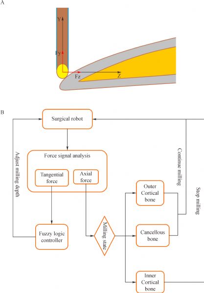
https://doi.org/10.5435/JAAOS-20-08-527
pmid: 22855855
|
| 3 |
J Lurie, C Tomkins-Lane. Management of lumbar spinal stenosis. BMJ 2016; 352: h6234
https://doi.org/10.1136/bmj.h6234
pmid: 26727925
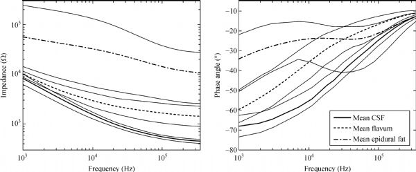
|
| 4 |
TD Koreckij, JS Fischgrund. Degenerative spondylolisthesis. J Spinal Disord Tech 2015; 28(7): 236–241
https://doi.org/10.1097/BSD.0000000000000298
pmid: 26172828
|
| 5 |
JL Melancia, AF Francisco, JL Antunes. Spinal stenosis. Handb Clin Neurol 2014; 119(119): 541–549
https://doi.org/10.1016/B978-0-7020-4086-3.00035-7
pmid: 24365318
|
| 6 |
SC Overley, JS Kim, BA Gogel, RK Merrill, AC Hecht. Tandem spinal stenosis: a systematic review. JBJS Rev 2017; 5(9): e2
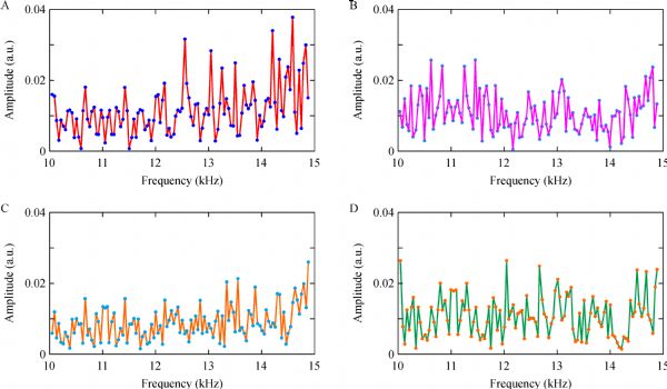
https://doi.org/10.2106/JBJS.RVW.17.00007
pmid: 28872572
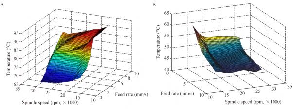
|
| 7 |
JH Lee, KH Choi, S Kang, DH Kim, DH Kim, BR Kim, W Kim, JH Kim, KH Do, JG Do, JS Ryu, K Min, SG Bahk, YH Park, HJ Bang, KH Shin, S Yang, HS Yang, SD Yoo, JS Yoo, KJ Yoon, SJ Yoon, GJ Lee, SY Lee, SC Lee, SY Lee, IS Lee, JS Lee, CH Lee, JY Lim, JY Han, SH Han, DH Sung, KH Cho, SY Kim, HJ Kim, W Ju. Nonsurgical treatments for patients with radicular pain from lumbosacral disc herniation. Spine J 2019; 19(9): 1478–1489
https://doi.org/10.1016/j.spinee.2019.06.004
pmid: 31201860
|
| 8 |
L Viezens, P Reer, A Strahl, L Weiser, M Schroeder, J Beyerlein, C Schaefer. Safety and efficacy of single-stage versus 2-stage spinal fusion via posterior instrumentation and anterior thoracoscopy: a retrospective matched-pair cohort study with 247 consecutive patients. World Neurosurg 2018; 109: e739–e747
https://doi.org/10.1016/j.wneu.2017.10.074
pmid: 29079258
|
| 9 |
OR Fjeld, L Grøvle, J Helgeland, MC Småstuen, TK Solberg, JA Zwart, M Grotle. Complications, reoperations, readmissions, and length of hospital stay in 34 639 surgical cases of lumbar disc herniation. Bone Joint J 2019; 101-B(4): 470–477
https://doi.org/10.1302/0301-620X.101B4.BJJ-2018-1184.R1
pmid: 30929479
|
| 10 |
H Inose, T Kato, M Yuasa, T Yamada, H Maehara, T Hirai, T Yoshii, S Kawabata, A Okawa. Comparison of decompression, decompression plus fusion, and decompression plus stabilization for degenerative spondylolisthesis: a prospective, randomized study. Clin Spine Surg 2018; 31(7): E347–E352
https://doi.org/10.1097/BSD.0000000000000659
pmid: 29877872
|
| 11 |
Z Chen, B Wu, X Zhai, Y Bai, X Zhu, B Luo, X Chen, C Li, M Yang, K Xu, C Liu, C Wang, Y Zhao, X Wei, K Chen, W Yang, D Ta, M Li. Basic study for ultrasound-based navigation for pedicle screw insertion using transmission and backscattered methods. PLoS One 2015; 10(4): e0122392
https://doi.org/10.1371/journal.pone.0122392
pmid: 25861053
|
| 12 |
T Fujishiro, Y Nakaya, S Fukumoto, S Adachi, A Nakano, K Fujiwara, I Baba, M Neo. Accuracy of pedicle screw placement with robotic guidance system: a cadaveric study. Spine 2015; 40(24): 1882–1889
https://doi.org/10.1097/BRS.0000000000001099
pmid: 26655804
|
| 13 |
M Galluzzo, S D’Adamio, R Pastorino, A Andreoli, S Servoli, L Bianchi, M Talamonti. Effect of anti IL-12/23 on body composition: results of bioelectrical impedance analysis in Caucasian psoriatic patients. Expert Opin Biol Ther 2018; 18(3): 229–235
https://doi.org/10.1080/14712598.2018.1419183
pmid: 29252034
|
| 14 |
S Wei, W Tao, H Zhu, Y Li. Three-dimensional intraoperative imaging with O-arm to establish a working trajectory in percutaneous endoscopic lumbar discectomy. Wideochir Inne Tech Maloinwazyjne 2016; 10(4): 555–560
pmid: 26865892
|
| 15 |
V Kosmopoulos, C Schizas. Pedicle screw placement accuracy: a meta-analysis. Spine 2007; 32(3): E111–E120
https://doi.org/10.1097/01.brs.0000254048.79024.8b
pmid: 17268254
|
| 16 |
LT Holly, KT Foley. Percutaneous placement of posterior cervical screws using three-dimensional fluoroscopy. Spine 2006; 31(5): 536–541
https://doi.org/10.1097/01.brs.0000201297.83920.a1
pmid: 16508547
|
| 17 |
J Hirayama, M Hashimoto. Percutaneous endoscopic diskectomy using an interlaminar approach based on 3D CT/MR fusion imaging. J Neurol Surg A Cent Eur Neurosurg 2019; 80(2): 88–95
https://doi.org/10.1055/s-0038-1673399
pmid: 30583302
|
| 18 |
Z Hu, X Li, J Cui, X He, C Li, Y Han, J Pan, M Yang, J Tan, L Li. Significance of preoperative planning software for puncture and channel establishment in percutaneous endoscopic lumbar DISCECTOMY: a study of 40 cases. Int J Surg 2017; 41: 97–103
https://doi.org/10.1016/j.ijsu.2017.03.059
pmid: 28344159
|
| 19 |
J Tang, Z Zhu, T Sui, D Kong, X Cao. Position and complications of pedicle screw insertion with or without image-navigation techniques in the thoracolumbar spine: a meta-analysis of comparative studies. J Biomed Res 2014; 28(3): 228–239
pmid: 25013406
|
| 20 |
Y Yang, F Wang, S Han, Y Wang, J Dong, L Li, D Zhou. Isocentric C-arm three-dimensional navigation versus conventional C-arm assisted C1-C2 transarticular screw fixation for atlantoaxial instability. Arch Orthop Trauma Surg 2015; 135(8): 1083–1092
https://doi.org/10.1007/s00402-015-2249-z
pmid: 26119707
|
| 21 |
JM Bledsoe, D Fenton, JL Fogelson, EW Nottmeier. Accuracy of upper thoracic pedicle screw placement using three-dimensional image guidance. Spine J 2009; 9(10): 817–821
https://doi.org/10.1016/j.spinee.2009.06.014
pmid: 19664966
|
| 22 |
MF Oertel, J Hobart, M Stein, V Schreiber, W Scharbrodt. Clinical and methodological precision of spinal navigation assisted by 3D intraoperative O-arm radiographic imaging. J Neurosurg Spine 2011; 14(4): 532–536
https://doi.org/10.3171/2010.10.SPINE091032
pmid: 21275555
|
| 23 |
Z Sun, D Yuan, Y Sun, Z Zhang, G Wang, Y Guo, G Wang, D Liu, P Chen, L Jing, F Yang, P Zhang, H Zhang, Y Wu, W Shi, J Wang. Application of intraoperative O-arm-assisted real-time navigation technique for spinal fixation. Translational Neuroence & Clinics 2017; 3(3): 135–146
https://doi.org/10.18679/CN11-6030_R.2017.022
|
| 24 |
S Bernhardt, SA Nicolau, V Agnus, L Soler, C Doignon, J Marescaux. Automatic localization of endoscope in intraoperative CT image: a simple approach to augmented reality guidance in laparoscopic surgery. Med Image Anal 2016; 30: 130–143
https://doi.org/10.1016/j.media.2016.01.008
pmid: 26925804
|
| 25 |
C, Li J, Zeng G, Ye W, Sun J, Hong J, Li C. ZhengDevelopment of a virtual reality preoperative planning system for postlateral endoscopic lumbar discectomy surgery and its clinical application. World Neurosurg 2019; 123: e1–e8
https://doi.org/10.1016/j.wneu.2018.08.082
pmid: 30144600
|
| 26 |
M Draelos, B Keller, C Viehland, OM Carrasco-Zevallos, A Kuo, J Izatt. Real-time visualization and interaction with static and live optical coherence tomography volumes in immersive virtual reality. Biomed Opt Express 2018; 9(6): 2825–2843
https://doi.org/10.1364/BOE.9.002825
pmid: 30258693
|
| 27 |
A Javaux, D Bouget, C Gruijthuijsen, D Stoyanov, T Vercauteren, S Ourselin, J Deprest, K Denis, E Vander Poorten. A mixed-reality surgical trainer with comprehensive sensing for fetal laser minimally invasive surgery. Int J CARS 2018; 13(12): 1949–1957
https://doi.org/10.1007/s11548-018-1822-7
pmid: 30054776
|
| 28 |
G Coelho, HLA Defino. The role of mixed reality simulation for surgical training in spine: phase 1 validation. Spine 2018; 43(22): 1609–1616
https://doi.org/10.1097/BRS.0000000000002856
pmid: 30180147
|
| 29 |
H Yu, Z Zhou, X Lei, H Liu, G Fan, S He. Mixed reality-based preoperative planning for training of percutaneous transforaminal endoscopic discectomy: a feasibility study. World Neurosurg 2019; 129: e767–e775
https://doi.org/10.1016/j.wneu.2019.06.020
pmid: 31203062
|
| 30 |
WY Lee, CL Shih. Control and breakthrough detection of a three-axis robotic bone drilling system. Mechatronics 2006; 16(2): 73–84
https://doi.org/10.1016/j.mechatronics.2005.11.002
|
| 31 |
MH Aziz, MA Ayub, R Jaafar. Real-time algorithm for detection of breakthrough bone drilling. Procedia Eng 2012; 41: 352–359
https://doi.org/10.1016/j.proeng.2012.07.184
|
| 32 |
Y Hu, H Jin, L Zhang, P Zhang, J Zhang. State recognition of pedicle drilling with force sensing in a robotic spinal surgical system. IEEE/ASME Trans Mechatron 2014; 19(1): 357–365
https://doi.org/10.1109/TMECH.2012.2237179
|
| 33 |
M Marco, M Rodríguez-Millán, C Santiuste, E Giner, M Henar Miguélez. A review on recent advances in numerical modelling of bone cutting. J Mech Behav Biomed Mater 2015; 44: 179–201
https://doi.org/10.1016/j.jmbbm.2014.12.006
pmid: 25676359
|
| 34 |
T Ortmaier, H Weiss, S Döbele, U Schreiber. Experiments on robot-assisted navigated drilling and milling of bones for pedicle screw placement. Int J Med Robot 2006; 2(4): 350–363
https://doi.org/10.1002/rcs.114
pmid: 17520654
|
| 35 |
WY Kim, SY Ko, JO Park, S Park. 6-DOF force feedback control of robot-assisted bone fracture reduction system using double F/T sensors and adjustable admittances to protect bones against damage. Mechatronics 2016; 35: 136–147
https://doi.org/10.1016/j.mechatronics.2016.02.005
|
| 36 |
Z Deng, H Jin, Y Hu, Y He, P Zhang, W Tian, J Zhang. Fuzzy force control and state detection in vertebral lamina milling. Mechatronics 2016; 35: 1–10
https://doi.org/10.1016/j.mechatronics.2016.02.004
|
| 37 |
L Fan, P Gao, B Zhao, Y Sun, X Xin, Y Hu, S Liu, J Zhang. Safety control strategy for vertebral lamina milling task. CAAI Trans Intell Technol 2016; 1(3): 249–258
https://doi.org/10.1016/j.trit.2016.10.005
|
| 38 |
Z Jiang, X Qi, Y Sun, Y Hu, Z Guillaume, J Zhang. Cutting depth monitoring based on milling force for robot-assisted laminectomy. IEEE Trans Autom Sci Eng 2020; 17(1): 2–14
https://doi.org/10.1109/TASE.2019.2920133
|
| 39 |
Y Kasahara, K Ohnishi, H Kawana. Analysis of drill wear based on torque and force sensorless cutting power estimation. IECON 2010—36th Annual Conference on IEEE Industrial Electronics Society. IEEE, 2010
|
| 40 |
T Osa, CF Abawi, N Sugita, H Chikuda, S Sugita, T Tanaka, H Oshima, T Moro, S Tanaka, M Mitsuishi. Hand-held bone cutting tool with autonomous penetration detection for spinal surgery. IEEE/ASME Trans Mechatron 2015; 20(6): 3018–3027
https://doi.org/10.1109/TMECH.2015.2410287
|
| 41 |
Y Dai, Y Xue, J Zhang. Vibration-based milling condition monitoring in robot-assisted spine surgery. IEEE/ASME Trans Mechatron 2015; 20(6): 3028–3039
https://doi.org/10.1109/TMECH.2015.2414177
|
| 42 |
Y Dai, Y Xue, J Zhang. A continuous wavelet transform approach for harmonic parameters estimation in the presence of impulsive noise. J Sound Vibrat 2016; 360: 300–314
https://doi.org/10.1016/j.jsv.2015.09.023
|
| 43 |
TJ Faes, HA van der Meij, JC de Munck, RM Heethaar. The electric resistivity of human tissues (100 Hz–10 MHz): a meta-analysis of review studies. Physiol Meas 1999; 20(4): R1–R10
https://doi.org/10.1088/0967-3334/20/4/201
pmid: 10593226
|
| 44 |
H Nakase, R Matsuda, Y Shin, YS Park, T Sakaki. The use of ultrasonic bone curettes in spinal surgery. Acta Neurochir (Wien) 2006; 148(2): 207–213
https://doi.org/10.1007/s00701-005-0655-7
pmid: 16311841
|
| 45 |
C Gabriel, S Gabriel, E Corthout. The dielectric properties of biological tissues: I. Literature survey. Phys Med Biol 1996; 41(11): 2231–2249
https://doi.org/10.1088/0031-9155/41/11/001
pmid: 8938024
|
| 46 |
DA Dean, T Ramanathan, D Machado, R Sundararajan. Electrical impedance spectroscopy study of biological tissues. J Electrost 2008; 66(3–4): 165–177
https://doi.org/10.1016/j.elstat.2007.11.005
pmid: 19255614
|
| 47 |
R Antakia, BH Brown, PE Highfield, TJ Stephenson, NJ Brown, SP Balasubramanian. Electrical impedance spectroscopy to aid parathyroid identification and preservation in central compartment neck surgery: a proof of concept in a rabbit model. Surg Innov 2016; 23(2): 176–182
https://doi.org/10.1177/1553350615607639
pmid: 26423912
|
| 48 |
Y Dai, Y Xue, J Zhang. Drilling electrode for real-time measurement of electrical impedance in bone tissues. Ann Biomed Eng 2014; 42(3): 579–588
https://doi.org/10.1007/s10439-013-0938-8
pmid: 24254254
|
| 49 |
F Shao, H Bai, M Tang, Y Xue, Y Dai, J Zhang. Tissue discrimination by bioelectrical impedance during PLL resection in anterior decompression surgery for treatment of cervical spondylotic myelopathy. J Orthop Surg Res 2019; 14(1): 341
https://doi.org/10.1186/s13018-019-1380-x
pmid: 31694719
|
| 50 |
T Wyss Balmer, J Ansó, E Muntane, K Gavaghan, S Weber, A Stahel, P Büchler. In-vivo electrical impedance measurement in mastoid bone. Ann Biomed Eng 2017; 45(4): 1122–1132
https://doi.org/10.1007/s10439-016-1758-4
pmid: 27830489
|
| 51 |
Y Turan, M Sayin, A Yurt, T Yilmaz, FD Ozer, C Temiz. Local tissue electrical resistances in transpedicular screw application in the thoracolumbar region. Turk Neurosurg 2016; 26(6): 937–943
pmid: 27560525
|
| 52 |
S Halonen, K Annala, J Kari, S Jokinen, A Lumme, K Kronström, A Yli-Hankala. Detection of spine structures with Bioimpedance Probe (BIP) Needle in clinical lumbar punctures. J Clin Monit Comput 2017; 31(5): 1065–1072
https://doi.org/10.1007/s10877-016-9915-8
pmid: 27492427
|
| 53 |
Z Li, C Chen, Y Lin, X Li, H Tan, MT Chan, WK Wu, S Zhan, Q Cao, J Shen. A novel probe for measuring tissue bioelectrical impedance to enhance pedicle screw placement in spinal surgery. Am J Transl Res 2018; 10(7): 2205–2212
pmid: 30093957
|
| 54 |
I Boesnach, M Hahn, J Moldenhauer, TH Beth, U Spetzger. Analysis of drill sound in spine surgery. Perspective in Image-guided Surgery—the Scientific Workshop on Medical Robotics, Navigation and Visualization. RheinAhrCampus Remagen, Germany, March 11–12, 2004
|
| 55 |
Z Liao, DA Axinte. On monitoring chip formation, penetration depth and cutting malfunctions in bone micro-drilling via acoustic emission. J Mater Process Technol 2016; 229: 82–93
https://doi.org/10.1016/j.jmatprotec.2015.09.016
|
| 56 |
Y Sun, H Jin, Y Hu, P Zhang, J Zhang. State recognition of bone drilling with audio signal in Robotic Orthopedics Surgery System. IEEE International Conference on Intelligent Robots and Systems. IEEE 2014: 3503–3508
|
| 57 |
F Guan, Y Sun, X Qi, Y Hu, G Yu, J Zhang. State recognition of bone drilling based on acoustic emission in pedicle screw operation. Sensors (Basel) 2018; 18(5): 1484
https://doi.org/10.3390/s18051484
pmid: 29747395
|
| 58 |
G Augustin, T Zigman, S Davila, T Udilljak, T Staroveski, D Brezak, S Babic. Cortical bone drilling and thermal osteonecrosis. Clin Biomech (Bristol, Avon) 2012; 27(4): 313–325
https://doi.org/10.1016/j.clinbiomech.2011.10.010
pmid: 22071428
|
| 59 |
HC Shin, YS Yoon. Bone temperature estimation during orthopaedic round bur milling operations. J Biomech 2006; 39(1): 33–39
https://doi.org/10.1016/j.jbiomech.2004.11.004
pmid: 16271585
|
| 60 |
L Wen, ZH Zhao, JB Song, DD Yu, M Chen, SGF Shen. Experimental study on thermal and force characteristics in the dry slotting of cortical bone. Adv Mat Res 2016; 1136: 233–238
https://doi.org/10.4028/www.scientific.net/AMR.1136.233
|
| 61 |
I Kais, AL Al-Abdullah, H Abdi, CP Lim. Force and temperature modelling of bone milling using artificial neural networks. Measurement 2018; 116: 25–37
https://doi.org/10.1016/j.measurement.2017.10.051
|
|
Viewed |
|
|
|
Full text
|
|
|
|
|
Abstract
|
|
|
|
|
Cited |
|
|
|
|
| |
Shared |
|
|
|
|
| |
Discussed |
|
|
|
|