|
|
|
Proteins moonlighting in tumor metabolism and epigenetics |
Lei Lv1( ), Qunying Lei2,3,4() ), Qunying Lei2,3,4() |
1. MOE Key Laboratory of Metabolism and Molecular Medicine, Department of Biochemistry and Molecular Biology, School of Basic Medical Sciences, Fudan University, Shanghai 200032, China
2. Fudan University Shanghai Cancer Center & Institutes of Biomedical Sciences; Cancer Institutes; Key Laboratory of Breast Cancer in Shanghai; The Shanghai Key Laboratory of Medical Epigenetics, Shanghai Medical College, Fudan University, Shanghai 200032, China
3. Department of Oncology, Shanghai Medical College, Fudan University, Shanghai 200032, China
4. State Key Laboratory of Medical Neurobiology, Fudan University, Shanghai 200032, China |
|
|
|
|
Abstract Cancer development is a complicated process controlled by the interplay of multiple signaling pathways and restrained by oxygen and nutrient accessibility in the tumor microenvironment. High plasticity in using diverse nutrients to adapt to metabolic stress is one of the hallmarks of cancer cells. To respond to nutrient stress and to meet the requirements for rapid cell proliferation, cancer cells reprogram metabolic pathways to take up more glucose and coordinate the production of energy and intermediates for biosynthesis. Such actions involve gene expression and activity regulation by the moonlighting function of oncoproteins and metabolic enzymes. The signal−moonlighting protein−metabolism axis facilitates the adaptation of tumor cells under varying environment conditions and can be therapeutically targeted for cancer treatment.
|
| Keywords
moonlighting function
tumor metabolism
epigenetics
|
|
Corresponding Author(s):
Lei Lv,Qunying Lei
|
|
Just Accepted Date: 20 November 2020
Online First Date: 22 December 2020
Issue Date: 18 June 2021
|
|
| 1 |
D Hanahan, RA Weinberg. Hallmarks of cancer: the next generation. Cell 2011; 144(5): 646–674
https://doi.org/10.1016/j.cell.2011.02.013
pmid: 21376230
|
| 2 |
O Warburg. On the origin of cancer cells. Science 1956; 123(3191): 309–314
https://doi.org/10.1126/science.123.3191.309
pmid: 13298683
|
| 3 |
KE Allison, BL Coomber, BW Bridle. Metabolic reprogramming in the tumour microenvironment: a hallmark shared by cancer cells and T lymphocytes. Immunology 2017; 152(2): 175–184
https://doi.org/10.1111/imm.12777
pmid: 28621843
|
| 4 |
MF Rodrigues, E Obre, FH de Melo, GC Santos Jr, A Galina, MG Jasiulionis, R Rossignol, FD Rumjanek, ND Amoêdo. Enhanced OXPHOS, glutaminolysis and β-oxidation constitute the metastatic phenotype of melanoma cells. Biochem J 2016; 473(6): 703–715
https://doi.org/10.1042/BJ20150645
pmid: 26699902
|
| 5 |
P Caro, AU Kishan, E Norberg, IA Stanley, B Chapuy, SB Ficarro, K Polak, D Tondera, J Gounarides, H Yin, F Zhou, MR Green, L Chen, S Monti, JA Marto, MA Shipp, NN Danial. Metabolic signatures uncover distinct targets in molecular subsets of diffuse large B cell lymphoma. Cancer Cell 2012; 22(4): 547–560
https://doi.org/10.1016/j.ccr.2012.08.014
pmid: 23079663
|
| 6 |
Y Liu, LS Zuckier, NV Ghesani. Dominant uptake of fatty acid over glucose by prostate cells: a potential new diagnostic and therapeutic approach. Anticancer Res 2010; 30(2): 369–374
pmid: 20332441
|
| 7 |
I Lazar, E Clement, S Dauvillier, D Milhas, M Ducoux-Petit, S LeGonidec, C Moro, V Soldan, S Dalle, S Balor, M Golzio, O Burlet-Schiltz, P Valet, C Muller, L Nieto. Adipocyte exosomes promote melanoma aggressiveness through fatty acid oxidation: a novel mechanism linking obesity and cancer. Cancer Res 2016; 76(14): 4051–4057
https://doi.org/10.1158/0008-5472.CAN-16-0651
pmid: 27216185
|
| 8 |
KM Nieman, HA Kenny, CV Penicka, A Ladanyi, R Buell-Gutbrod, MR Zillhardt, IL Romero, MS Carey, GB Mills, GS Hotamisligil, SD Yamada, ME Peter, K Gwin, E Lengyel. Adipocytes promote ovarian cancer metastasis and provide energy for rapid tumor growth. Nat Med 2011; 17(11): 1498–1503
https://doi.org/10.1038/nm.2492
pmid: 22037646
|
| 9 |
Y Cao. Adipocyte and lipid metabolism in cancer drug resistance. J Clin Invest 2019; 129(8): 3006–3017
https://doi.org/10.1172/JCI127201
pmid: 31264969
|
| 10 |
H Iwamoto, M Abe, Y Yang, D Cui, T Seki, M Nakamura, K Hosaka, S Lim, J Wu, X He, X Sun, Y Lu, Q Zhou, W Shi, T Torimura, G Nie, Q Li, Y Cao. Cancer lipid metabolism confers antiangiogenic drug resistance. Cell Metab 2018; 28(1): 104–117.e5
https://doi.org/DOI: 10.1016/j.cmet.2018.05.005
pmid: 29861385
|
| 11 |
PS Ward, CB Thompson. Metabolic reprogramming: a cancer hallmark even Warburg did not anticipate. Cancer Cell 2012; 21(3): 297–308
https://doi.org/10.1016/j.ccr.2012.02.014
pmid: 22439925
|
| 12 |
M Reina-Campos, J Moscat, M Diaz-Meco. Metabolism shapes the tumor microenvironment. Curr Opin Cell Biol 2017; 48: 47–53
https://doi.org/10.1016/j.ceb.2017.05.006
pmid: 28605656
|
| 13 |
AE Boukouris, SD Zervopoulos, ED Michelakis. Metabolic enzymes moonlighting in the nucleus: metabolic regulation of gene transcription. Trends Biochem Sci 2016; 41(8): 712–730
https://doi.org/10.1016/j.tibs.2016.05.013
pmid: 27345518
|
| 14 |
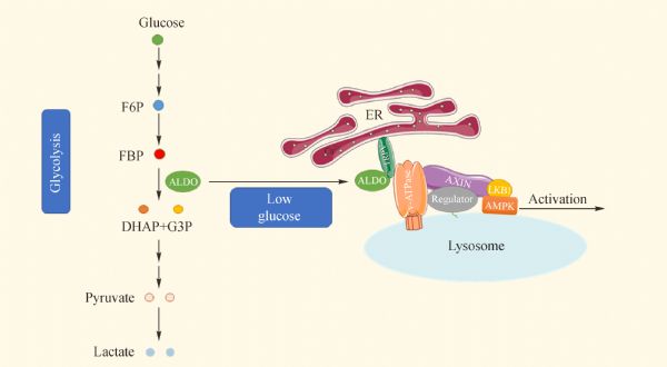
M Li, CS Zhang, Y Zong, JW Feng, T Ma, M Hu, Z Lin, X Li, C Xie, Y Wu, D Jiang, Y Li, C Zhang, X Tian, W Wang, Y Yang, J Chen, J Cui, YQ Wu, X Chen, QF Liu, J Wu, SY Lin, Z Ye, Y Liu, HL Piao, L Yu, Z Zhou, XS Xie, DG Hardie, SC Lin. Transient receptor potential V channels are essential for glucose sensing by aldolase and AMPK. Cell Metab 2019; 30(3): 508–524.e12
https://doi.org/10.1016/j.cmet.2019.05.018
|
| 15 |
P Huangyang, F Li, P Lee, I Nissim, AM Weljie, A Mancuso, B Li, B Keith, SS Yoon, MC Simon. Fructose-1,6-bisphosphatase 2 inhibits sarcoma progression by restraining mitochondrial biogenesis. Cell Metab 2020; 31(1): 174–188.e7
https://doi.org/DOI: 10.1016/j.cmet.2019.10.012
pmid: 31761563
|
| 16 |
D Xu, Z Wang, Y Xia, F Shao, W Xia, Y Wei, X Li, X Qian, JH Lee, L Du, Y Zheng, G Lv, JS Leu, H Wang, D Xing, T Liang, MC Hung, Z Lu. The gluconeogenic enzyme PCK1 phosphorylates INSIG1/2 for lipogenesis. Nature 2020; 580(7804): 530–535
https://doi.org/10.1038/s41586-020-2183-2
pmid: 32322062
|
| 17 |
A Fernández-Medarde, E Santos. Ras in cancer and developmental diseases. Genes Cancer 2011; 2(3): 344–358
https://doi.org/10.1177/1947601911411084
pmid: 21779504
|
| 18 |
EM Kerr, E Gaude, FK Turrell, C Frezza, CP Martins. Mutant Kras copy number defines metabolic reprogramming and therapeutic susceptibilities. Nature 2016; 531(7592): 110–113
https://doi.org/10.1038/nature16967
pmid: 26909577
|
| 19 |
AD Cox, CJ Der. Ras history: the saga continues. Small GTPases 2010; 1(1): 2–27
https://doi.org/10.4161/sgtp.1.1.12178
pmid: 21686117
|
| 20 |
DK Simanshu, DV Nissley, F McCormick. RAS proteins and their regulators in human disease. Cell 2017; 170(1): 17–33
https://doi.org/10.1016/j.cell.2017.06.009
pmid: 28666118
|
| 21 |
J Hallin, LD Engstrom, L Hargis, A Calinisan, R Aranda, DM Briere, N Sudhakar, V Bowcut, BR Baer, JA Ballard, MR Burkard, JB Fell, JP Fischer, GP Vigers, Y Xue, S Gatto, J Fernandez-Banet, A Pavlicek, K Velastagui, RC Chao, J Barton, M Pierobon, E Baldelli, EF Patricoin 3rd, DP Cassidy, MA Marx, II Rybkin, ML Johnson, SI Ou, P Lito, KP Papadopoulos, PA Jänne, P Olson, JG Christensen. The KRASG12C inhibitor MRTX849 provides insight toward therapeutic susceptibility of KRAS-mutant cancers in mouse models and patients. Cancer Discov 2020; 10(1): 54–71
https://doi.org/10.1158/2159-8290.CD-19-1167
pmid: 31658955
|
| 22 |
AC Kimmelman. Metabolic dependencies in RAS-driven cancers. Clin Cancer Res 2015; 21(8): 1828–1834
https://doi.org/10.1158/1078-0432.CCR-14-2425
pmid: 25878364
|
| 23 |
H Ying, AC Kimmelman, CA Lyssiotis, S Hua, GC Chu, E Fletcher-Sananikone, JW Locasale, J Son, H Zhang, JL Coloff, H Yan, W Wang, S Chen, A Viale, H Zheng, JH Paik, C Lim, AR Guimaraes, ES Martin, J Chang, AF Hezel, SR Perry, J Hu, B Gan, Y Xiao, JM Asara, R Weissleder, YA Wang, L Chin, LC Cantley, RA DePinho. Oncogenic Kras maintains pancreatic tumors through regulation of anabolic glucose metabolism. Cell 2012; 149(3): 656–670
https://doi.org/10.1016/j.cell.2012.01.058
pmid: 22541435
|
| 24 |
CR Amendola, JP Mahaffey, SJ Parker, IM Ahearn, WC Chen, M Zhou, H Court, J Shi, SL Mendoza, MJ Morten, E Rothenberg, E Gottlieb, YZ Wadghiri, R Possemato, SR Hubbard, A Balmain, AC Kimmelman, MR Philips. KRAS4A directly regulates hexokinase 1. Nature 2019; 576(7787): 482–486
https://doi.org/10.1038/s41586-019-1832-9
pmid: 31827279
|
| 25 |
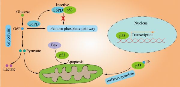
KH Vousden, KM Ryan. p53 and metabolism. Nat Rev Cancer 2009; 9(10): 691–700
https://doi.org/10.1038/nrc2715
pmid: 19759539
|
| 26 |
K Kawauchi, K Araki, K Tobiume, N Tanaka. p53 regulates glucose metabolism through an IKK-NF-κB pathway and inhibits cell transformation. Nat Cell Biol 2008; 10(5): 611–618
https://doi.org/10.1038/ncb1724
pmid: 18391940
|
| 27 |
JE Chipuk, T Kuwana, L Bouchier-Hayes, NM Droin, DD Newmeyer, M Schuler, DR Green. Direct activation of Bax by p53 mediates mitochondrial membrane permeabilization and apoptosis. Science 2004; 303(5660): 1010–1014
https://doi.org/10.1126/science.1092734
pmid: 14963330
|
| 28 |
ND Marchenko, S Wolff, S Erster, K Becker, UM Moll. Monoubiquitylation promotes mitochondrial p53 translocation. EMBO J 2007; 26(4): 923–934
https://doi.org/10.1038/sj.emboj.7601560
pmid: 17268548
|
| 29 |
JH Park, J Zhuang, J Li, PM Hwang. p53 as guardian of the mitochondrial genome. FEBS Lett 2016; 590(7): 924–934
https://doi.org/10.1002/1873-3468.12061
pmid: 26780878
|
| 30 |
P Jiang, W Du, X Wang, A Mancuso, X Gao, M Wu, X Yang. p53 regulates biosynthesis through direct inactivation of glucose-6-phosphate dehydrogenase. Nat Cell Biol 2011; 13(3): 310–316
https://doi.org/10.1038/ncb2172
pmid: 21336310
|
| 31 |
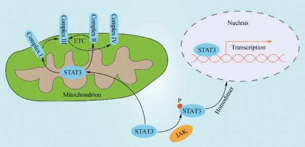
EJ Hillmer, H Zhang, HS Li, SS Watowich. STAT3 signaling in immunity. Cytokine Growth Factor Rev 2016; 31: 1–15
https://doi.org/10.1016/j.cytogfr.2016.05.001
pmid: 27185365
|
| 32 |
JF Bromberg, MH Wrzeszczynska, G Devgan, Y Zhao, RG Pestell, C Albanese, JE Darnell Jr. Stat3 as an oncogene. Cell 1999; 98(3): 295–303
https://doi.org/10.1016/S0092-8674(00)81959-5
pmid: 10458605
|
| 33 |
DE Levy, CK Lee. What does Stat3 do? J Clin Invest 2002; 109(9): 1143–1148
https://doi.org/10.1172/JCI0215650
pmid: 11994402
|
| 34 |
AC Guanizo, CD Fernando, DJ Garama, DJ Gough. STAT3: a multifaceted oncoprotein. Growth Factors 2018; 36(1-2): 1–14
https://doi.org/10.1080/08977194.2018.1473393
pmid: 29873274
|
| 35 |
S Zhao, K Venkatasubbarao, JW Lazor, J Sperry, C Jin, L Cao, JW Freeman. Inhibition of STAT3 Tyr705 phosphorylation by Smad4 suppresses transforming growth factor beta-mediated invasion and metastasis in pancreatic cancer cells. Cancer Res 2008; 68(11): 4221–4228
https://doi.org/10.1158/0008-5472.CAN-07-5123
pmid: 18519681
|
| 36 |
J Bollrath, TJ Phesse, VA von Burstin, T Putoczki, M Bennecke, T Bateman, T Nebelsiek, T Lundgren-May, O Canli, S Schwitalla, V Matthews, RM Schmid, T Kirchner, MC Arkan, M Ernst, FR Greten. gp130-mediated Stat3 activation in enterocytes regulates cell survival and cell-cycle progression during colitis-associated tumorigenesis. Cancer Cell 2009; 15(2): 91–102
https://doi.org/10.1016/j.ccr.2009.01.002
pmid: 19185844
|
| 37 |
J Wegrzyn, R Potla, YJ Chwae, NB Sepuri, Q Zhang, T Koeck, M Derecka, K Szczepanek, M Szelag, A Gornicka, A Moh, S Moghaddas, Q Chen, S Bobbili, J Cichy, J Dulak, DP Baker, A Wolfman, D Stuehr, MO Hassan, XY Fu, N Avadhani, JI Drake, P Fawcett, EJ Lesnefsky, AC Larner. Function of mitochondrial Stat3 in cellular respiration. Science 2009; 323(5915): 793–797
https://doi.org/10.1126/science.1164551
pmid: 19131594
|
| 38 |
DJ Garama, CL White, JJ Balic, DJ Gough. Mitochondrial STAT3: powering up a potent factor. Cytokine 2016; 87: 20–25
https://doi.org/10.1016/j.cyto.2016.05.019
pmid: 27269970
|
| 39 |
D Genini, L Brambilla, E Laurini, J Merulla, G Civenni, S Pandit, R D’Antuono, L Perez, DE Levy, S Pricl, GM Carbone, CV Catapano. Mitochondrial dysfunction induced by a SH2 domain-targeting STAT3 inhibitor leads to metabolic synthetic lethality in cancer cells. Proc Natl Acad Sci USA 2017; 114(25): E4924–E4933
https://doi.org/10.1073/pnas.1615730114
pmid: 28584133
|
| 40 |
S Pelengaris, M Khan. The many faces of c-MYC. Arch Biochem Biophys 2003; 416(2): 129–136
https://doi.org/10.1016/S0003-9861(03)00294-7
pmid: 12893289
|
| 41 |
A Kuzyk, S Mai. c-MYC-induced genomic instability. Cold Spring Harb Perspect Med 2014; 4(4): a014373
https://doi.org/10.1101/cshperspect.a014373
pmid: 24692190
|
| 42 |
CV Dang. MYC on the path to cancer. Cell 2012; 149(1): 22–35
https://doi.org/10.1016/j.cell.2012.03.003
pmid: 22464321
|
| 43 |
A Kumari, WP Folk, D Sakamuro. The dual roles of MYC in genomic instability and cancer chemoresistance. Genes (Basel) 2017; 8(6): E158
https://doi.org/10.3390/genes8060158
pmid: 28590415
|
| 44 |
FR Dejure, M Eilers. MYC and tumor metabolism: chicken and egg. EMBO J 2017; 36(23): 3409–3420
https://doi.org/10.15252/embj.201796438
pmid: 29127156
|
| 45 |
NN Pavlova, CB Thompson. The emerging hallmarks of cancer metabolism. Cell Metab 2016; 23(1): 27–47
https://doi.org/10.1016/j.cmet.2015.12.006
pmid: 26771115
|
| 46 |
H Shim, C Dolde, BC Lewis, CS Wu, G Dang, RA Jungmann, R Dalla-Favera, CV Dang. c-Myc transactivation of LDH-A: implications for tumor metabolism and growth. Proc Natl Acad Sci USA 1997; 94(13): 6658–6663
https://doi.org/10.1073/pnas.94.13.6658
pmid: 9192621
|
| 47 |
Y Fang, ZY Shen, YZ Zhan, XC Feng, KL Chen, YS Li, HJ Deng, SM Pan, DH Wu, Y Ding. CD36 inhibits β-catenin/c-myc-mediated glycolysis through ubiquitination of GPC4 to repress colorectal tumorigenesis. Nat Commun 2019; 10(1): 3981
https://doi.org/10.1038/s41467-019-11662-3
pmid: 31484922
|
| 48 |
CJ David, M Chen, M Assanah, P Canoll, JL Manley. HnRNP proteins controlled by c-Myc deregulate pyruvate kinase mRNA splicing in cancer. Nature 2010; 463(7279): 364–368
https://doi.org/10.1038/nature08697
pmid: 20010808
|
| 49 |
KM Lee, JM Giltnane, JM Balko, LJ Schwarz, AL Guerrero-Zotano, KE Hutchinson, MJ Nixon, MV Estrada, V Sánchez, ME Sanders, T Lee, H Gómez, A Lluch, JA Pérez-Fidalgo, MM Wolf, G Andrejeva, JC Rathmell, SW Fesik, CL Arteaga. MYC and MCL1 cooperatively promote chemotherapy-resistant breast cancer stem cells via regulation of mitochondrial oxidative phosphorylation. Cell Metab 2017; 26(4): 633–647.e7
https://doi.org/10.1016/j.cmet.2017.09.009
pmid: 28978427
|
| 50 |
H Pelicano, DS Martin, RH Xu, P Huang. Glycolysis inhibition for anticancer treatment. Oncogene 2006; 25(34): 4633–4646
https://doi.org/10.1038/sj.onc.1209597
pmid: 16892078
|
| 51 |
M Ali, P Rellos, TM Cox. Hereditary fructose intolerance. J Med Genet 1998; 35(5): 353–365
https://doi.org/10.1136/jmg.35.5.353
pmid: 9610797
|
| 52 |
YC Chang, YC Yang, CP Tien, CJ Yang, M Hsiao. Roles of aldolase family genes in human cancers and diseases. Trends Endocrinol Metab 2018; 29(8): 549–559
https://doi.org/10.1016/j.tem.2018.05.003
pmid: 29907340
|
| 53 |
IA Rose, EL O’Connell. Studies on the interaction of aldolase with substrate analogues. J Biol Chem 1969; 244(1): 126–134
pmid: 5773276
|
| 54 |
CS Zhang, SA Hawley, Y Zong, M Li, Z Wang, A Gray, T Ma, J Cui, JW Feng, M Zhu, YQ Wu, TY Li, Z Ye, SY Lin, H Yin, HL Piao, DG Hardie, SC Lin. Fructose-1,6-bisphosphate and aldolase mediate glucose sensing by AMPK. Nature 2017; 548(7665): 112–116
https://doi.org/10.1038/nature23275
pmid: 28723898
|
| 55 |
S Herzig, RJ Shaw. AMPK: guardian of metabolism and mitochondrial homeostasis. Nat Rev Mol Cell Biol 2018; 19(2): 121–135
https://doi.org/10.1038/nrm.2017.95
pmid: 28974774
|
| 56 |
C Tristan, N Shahani, TW Sedlak, A Sawa. The diverse functions of GAPDH: views from different subcellular compartments. Cell Signal 2011; 23(2): 317–323
https://doi.org/10.1016/j.cellsig.2010.08.003
pmid: 20727968
|
| 57 |
J Yun, E Mullarky, C Lu, KN Bosch, A Kavalier, K Rivera, J Roper, II Chio, EG Giannopoulou, C Rago, A Muley, JM Asara, J Paik, O Elemento, Z Chen, DJ Pappin, LE Dow, N Papadopoulos, SS Gross, LC Cantley. Vitamin C selectively kills KRAS and BRAF mutant colorectal cancer cells by targeting GAPDH. Science 2015; 350(6266): 1391–1396
https://doi.org/10.1126/science.aaa5004
pmid: 26541605
|
| 58 |
F Rodríguez-Pascual, M Redondo-Horcajo, N Magán-Marchal, D Lagares, A Martínez-Ruiz, H Kleinert, S Lamas. Glyceraldehyde-3-phosphate dehydrogenase regulates endothelin-1 expression by a novel, redox-sensitive mechanism involving mRNA stability. Mol Cell Biol 2008; 28(23): 7139–7155
https://doi.org/10.1128/MCB.01145-08
pmid: 18809573
|
| 59 |
MR Hara, N Agrawal, SF Kim, MB Cascio, M Fujimuro, Y Ozeki, M Takahashi, JH Cheah, SK Tankou, LD Hester, CD Ferris, SD Hayward, SH Snyder, A Sawa. S-nitrosylated GAPDH initiates apoptotic cell death by nuclear translocation following Siah1 binding. Nat Cell Biol 2005; 7(7): 665–674
https://doi.org/10.1038/ncb1268
pmid: 15951807
|
| 60 |
MD Kornberg, N Sen, MR Hara, KR Juluri, JV Nguyen, AM Snowman, L Law, LD Hester, SH Snyder. GAPDH mediates nitrosylation of nuclear proteins. Nat Cell Biol 2010; 12(11): 1094–1100
https://doi.org/10.1038/ncb2114
pmid: 20972425
|
| 61 |
N Sen, MR Hara, MD Kornberg, MB Cascio, BI Bae, N Shahani, B Thomas, TM Dawson, VL Dawson, SH Snyder, A Sawa. Nitric oxide-induced nuclear GAPDH activates p300/CBP and mediates apoptosis. Nat Cell Biol 2008; 10(7): 866–873
https://doi.org/10.1038/ncb1747
pmid: 18552833
|
| 62 |
A Colell, JE Ricci, S Tait, S Milasta, U Maurer, L Bouchier-Hayes, P Fitzgerald, A Guio-Carrion, NJ Waterhouse, CW Li, B Mari, P Barbry, DD Newmeyer, HM Beere, DR Green. GAPDH and autophagy preserve survival after apoptotic cytochrome c release in the absence of caspase activation. Cell 2007; 129(5): 983–997
https://doi.org/10.1016/j.cell.2007.03.045
pmid: 17540177
|
| 63 |
C Chang, H Su, D Zhang, Y Wang, Q Shen, B Liu, R Huang, T Zhou, C Peng, CC Wong, HM Shen, J Lippincott-Schwartz, W Liu. AMPK-dependent phosphorylation of GAPDH triggers Sirt1 activation and is necessary for autophagy upon glucose starvation. Mol Cell 2015; 60(6): 930–940
https://doi.org/10.1016/j.molcel.2015.10.037
pmid: 26626483
|
| 64 |
L Zheng, RG Roeder, Y Luo. S phase activation of the histone H2B promoter by OCA-S, a coactivator complex that contains GAPDH as a key component. Cell 2003; 114(2): 255–266
https://doi.org/10.1016/S0092-8674(03)00552-X
pmid: 12887926
|
| 65 |
MA Sirover. Subcellular dynamics of multifunctional protein regulation: mechanisms of GAPDH intracellular translocation. J Cell Biochem 2012; 113(7): 2193–2200
https://doi.org/10.1002/jcb.24113
pmid: 22388977
|
| 66 |
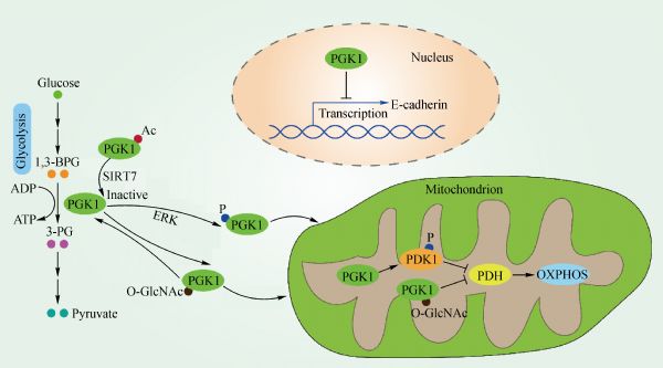
Y Zhang, G Yu, H Chu, X Wang, L Xiong, G Cai, R Liu, H Gao, B Tao, W Li, G Li, J Liang, W Yang. Macrophage-associated PGK1 phosphorylation promotes aerobic glycolysis and tumorigenesis. Mol Cell 2018; 71(2): 201–215.e7
https://doi.org/DOI: 10.1016/j.molcel.2018.06.023
pmid: 30029001
|
| 67 |
H Hu, W Zhu, J Qin, M Chen, L Gong, L Li, X Liu, Y Tao, H Yin, H Zhou, L Zhou, D Ye, Q Ye, D Gao. Acetylation of PGK1 promotes liver cancer cell proliferation and tumorigenesis. Hepatology 2017; 65(2): 515–528
https://doi.org/10.1002/hep.28887
pmid: 27774669
|
| 68 |
H Nie, H Ju, J Fan, X Shi, Y Cheng, X Cang, Z Zheng, X Duan, W Yi. O-GlcNAcylation of PGK1 coordinates glycolysis and TCA cycle to promote tumor growth. Nat Commun 2020; 11(1): 36
https://doi.org/10.1038/s41467-019-13601-8
pmid: 31911580
|
| 69 |
X Li, Y Jiang, J Meisenhelder, W Yang, DH Hawke, Y Zheng, Y Xia, K Aldape, J He, T Hunter, L Wang, Z Lu. Mitochondria-translocated PGK1 functions as a protein kinase to coordinate glycolysis and the TCA cycle in tumorigenesis. Mol Cell 2016; 61(5): 705–719
https://doi.org/10.1016/j.molcel.2016.02.009
pmid: 26942675
|
| 70 |
X Qian, X Li, Z Shi, Y Xia, Q Cai, D Xu, L Tan, L Du, Y Zheng, D Zhao, C Zhang, PL Lorenzi, Y You, BH Jiang, T Jiang, H Li, Z Lu. PTEN suppresses glycolysis by dephosphorylating and inhibiting autophosphorylated PGK1. Mol Cell 2019; 76(3): 516–527.e7
https://doi.org/DOI: 10.1016/j.molcel.2019.08.006
pmid: 31492635
|
| 71 |
X Qian, X Li, Q Cai, C Zhang, Q Yu, Y Jiang, JH Lee, D Hawke, Y Wang, Y Xia, Y Zheng, BH Jiang, DX Liu, T Jiang, Z Lu. Phosphoglycerate kinase 1 phosphorylates beclin1 to induce autophagy. Mol Cell 2017; 65(5): 917–931.e6
https://doi.org/DOI: 10.1016/j.molcel.2017.01.027
pmid: 28238651
|
| 72 |
X Qian, X Li, Z Lu. Protein kinase activity of the glycolytic enzyme PGK1 regulates autophagy to promote tumorigenesis. Autophagy 2017; 13(7): 1246–1247
https://doi.org/10.1080/15548627.2017.1313945
pmid: 28486006
|
| 73 |
C Liang, S Shi, Y Qin, Q Meng, J Hua, Q Hu, S Ji, B Zhang, J Xu, XJ Yu. Localisation of PGK1 determines metabolic phenotype to balance metastasis and proliferation in patients with SMAD4-negative pancreatic cancer. Gut 2020; 69(5): 888–900
https://doi.org/10.1136/gutjnl-2018-317163
pmid: 31611300
|
| 74 |
TL Dayton, T Jacks, MG Vander Heiden. PKM2, cancer metabolism, and the road ahead. EMBO Rep 2016; 17(12): 1721–1730
https://doi.org/10.15252/embr.201643300
pmid: 27856534
|
| 75 |
S Mazurek. Pyruvate kinase type M2: a key regulator of the metabolic budget system in tumor cells. Int J Biochem Cell Biol 2011; 43(7): 969–980
https://doi.org/10.1016/j.biocel.2010.02.005
pmid: 20156581
|
| 76 |
Q Sun, X Chen, J Ma, H Peng, F Wang, X Zha, Y Wang, Y Jing, H Yang, R Chen, L Chang, Y Zhang, J Goto, H Onda, T Chen, MR Wang, Y Lu, H You, D Kwiatkowski, H Zhang. Mammalian target of rapamycin up-regulation of pyruvate kinase isoenzyme type M2 is critical for aerobic glycolysis and tumor growth. Proc Natl Acad Sci USA 2011; 108(10): 4129–4134
https://doi.org/10.1073/pnas.1014769108
pmid: 21325052
|
| 77 |
HR Christofk, MG Vander Heiden, MH Harris, A Ramanathan, RE Gerszten, R Wei, MD Fleming, SL Schreiber, LC Cantley. The M2 splice isoform of pyruvate kinase is important for cancer metabolism and tumour growth. Nature 2008; 452(7184): 230–233
https://doi.org/10.1038/nature06734
pmid: 18337823
|
| 78 |
HR Christofk, MG Vander Heiden, N Wu, JM Asara, LC Cantley. Pyruvate kinase M2 is a phosphotyrosine-binding protein. Nature 2008; 452(7184): 181–186
https://doi.org/10.1038/nature06667
pmid: 18337815
|
| 79 |
L Lv, D Li, D Zhao, R Lin, Y Chu, H Zhang, Z Zha, Y Liu, Z Li, Y Xu, G Wang, Y Huang, Y Xiong, KL Guan, QY Lei. Acetylation targets the M2 isoform of pyruvate kinase for degradation through chaperone-mediated autophagy and promotes tumor growth. Mol Cell 2011; 42(6): 719–730
https://doi.org/10.1016/j.molcel.2011.04.025
pmid: 21700219
|
| 80 |
AN Macintyre, JC Rathmell. PKM2 and the tricky balance of growth and energy in cancer. Mol Cell 2011; 42(6): 713–714
https://doi.org/10.1016/j.molcel.2011.06.003
pmid: 21700216
|
| 81 |
L Lv, YP Xu, D Zhao, FL Li, W Wang, N Sasaki, Y Jiang, X Zhou, TT Li, KL Guan, QY Lei, Y Xiong. Mitogenic and oncogenic stimulation of K433 acetylation promotes PKM2 protein kinase activity and nuclear localization. Mol Cell 2013; 52(3): 340–352
https://doi.org/10.1016/j.molcel.2013.09.004
pmid: 24120661
|
| 82 |
W Yang, Y Zheng, Y Xia, H Ji, X Chen, F Guo, CA Lyssiotis, K Aldape, LC Cantley, Z Lu. ERK1/2-dependent phosphorylation and nuclear translocation of PKM2 promotes the Warburg effect. Nat Cell Biol 2012; 14(12): 1295–1304
https://doi.org/10.1038/ncb2629
pmid: 23178880
|
| 83 |
W Yang, Y Xia, H Ji, Y Zheng, J Liang, W Huang, X Gao, K Aldape, Z Lu. Nuclear PKM2 regulates β-catenin transactivation upon EGFR activation. Nature 2011; 480(7375): 118–122
https://doi.org/10.1038/nature10598
pmid: 22056988
|
| 84 |
S Li, SK Swanson, M Gogol, L Florens, MP Washburn, JL Workman, T Suganuma. Serine and SAM responsive complex SESAME regulates histone modification crosstalk by sensing cellular metabolism. Mol Cell 2015; 60(3): 408–421
https://doi.org/10.1016/j.molcel.2015.09.024
pmid: 26527276
|
| 85 |
W Yang, Y Xia, D Hawke, X Li, J Liang, D Xing, K Aldape, T Hunter, WK Alfred Yung, Z Lu. PKM2 phosphorylates histone H3 and promotes gene transcription and tumorigenesis. Cell 2012; 150(4): 685–696
https://doi.org/10.1016/j.cell.2012.07.018
pmid: 22901803
|
| 86 |
MC Hsu, WC Hung. Pyruvate kinase M2 fuels multiple aspects of cancer cells: from cellular metabolism, transcriptional regulation to extracellular signaling. Mol Cancer 2018; 17(1): 35
https://doi.org/10.1186/s12943-018-0791-3
pmid: 29455645
|
| 87 |
X Gao, H Wang, JJ Yang, X Liu, ZR Liu. Pyruvate kinase M2 regulates gene transcription by acting as a protein kinase. Mol Cell 2012; 45(5): 598–609
https://doi.org/10.1016/j.molcel.2012.01.001
pmid: 22306293
|
| 88 |
KE Keller, ZM Doctor, ZW Dwyer, YS Lee. SAICAR induces protein kinase activity of PKM2 that is necessary for sustained proliferative signaling of cancer cells. Mol Cell 2014; 53(5): 700–709
https://doi.org/10.1016/j.molcel.2014.02.015
pmid: 24606918
|
| 89 |
W Luo, H Hu, R Chang, J Zhong, M Knabel, R O’Meally, RN Cole, A Pandey, GL Semenza. Pyruvate kinase M2 is a PHD3-stimulated coactivator for hypoxia-inducible factor 1. Cell 2011; 145(5): 732–744
https://doi.org/10.1016/j.cell.2011.03.054
pmid: 21620138
|
| 90 |
HJ Wang, YJ Hsieh, WC Cheng, CP Lin, YS Lin, SF Yang, CC Chen, Y Izumiya, JS Yu, HJ Kung, WC Wang. JMJD5 regulates PKM2 nuclear translocation and reprograms HIF-1α-mediated glucose metabolism. Proc Natl Acad Sci USA 2014; 111(1): 279–284
https://doi.org/10.1073/pnas.1311249111
pmid: 24344305
|
| 91 |
M Demaria, V Poli. PKM2, STAT3 and HIF-1α: the Warburg’s vicious circle. JAK-STAT 2012; 1(3): 194–196
https://doi.org/10.4161/jkst.20662
pmid: 24058770
|
| 92 |
N Azoitei, A Becher, K Steinestel, A Rouhi, K Diepold, F Genze, T Simmet, T Seufferlein. PKM2 promotes tumor angiogenesis by regulating HIF-1α through NF-kB activation. Mol Cancer 2016; 15(1): 3
https://doi.org/10.1186/s12943-015-0490-2
pmid: 26739387
|
| 93 |
AM Hosios, BP Fiske, DY Gui, MG Vander Heiden. Lack of evidence for PKM2 protein kinase activity. Mol Cell 2015; 59(5): 850–857
https://doi.org/10.1016/j.molcel.2015.07.013
pmid: 26300261
|
| 94 |
MG Vander Heiden, HR Christofk, E Schuman, AO Subtelny, H Sharfi, EE Harlow, J Xian, LC Cantley. Identification of small molecule inhibitors of pyruvate kinase M2. Biochem Pharmacol 2010; 79(8): 1118–1124
https://doi.org/10.1016/j.bcp.2009.12.003
pmid: 20005212
|
| 95 |
Y Wang, F Hao, Y Nan, L Qu, W Na, C Jia, X Chen. PKM2 inhibitor shikonin overcomes the cisplatin resistance in bladder cancer by inducing necroptosis. Int J Biol Sci 2018; 14(13): 1883–1891
https://doi.org/10.7150/ijbs.27854
pmid: 30443191
|
| 96 |
J Chen, J Xie, Z Jiang, B Wang, Y Wang, X Hu. Shikonin and its analogs inhibit cancer cell glycolysis by targeting tumor pyruvate kinase-M2. Oncogene 2011; 30(42): 4297–4306
https://doi.org/10.1038/onc.2011.137
pmid: 21516121
|
| 97 |
C Kung, J Hixon, S Choe, K Marks, S Gross, E Murphy, B DeLaBarre, G Cianchetta, S Sethumadhavan, X Wang, S Yan, Y Gao, C Fang, W Wei, F Jiang, S Wang, K Qian, J Saunders, E Driggers, HK Woo, K Kunii, S Murray, H Yang, K Yen, W Liu, LC Cantley, MG Vander Heiden, SM Su, S Jin, FG Salituro, L Dang. Small molecule activation of PKM2 in cancer cells induces serine auxotrophy. Chem Biol 2012; 19(9): 1187–1198
https://doi.org/10.1016/j.chembiol.2012.07.021
pmid: 22999886
|
| 98 |
D Anastasiou, Y Yu, WJ Israelsen, JK Jiang, MB Boxer, BS Hong, W Tempel, S Dimov, M Shen, A Jha, H Yang, KR Mattaini, CM Metallo, BP Fiske, KD Courtney, S Malstrom, TM Khan, C Kung, AP Skoumbourdis, H Veith, N Southall, MJ Walsh, KR Brimacombe, W Leister, SY Lunt, ZR Johnson, KE Yen, K Kunii, SM Davidson, HR Christofk, CP Austin, J Inglese, MH Harris, JM Asara, G Stephanopoulos, FG Salituro, S Jin, L Dang, DS Auld, HW Park, LC Cantley, CJ Thomas, MG Vander Heiden. Pyruvate kinase M2 activators promote tetramer formation and suppress tumorigenesis. Nat Chem Biol 2012; 8(10): 839–847
https://doi.org/10.1038/nchembio.1060
pmid: 22922757
|
| 99 |
KM Parnell, JM Foulks, RN Nix, A Clifford, J Bullough, B Luo, A Senina, D Vollmer, J Liu, V McCarthy, Y Xu, M Saunders, XH Liu, S Pearce, K Wright, M O’Reilly, MV McCullar, KK Ho, SB Kanner. Pharmacologic activation of PKM2 slows lung tumor xenograft growth. Mol Cancer Ther 2013; 12(8): 1453–1460
https://doi.org/10.1158/1535-7163.MCT-13-0026
pmid: 23720766
|
| 100 |
C Dong, T Yuan, Y Wu, Y Wang, TW Fan, S Miriyala, Y Lin, J Yao, J Shi, T Kang, P Lorkiewicz, D St Clair, MC Hung, BM Evers, BP Zhou. Loss of FBP1 by Snail-mediated repression provides metabolic advantages in basal-like breast cancer. Cancer Cell 2013; 23(3): 316–331
https://doi.org/10.1016/j.ccr.2013.01.022
pmid: 23453623
|
| 101 |
J Zhang, J Wang, H Xing, Q Li, Q Zhao, J Li. Down-regulation of FBP1 by ZEB1-mediated repression confers to growth and invasion in lung cancer cells. Mol Cell Biochem 2016; 411(1–2): 331–340
https://doi.org/10.1007/s11010-015-2595-8
pmid: 26546081
|
| 102 |
H Hirata, K Sugimachi, H Komatsu, M Ueda, T Masuda, R Uchi, S Sakimura, S Nambara, T Saito, Y Shinden, T Iguchi, H Eguchi, S Ito, K Terashima, K Sakamoto, M Hirakawa, H Honda, K Mimori. Decreased expression of fructose-1,6-bisphosphatase associates with glucose metabolism and tumor progression in hepatocellular carcinoma. Cancer Res 2016; 76(11): 3265–3276
https://doi.org/10.1158/0008-5472.CAN-15-2601
pmid: 27197151
|
| 103 |
B Son, S Lee, H Kim, H Kang, J Jeon, S Jo, KM Seong, SJ Lee, H Youn, B Youn. Decreased FBP1 expression rewires metabolic processes affecting aggressiveness of glioblastoma. Oncogene 2020; 39(1): 36–49
https://doi.org/10.1038/s41388-019-0974-4
pmid: 31444412
|
| 104 |
B Li, B Qiu, DS Lee, ZE Walton, JD Ochocki, LK Mathew, A Mancuso, TP Gade, B Keith, I Nissim, MC Simon. Fructose-1,6-bisphosphatase opposes renal carcinoma progression. Nature 2014; 513(7517): 251–255
https://doi.org/10.1038/nature13557
pmid: 25043030
|
| 105 |
C Lu, C Ren, T Yang, Y Sun, P Qiao, D Wang, S Lv, Z Yu. A noncanonical role of fructose-1, 6-bisphosphatase 1 is essential for inhibition of Notch1 in breast cancer. Mol Cancer Res 2020; 18(5): 787–796
https://doi.org/10.1158/1541-7786.MCR-19-0842
pmid: 32041737
|
| 106 |
J Cong, X Wang, X Zheng, D Wang, B Fu, R Sun, Z Tian, H Wei. Dysfunction of natural killer cells by FBP1-induced inhibition of glycolysis during lung cancer progression. Cell Metab 2018; 28(2): 243–255.e5
https://doi.org/DOI: 10.1016/j.cmet.2018.06.021
pmid: 30033198
|
| 107 |
SC Burgess, T He, Z Yan, J Lindner, AD Sherry, CR Malloy, JD Browning, MA Magnuson. Cytosolic phosphoenolpyruvate carboxykinase does not solely control the rate of hepatic gluconeogenesis in the intact mouse liver. Cell Metab 2007; 5(4): 313–320
https://doi.org/10.1016/j.cmet.2007.03.004
pmid: 17403375
|
| 108 |
A Méndez-Lucas, P Hyroššová, L Novellasdemunt, F Viñals, JC Perales. Mitochondrial phosphoenolpyruvate carboxykinase (PEPCK-M) is a pro-survival, endoplasmic reticulum (ER) stress response gene involved in tumor cell adaptation to nutrient availability. J Biol Chem 2014; 289(32): 22090–22102
https://doi.org/10.1074/jbc.M114.566927
pmid: 24973213
|
| 109 |
JM Matés, JA Campos-Sandoval, JL Santos-Jiménez, J Márquez. Dysregulation of glutaminase and glutamine synthetase in cancer. Cancer Lett 2019; 467: 29–39
https://doi.org/10.1016/j.canlet.2019.09.011
pmid: 31574293
|
| 110 |
P Mishra, DC Chan. Metabolic regulation of mitochondrial dynamics. J Cell Biol 2016; 212(4): 379–387
https://doi.org/10.1083/jcb.201511036
pmid: 26858267
|
| 111 |
P Maycotte, A Marín-Hernández, M Goyri-Aguirre, M Anaya-Ruiz, J Reyes-Leyva, P Cortés-Hernández. Mitochondrial dynamics and cancer. Tumour Biol 2017; 39(5): 1010428317698391
https://doi.org/10.1177/1010428317698391
pmid: 28468591
|
| 112 |
WF Cai, C Zhang, YQ Wu, G Zhuang, Z Ye, CS Zhang, SC Lin. Glutaminase GLS1 senses glutamine availability in a non-enzymatic manner triggering mitochondrial fusion. Cell Res 2018; 28(8): 865–867
https://doi.org/10.1038/s41422-018-0057-z
pmid: 29934617
|
| 113 |
TJ Stillman, PJ Baker, KL Britton, DW Rice. Conformational flexibility in glutamate dehydrogenase: role of water in substrate recognition and catalysis. J Mol Biol 1993; 234(4): 1131–1139
https://doi.org/10.1006/jmbi.1993.1665
pmid: 8263917
|
| 114 |
I Zaganas, C Spanaki, A Plaitakis. Expression of human GLUD2 glutamate dehydrogenase in human tissues: functional implications. Neurochem Int 2012; 61(4): 455–462
https://doi.org/10.1016/j.neuint.2012.06.007
pmid: 22709674
|
| 115 |
TM Michaelidis, G Tzimagiorgis, NK Moschonas, J Papamatheakis. The human glutamate dehydrogenase gene family: gene organization and structural characterization. Genomics 1993; 16(1): 150–160
https://doi.org/10.1006/geno.1993.1152
pmid: 8486350
|
| 116 |
L Jin, D Li, GN Alesi, J Fan, HB Kang, Z Lu, TJ Boggon, P Jin, H Yi, ER Wright, D Duong, NT Seyfried, R Egnatchik, RJ DeBerardinis, KR Magliocca, C He, ML Arellano, HJ Khoury, DM Shin, FR Khuri, S Kang. Glutamate dehydrogenase 1 signals through antioxidant glutathione peroxidase 1 to regulate redox homeostasis and tumor growth. Cancer Cell 2015; 27(2): 257–270
https://doi.org/10.1016/j.ccell.2014.12.006
pmid: 25670081
|
| 117 |
BI Fedeles, V Singh, JC Delaney, D Li, JM Essigmann. The AlkB family of Fe(II)/α-ketoglutarate-dependent dioxygenases: repairing nucleic acid alkylation damage and beyond. J Biol Chem 2015; 290(34): 20734–20742
https://doi.org/10.1074/jbc.R115.656462
pmid: 26152727
|
| 118 |
C Yang, B Ko, CT Hensley, L Jiang, AT Wasti, J Kim, J Sudderth, MA Calvaruso, L Lumata, M Mitsche, J Rutter, ME Merritt, RJ DeBerardinis. Glutamine oxidation maintains the TCA cycle and cell survival during impaired mitochondrial pyruvate transport. Mol Cell 2014; 56(3): 414–424
https://doi.org/10.1016/j.molcel.2014.09.025
pmid: 25458842
|
| 119 |
L Jin, J Chun, C Pan, A Kumar, G Zhang, Y Ha, D Li, GN Alesi, Y Kang, L Zhou, WM Yu, KR Magliocca, FR Khuri, CK Qu, C Metallo, TK Owonikoko, S Kang. The PLAG1–GDH1 axis promotes anoikis resistance and tumor metastasis through CamKK2–AMPK signaling in LKB1-deficient lung cancer. Mol Cell 2018; 69(1): 87–99.e7
https://doi.org/DOI: 10.1016/j.molcel.2017.11.025
pmid: 29249655
|
| 120 |
X Wang, R Liu, X Qu, H Yu, H Chu, Y Zhang, W Zhu, X Wu, H Gao, B Tao, W Li, J Liang, G Li, W Yang. α-ketoglutarate-activated NF-κB signaling promotes compensatory glucose uptake and brain tumor development. Mol Cell 2019; 76(1): 148–162.e7
https://doi.org/DOI: 10.1016/j.molcel.2019.07.007
pmid: 31447391
|
| 121 |
G Di Conza, CH Tsai, PC Ho. Fifty shades of α-ketoglutarate on cellular programming. Mol Cell 2019; 76(1): 1–3
https://doi.org/10.1016/j.molcel.2019.09.002
pmid: 31585100
|
| 122 |
CP Diggle, M Shires, D Leitch, D Brooke, IM Carr, AF Markham, BE Hayward, A Asipu, DT Bonthron. Ketohexokinase: expression and localization of the principal fructose-metabolizing enzyme. J Histochem Cytochem 2009; 57(8): 763–774
https://doi.org/10.1369/jhc.2009.953190
pmid: 19365088
|
| 123 |
T Ishimoto, MA Lanaspa, MT Le, GE Garcia, CP Diggle, PS Maclean, MR Jackman, A Asipu, CA Roncal-Jimenez, T Kosugi, CJ Rivard, S Maruyama, B Rodriguez-Iturbe, LG Sánchez-Lozada, DT Bonthron, YY Sautin, RJ Johnson. Opposing effects of fructokinase C and A isoforms on fructose-induced metabolic syndrome in mice. Proc Natl Acad Sci USA 2012; 109(11): 4320–4325
https://doi.org/10.1073/pnas.1119908109
pmid: 22371574
|
| 124 |
X Li, X Qian, Z Lu. Fructokinase A acts as a protein kinase to promote nucleotide synthesis. Cell Cycle 2016; 15(20): 2689–2690
https://doi.org/10.1080/15384101.2016.1204861
pmid: 27356213
|
| 125 |
X Li, X Qian, LX Peng, Y Jiang, DH Hawke, Y Zheng, Y Xia, JH Lee, G Cote, H Wang, L Wang, CN Qian, Z Lu. A splicing switch from ketohexokinase-C to ketohexokinase-A drives hepatocellular carcinoma formation. Nat Cell Biol 2016; 18(5): 561–571
https://doi.org/10.1038/ncb3338
pmid: 27088854
|
| 126 |
D Xu, X Li, F Shao, G Lv, H Lv, JH Lee, X Qian, Z Wang, Y Xia, L Du, Y Zheng, H Wang, J Lyu, Z Lu. The protein kinase activity of fructokinase A specifies the antioxidant responses of tumor cells by phosphorylating p62. Sci Adv 2019; 5(4): eaav4570
https://doi.org/10.1126/sciadv.aav4570
pmid: 31032410
|
| 127 |
L Ippolito, A Morandi, E Giannoni, P Chiarugi. Lactate: a metabolic driver in the tumour landscape. Trends Biochem Sci 2019; 44(2): 153–166
https://doi.org/10.1016/j.tibs.2018.10.011
pmid: 30473428
|
| 128 |
D Zhao, SW Zou, Y Liu, X Zhou, Y Mo, P Wang, YH Xu, B Dong, Y Xiong, QY Lei, KL Guan. Lysine-5 acetylation negatively regulates lactate dehydrogenase A and is decreased in pancreatic cancer. Cancer Cell 2013; 23(4): 464–476
https://doi.org/10.1016/j.ccr.2013.02.005
pmid: 23523103
|
| 129 |
VR Fantin, J St-Pierre, P Leder. Attenuation of LDH-A expression uncovers a link between glycolysis, mitochondrial physiology, and tumor maintenance. Cancer Cell 2006; 9(6): 425–434
https://doi.org/10.1016/j.ccr.2006.04.023
pmid: 16766262
|
| 130 |
SB Rosalki, JH Wilkinson. Reduction of α-ketobutyrate by human serum. Nature 1960; 188(4756): 1110–1111
https://doi.org/10.1038/1881110a0
pmid: 13743238
|
| 131 |
Y Liu, JZ Guo, Y Liu, K Wang, W Ding, H Wang, X Liu, S Zhou, XC Lu, HB Yang, C Xu, W Gao, L Zhou, YP Wang, W Hu, Y Wei, C Huang, QY Lei. Nuclear lactate dehydrogenase A senses ROS to produce α-hydroxybutyrate for HPV-induced cervical tumor growth. Nat Commun 2018; 9(1): 4429
https://doi.org/10.1038/s41467-018-06841-7
pmid: 30356100
|
| 132 |
AM Intlekofer, B Wang, H Liu, H Shah, C Carmona-Fontaine, AS Rustenburg, S Salah, MR Gunner, JD Chodera, JR Cross, CB Thompson. L-2-Hydroxyglutarate production arises from noncanonical enzyme function at acidic pH. Nat Chem Biol 2017; 13(5): 494–500
https://doi.org/10.1038/nchembio.2307
pmid: 28263965
|
| 133 |
H Yan, DW Parsons, G Jin, R McLendon, BA Rasheed, W Yuan, I Kos, I Batinic-Haberle, S Jones, GJ Riggins, H Friedman, A Friedman, D Reardon, J Herndon, KW Kinzler, VE Velculescu, B Vogelstein, DD Bigner. IDH1 and IDH2 mutations in gliomas. N Engl J Med 2009; 360(8): 765–773
https://doi.org/10.1056/NEJMoa0808710
pmid: 19228619
|
| 134 |
MR Kang, MS Kim, JE Oh, YR Kim, SY Song, SI Seo, JY Lee, NJ Yoo, SH Lee. Mutational analysis of IDH1 codon 132 in glioblastomas and other common cancers. Int J Cancer 2009; 125(2): 353–355
https://doi.org/10.1002/ijc.24379
pmid: 19378339
|
| 135 |
S Nobusawa, H Yokoo. IDH1/2 mutations in gliomas. Brain Nerve 2011; 63(12): 1378–1386
pmid: 22147457
|
| 136 |
ER Mardis, L Ding, DJ Dooling, DE Larson, MD McLellan, K Chen, DC Koboldt, RS Fulton, KD Delehaunty, SD McGrath, LA Fulton, DP Locke, VJ Magrini, RM Abbott, TL Vickery, JS Reed, JS Robinson, T Wylie, SM Smith, L Carmichael, JM Eldred, CC Harris, J Walker, JB Peck, F Du, AF Dukes, GE Sanderson, AM Brummett, E Clark, JF McMichael, RJ Meyer, JK Schindler, CS Pohl, JW Wallis, X Shi, L Lin, H Schmidt, Y Tang, C Haipek, ME Wiechert, JV Ivy, J Kalicki, G Elliott, RE Ries, JE Payton, P Westervelt, MH Tomasson, MA Watson, J Baty, S Heath, WD Shannon, R Nagarajan, DC Link, MJ Walter, TA Graubert, JF DiPersio, RK Wilson, TJ Ley. Recurring mutations found by sequencing an acute myeloid leukemia genome. N Engl J Med 2009; 361(11): 1058–1066
https://doi.org/10.1056/NEJMoa0903840
pmid: 19657110
|
| 137 |
DR Borger, KK Tanabe, KC Fan, HU Lopez, VR Fantin, KS Straley, DP Schenkein, AF Hezel, M Ancukiewicz, HM Liebman, EL Kwak, JW Clark, DP Ryan, V Deshpande, D Dias-Santagata, LW Ellisen, AX Zhu, AJ Iafrate. Frequent mutation of isocitrate dehydrogenase (IDH)1 and IDH2 in cholangiocarcinoma identified through broad-based tumor genotyping. Oncologist 2012; 17(1): 72–79
https://doi.org/10.1634/theoncologist.2011-0386
pmid: 22180306
|
| 138 |
H Yang, D Ye, KL Guan, Y Xiong. IDH1 and IDH2 mutations in tumorigenesis: mechanistic insights and clinical perspectives. Clin Cancer Res 2012; 18(20): 5562–5571
https://doi.org/10.1158/1078-0432.CCR-12-1773
pmid: 23071358
|
| 139 |
PS Ward, J Patel, DR Wise, O Abdel-Wahab, BD Bennett, HA Coller, JR Cross, VR Fantin, CV Hedvat, AE Perl, JD Rabinowitz, M Carroll, SM Su, KA Sharp, RL Levine, CB Thompson. The common feature of leukemia-associated IDH1 and IDH2 mutations is a neomorphic enzyme activity converting α-ketoglutarate to 2-hydroxyglutarate. Cancer Cell 2010; 17(3): 225–234
https://doi.org/10.1016/j.ccr.2010.01.020
pmid: 20171147
|
| 140 |
ZJ Reitman, DW Parsons, H Yan. IDH1 and IDH2: not your typical oncogenes. Cancer Cell 2010; 17(3): 215–216
https://doi.org/10.1016/j.ccr.2010.02.024
pmid: 20227034
|
| 141 |
L Dang, DW White, S Gross, BD Bennett, MA Bittinger, EM Driggers, VR Fantin, HG Jang, S Jin, MC Keenan, KM Marks, RM Prins, PS Ward, KE Yen, LM Liau, JD Rabinowitz, LC Cantley, CB Thompson, MG Vander Heiden, SM Su. Cancer-associated IDH1 mutations produce 2-hydroxyglutarate. Nature 2009; 462(7274): 739–744
https://doi.org/10.1038/nature08617
pmid: 19935646
|
| 142 |
W Xu, H Yang, Y Liu, Y Yang, P Wang, SH Kim, S Ito, C Yang, P Wang, MT Xiao, LX Liu, WQ Jiang, J Liu, JY Zhang, B Wang, S Frye, Y Zhang, YH Xu, QY Lei, KL Guan, SM Zhao, Y Xiong. Oncometabolite 2-hydroxyglutarate is a competitive inhibitor of α-ketoglutarate-dependent dioxygenases. Cancer Cell 2011; 19(1): 17–30
https://doi.org/10.1016/j.ccr.2010.12.014
pmid: 21251613
|
| 143 |
C Lu, PS Ward, GS Kapoor, D Rohle, S Turcan, O Abdel-Wahab, CR Edwards, R Khanin, ME Figueroa, A Melnick, KE Wellen, DM O’Rourke, SL Berger, TA Chan, RL Levine, IK Mellinghoff, CB Thompson. IDH mutation impairs histone demethylation and results in a block to cell differentiation. Nature 2012; 483(7390): 474–478
https://doi.org/10.1038/nature10860
pmid: 22343901
|
| 144 |
H Zhu, Y Zhang, J Chen, J Qiu, K Huang, M Wu, C Xia. IDH1 R132H mutation enhances cell migration by activating AKT− mTOR signaling pathway, but sensitizes cells to 5-FU treatment as NADPH and GSH are reduced. PLoS One 2017; 12(1): e0169038
https://doi.org/10.1371/journal.pone.0169038
pmid: 28052098
|
| 145 |
DW Parsons, S Jones, X Zhang, JC Lin, RJ Leary, P Angenendt, P Mankoo, H Carter, IM Siu, GL Gallia, A Olivi, R McLendon, BA Rasheed, S Keir, T Nikolskaya, Y Nikolsky, DA Busam, H Tekleab, LA Diaz Jr, J Hartigan, DR Smith, RL Strausberg, SK Marie, SM Shinjo, H Yan, GJ Riggins, DD Bigner, R Karchin, N Papadopoulos, G Parmigiani, B Vogelstein, VE Velculescu, KW Kinzler. An integrated genomic analysis of human glioblastoma multiforme. Science 2008; 321(5897): 1807–1812
https://doi.org/10.1126/science.1164382
pmid: 18772396
|
| 146 |
C Loenarz, CJ Schofield. Expanding chemical biology of 2-oxoglutarate oxygenases. Nat Chem Biol 2008; 4(3): 152–156
https://doi.org/10.1038/nchembio0308-152
pmid: 18277970
|
| 147 |
C Reiter-Brennan, L Semmler, A Klein. The effects of 2-hydroxyglutarate on the tumorigenesis of gliomas. Contemp Oncol (Pozn) 2018; 22(4): 215–222
https://doi.org/10.5114/wo.2018.82642
pmid: 30783384
|
| 148 |
P Wang, J Wu, S Ma, L Zhang, J Yao, KA Hoadley, MD Wilkerson, CM Perou, KL Guan, D Ye, Y Xiong. Oncometabolite D-2-hydroxyglutarate inhibits ALKBH DNA repair enzymes and sensitizes IDH mutant cells to alkylating agents. Cell Rep 2015; 13(11): 2353–2361
https://doi.org/10.1016/j.celrep.2015.11.029
pmid: 26686626
|
| 149 |
O Yogev, O Yogev, E Singer, E Shaulian, M Goldberg, TD Fox, O Pines. Fumarase: a mitochondrial metabolic enzyme and a cytosolic/nuclear component of the DNA damage response. PLoS Biol 2010; 8(3): e1000328
https://doi.org/10.1371/journal.pbio.1000328
pmid: 20231875
|
| 150 |
M Leshets, YBH Silas, N Lehming, O Pines. Fumarase: from the TCA cycle to DNA damage response and tumor suppression. Front Mol Biosci 2018; 5: 68
https://doi.org/10.3389/fmolb.2018.00068
pmid: 30090811
|
| 151 |
M Xiao, H Yang, W Xu, S Ma, H Lin, H Zhu, L Liu, Y Liu, C Yang, Y Xu, S Zhao, D Ye, Y Xiong, KL Guan. Inhibition of α-KG-dependent histone and DNA demethylases by fumarate and succinate that are accumulated in mutations of FH and SDH tumor suppressors. Genes Dev 2012; 26(12): 1326–1338
https://doi.org/10.1101/gad.191056.112
pmid: 22677546
|
| 152 |
Y Jiang, X Qian, J Shen, Y Wang, X Li, R Liu, Y Xia, Q Chen, G Peng, SY Lin, Z Lu. Local generation of fumarate promotes DNA repair through inhibition of histone H3 demethylation. Nat Cell Biol 2015; 17(9): 1158–1168
https://doi.org/10.1038/ncb3209
pmid: 26237645
|
| 153 |
T Wang, Q Yu, J Li, B Hu, Q Zhao, C Ma, W Huang, L Zhuo, H Fang, L Liao, Y Eugene Chin, Y Jiang. O-GlcNAcylation of fumarase maintains tumour growth under glucose deficiency. Nat Cell Biol 2017; 19(7): 833–843
https://doi.org/10.1038/ncb3562
pmid: 28628081
|
| 154 |
R Lin, R Tao, X Gao, T Li, X Zhou, KL Guan, Y Xiong, QY Lei. Acetylation stabilizes ATP-citrate lyase to promote lipid biosynthesis and tumor growth. Mol Cell 2013; 51(4): 506–518
https://doi.org/10.1016/j.molcel.2013.07.002
pmid: 23932781
|
| 155 |
KE Wellen, G Hatzivassiliou, UM Sachdeva, TV Bui, JR Cross, CB Thompson. ATP-citrate lyase links cellular metabolism to histone acetylation. Science 2009; 324(5930): 1076–1080
https://doi.org/10.1126/science.1164097
pmid: 19461003
|
| 156 |
JV Lee, A Carrer, S Shah, NW Snyder, S Wei, S Venneti, AJ Worth, ZF Yuan, HW Lim, S Liu, E Jackson, NM Aiello, NB Haas, TR Rebbeck, A Judkins, KJ Won, LA Chodosh, BA Garcia, BZ Stanger, MD Feldman, IA Blair, KE Wellen. Akt-dependent metabolic reprogramming regulates tumor cell histone acetylation. Cell Metab 2014; 20(2): 306–319
https://doi.org/10.1016/j.cmet.2014.06.004
pmid: 24998913
|
| 157 |
B Henderson, AC Martin. Protein moonlighting: a new factor in biology and medicine. Biochem Soc Trans 2014; 42(6): 1671–1678
https://doi.org/10.1042/BST20140273
pmid: 25399588
|
| 158 |
EM Palsson-McDermott, L Dyck, Z Zasłona, D Menon, AF McGettrick, KHG Mills, LA O’Neill. Pyruvate kinase M2 is required for the expression of the immune checkpoint PD-L1 in immune cells and tumors. Front Immunol 2017; 8: 1300
https://doi.org/10.3389/fimmu.2017.01300
pmid: 29081778
|
|
Viewed |
|
|
|
Full text
|
|
|
|
|
Abstract
|
|
|
|
|
Cited |
|
|
|
|
| |
Shared |
|
|
|
|
| |
Discussed |
|
|
|
|