|
|
|
Quantitative proteomics revealed extensive microenvironmental changes after stem cell transplantation in ischemic stroke |
Yao Chen1,2,3,7, Fahuan Song1,2,3, Mengjiao Tu1,2,3,8, Shuang Wu1,2,3, Xiao He1,2,3, Hao Liu1,2,3, Caiyun Xu1,2,3, Kai Zhang1,2,3, Yuankai Zhu1,2,3, Rui Zhou1,2,3, Chentao Jin1,2,3, Ping Wang5,6, Hong Zhang1,2,3,4,5,6( ), Mei Tian1,2,3() ), Mei Tian1,2,3() |
1. Department of Nuclear Medicine and Medical PET Center, The Second Affiliated Hospital, Zhejiang University School of Medicine, Hangzhou 310009, China
2. Institute of Nuclear Medicine and Molecular Imaging, Zhejiang University, Hangzhou 310009, China
3. Key Laboratory of Medical Molecular Imaging of Zhejiang Province, Hangzhou 310009, China
4. Shanxi Medical University, Taiyuan 030001, China
5. Key Laboratory for Biomedical Engineering of Ministry of Education, Zhejiang University, Hangzhou 310027, China
6. College of Biomedical Engineering and Instrument Science, Zhejiang University, Hangzhou 310027, China
7. Department of Radiology, Zhejiang Hospital, Hangzhou 310030, China
8. Department of PET Center, The First Affiliated Hospital, Zhejiang University School of Medicine, Hangzhou 310006, China |
|
|
|
|
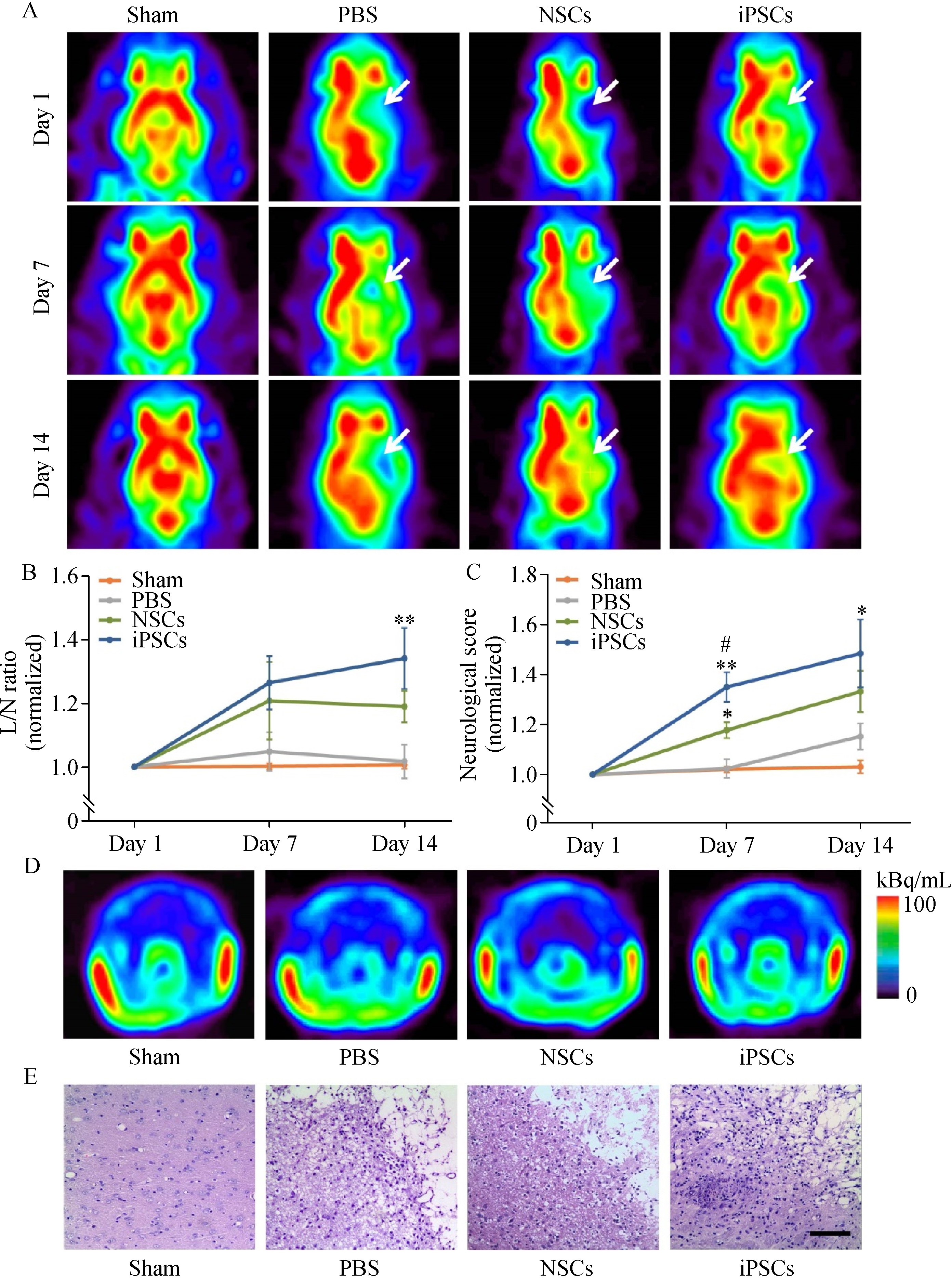
Abstract The local microenvironment is essential to stem cell-based therapy for ischemic stroke, and spatiotemporal changes of the microenvironment in the pathological process provide vital clues for understanding the therapeutic mechanisms. However, relevant studies on microenvironmental changes were mainly confined in the acute phase of stroke, and long-term changes remain unclear. This study aimed to investigate the microenvironmental changes in the subacute and chronic phases of ischemic stroke after stem cell transplantation. Herein, induced pluripotent stem cells (iPSCs) and neural stem cells (NSCs) were transplanted into the ischemic brain established by middle cerebral artery occlusion surgery. Positron emission tomography imaging and neurological tests were applied to evaluate the metabolic and neurofunctional alterations of rats transplanted with stem cells. Quantitative proteomics was employed to investigate the protein expression profiles in iPSCs-transplanted brain in the subacute and chronic phases of stroke. Compared with NSCs-transplanted rats, significantly increased glucose metabolism and neurofunctional scores were observed in iPSCs-transplanted rats. Subsequent proteomic data of iPSCs-transplanted rats identified a total of 39 differentially expressed proteins in the subacute and chronic phases, which are involved in various ischemic stroke-related biological processes, including neuronal survival, axonal remodeling, antioxidative stress, and mitochondrial function restoration. Taken together, our study indicated that iPSCs have a positive therapeutic effect in ischemic stroke and emphasized the wide-ranging microenvironmental changes in the subacute and chronic phases.
|
| Keywords
ischemic stroke
microenvironment
induced pluripotent stem cells (iPSCs)
positron emission tomography (PET)
quantitative proteomics
|
|
Corresponding Author(s):
Hong Zhang,Mei Tian
|
|
Just Accepted Date: 16 April 2021
Online First Date: 13 July 2021
Issue Date: 18 July 2022
|
|
| 1 |
GBD 2017 DALYs and HALE Collaborators. Global, regional, and national disability-adjusted life-years (DALYs) for 359 diseases and injuries and healthy life expectancy (HALE) for 195 countries and territories, 1990−2017: a systematic analysis for the Global Burden of Disease Study 2017. Lancet 2018; 392(10159): 1859–1922
https://doi.org/10.1016/S0140-6736(18)32335-3
pmid: 30415748
|
| 2 |
WJ Powers, AA Rabinstein, T Ackerson, OM Adeoye, NC Bambakidis, K Becker, J Biller, M Brown, BM Demaerschalk, B Hoh, EC Jauch, CS Kidwell, TM Leslie-Mazwi, B Ovbiagele, PA Scott, KN Sheth, AM Southerland, DV Summers, DL Tirschwell. Guidelines for the early management of patients with acute ischemic stroke: 2019 update to the 2018 guidelines for the early management of acute ischemic stroke: a guideline for healthcare professionals from the American Heart Association/American Stroke Association. Stroke 2019; 50(12): e344–e418
https://doi.org/10.1161/STR.0000000000000211
pmid: 31662037
|
| 3 |
C Stonesifer, S Corey, S Ghanekar, Z Diamandis, SA Acosta, CV Borlongan. Stem cell therapy for abrogating stroke-induced neuroinflammation and relevant secondary cell death mechanisms. Prog Neurobiol 2017; 158: 94–131
https://doi.org/10.1016/j.pneurobio.2017.07.004
pmid: 28743464
|
| 4 |
L Wei, ZZ Wei, MQ Jiang, O Mohamad, SP Yu. Stem cell transplantation therapy for multifaceted therapeutic benefits after stroke. Prog Neurobiol 2017; 157: 49–78
https://doi.org/10.1016/j.pneurobio.2017.03.003
pmid: 28322920
|
| 5 |
K Oki, J Tatarishvili, J Wood, P Koch, S Wattananit, Y Mine, E Monni, D Tornero, H Ahlenius, J Ladewig, O Brüstle, O Lindvall, Z Kokaia. Human-induced pluripotent stem cells form functional neurons and improve recovery after grafting in stroke-damaged brain. Stem Cells 2012; 30(6): 1120–1133
https://doi.org/10.1002/stem.1104
pmid: 22495829
|
| 6 |
H Zhang, F Song, C Xu, H Liu, Z Wang, J Li, S Wu, Y Shen, Y Chen, Y Zhu, R Du, M Tian. Spatiotemporal PET imaging of dynamic metabolic changes after therapeutic approaches of induced pluripotent stem cells, neuronal stem cells, and a Chinese patent medicine in stroke. J Nucl Med 2015; 56(11): 1774–1779 PMID: 26359258
https://doi.org/DOI: 10.2967/jnumed.115.163170
|
| 7 |
RH Andres, N Horie, W Slikker, H Keren-Gill, K Zhan, G Sun, NC Manley, MP Pereira, LA Sheikh, EL McMillan, BT Schaar, CN Svendsen, TM Bliss, GK Steinberg. Human neural stem cells enhance structural plasticity and axonal transport in the ischaemic brain. Brain 2011; 134(6): 1777–1789
https://doi.org/10.1093/brain/awr094
pmid: 21616972
|
| 8 |
A Alvarez-Buylla, JM Garcia-Verdugo. Neurogenesis in adult subventricular zone. J Neurosci 2002; 22(3): 629–634
https://doi.org/10.1523/JNEUROSCI.22-03-00629.2002
pmid: 11826091
|
| 9 |
C Reis, M Wilkinson, H Reis, O Akyol, V Gospodarev, C Araujo, S Chen, JH Zhang. A look into stem cell therapy: exploring the options for treatment of ischemic stroke. Stem Cells Int 2017; 2017: 3267352
https://doi.org/10.1155/2017/3267352
pmid: 29201059
|
| 10 |
M Bacigaluppi, GL Russo, L Peruzzotti-Jametti, S Rossi, S Sandrone, E Butti, R De Ceglia, A Bergamaschi, C Motta, M Gallizioli, V Studer, E Colombo, C Farina, G Comi, LS Politi, L Muzio, C Villani, RW Invernizzi, DM Hermann, D Centonze, G Martino. Neural stem cell transplantation induces stroke recovery by upregulating glutamate transporter GLT-1 in astrocytes. J Neurosci 2016; 36(41): 10529–10544
https://doi.org/10.1523/JNEUROSCI.1643-16.2016
pmid: 27733606
|
| 11 |
K Takahashi, S Yamanaka. Induction of pluripotent stem cells from mouse embryonic and adult fibroblast cultures by defined factors. Cell 2006; 126(4): 663–676
https://doi.org/10.1016/j.cell.2006.07.024
pmid: 16904174
|
| 12 |
DK Smith, M He, CL Zhang, JC Zheng. The therapeutic potential of cell identity reprogramming for the treatment of aging-related neurodegenerative disorders. Prog Neurobiol 2017; 157: 212–229
https://doi.org/10.1016/j.pneurobio.2016.01.006
pmid: 26844759
|
| 13 |
MJ Chau, TC Deveau, M Song, X Gu, D Chen, L Wei. iPSC transplantation increases regeneration and functional recovery after ischemic stroke in neonatal rats. Stem Cells 2014; 32(12): 3075–3087
https://doi.org/10.1002/stem.1802
pmid: 25132189
|
| 14 |
E Sánchez-Mendoza, V Bellver-Landete, JJ Merino, MP González, R Martínez-Murillo, MJ Oset-Gasque. Review: Could neurotransmitters influence neurogenesis and neurorepair after stroke? Neuropathol Appl Neurobiol 2013; 39(7): 722–735
https://doi.org/10.1111/nan.12082
pmid: 23941684
|
| 15 |
JD Bernstock, L Peruzzotti-Jametti, D Ye, FA Gessler, D Maric, N Vicario, YJ Lee, S Pluchino, JM Hallenbeck. Neural stem cell transplantation in ischemic stroke: a role for preconditioning and cellular engineering. J Cereb Blood Flow Metab 2017; 37(7): 2314–2319
https://doi.org/10.1177/0271678X17700432
pmid: 28303738
|
| 16 |
U Dirnagl, C Iadecola, MA Moskowitz. Pathobiology of ischaemic stroke: an integrated view. Trends Neurosci 1999; 22(9): 391–397
https://doi.org/10.1016/S0166-2236(99)01401-0
pmid: 10441299
|
| 17 |
A ElAli, P Thériault, S Rivest. The role of pericytes in neurovascular unit remodeling in brain disorders. Int J Mol Sci 2014; 15(4): 6453–6474
https://doi.org/10.3390/ijms15046453
pmid: 24743889
|
| 18 |
H Li, W You, X Li, H Shen, G Chen. Proteomic-based approaches for the study of ischemic stroke. Transl Stroke Res 2019; 10(6): 601–606
https://doi.org/10.1007/s12975-019-00716-9
pmid: 31278685
|
| 19 |
M Wen, Y Jin, H Zhang, X Sun, Y Kuai, W Tan. Proteomic analysis of rat cerebral cortex in the subacute to long-term phases of focal cerebral ischemia-reperfusion injury. J Proteome Res 2019; 18(8): 3099–3118
https://doi.org/10.1021/acs.jproteome.9b00220
pmid: 31265301
|
| 20 |
A Datta, Q Jingru, TH Khor, MT Teo, K Heese, SK Sze. Quantitative neuroproteomics of an in vivo rodent model of focal cerebral ischemia/reperfusion injury reveals a temporal regulation of novel pathophysiological molecular markers. J Proteome Res 2011; 10(11): 5199–5213
https://doi.org/10.1021/pr200673y
pmid: 21950801
|
| 21 |
M Ning, DA Sarracino, AT Kho, S Guo, SR Lee, B Krastins, FS Buonanno, JA Vizcaíno, S Orchard, D McMullin, X Wang, EH Lo. Proteomic temporal profile of human brain endothelium after oxidative stress. Stroke 2011; 42(1): 37–43
https://doi.org/10.1161/STROKEAHA.110.585703
pmid: 21164131
|
| 22 |
D He, Z Zhang, J Lao, H Meng, L Han, F chen, D Ye, H Zhang, Y Xun. Proteomic analysis of the peri-infarct area after human umbilical cord mesenchymal stem cell transplantation in experimental stroke. Aging Dis 2016; 7(5): 623–634
https://doi.org/10.14336/AD.2016.0121
pmid: 27699085
|
| 23 |
JH Sung, EH Cho, MO Kim, PO Koh. Identification of proteins differentially expressed by melatonin treatment in cerebral ischemic injury—a proteomics approach. J Pineal Res 2009; 46(3): 300–306
https://doi.org/10.1111/j.1600-079X.2008.00661.x
pmid: 19196433
|
| 24 |
JH Garcia, S Wagner, KF Liu, XJ Hu. Neurological deficit and extent of neuronal necrosis attributable to middle cerebral artery occlusion in rats. Statistical validation. Stroke 1995; 26(4): 627–635
https://doi.org/10.1161/01.STR.26.4.627
pmid: 7709410
|
| 25 |
J Wang, F Chao, F Han, G Zhang, Q Xi, J Li, H Jiang, J Wang, G Yu, M Tian, H Zhang. PET demonstrates functional recovery after transplantation of induced pluripotent stem cells in a rat model of cerebral ischemic injury. J Nucl Med 2013; 54(5): 785–792
https://doi.org/10.2967/jnumed.112.111112
pmid: 23503731
|
| 26 |
ZH Taxin, SA Neymotin, A Mohan, P Lipton, WW Lytton. Modeling molecular pathways of neuronal ischemia. Prog Mol Biol Transl Sci 2014; 123: 249–275
https://doi.org/10.1016/B978-0-12-397897-4.00014-0
pmid: 24560148
|
| 27 |
H Yuan, JE Frank, Y Hong, H An, C Eldeniz, J Nie, A Bunevicius, D Shen, W Lin. Spatiotemporal uptake characteristics of [18]F-2-fluoro-2-deoxy-D-glucose in a rat middle cerebral artery occlusion model. Stroke 2013; 44(8): 2292–2299
https://doi.org/10.1161/STROKEAHA.113.000903
pmid: 23743978
|
| 28 |
N Kosi, I Alić, I Salamon, D Mitrečić. Stroke promotes survival of nearby transplanted neural stem cells by decreasing their activation of caspase 3 while not affecting their differentiation. Neurosci Lett 2018; 666: 111–119
https://doi.org/10.1016/j.neulet.2017.12.040
pmid: 29278729
|
| 29 |
B Zhao, QJ Shi, ZZ Zhang, SY Wang, X Wang, H Wang. Protective effects of paeonol on subacute/chronic brain injury during cerebral ischemia in rats. Exp Ther Med 2018; 15(4): 3836–3846
https://doi.org/10.3892/etm.2018.5893
pmid: 29563983
|
| 30 |
S Boulos, BP Meloni, PG Arthur, B Majda, C Bojarski, NW Knuckey. Evidence that intracellular cyclophilin A and cyclophilin A/CD147 receptor-mediated ERK1/2 signalling can protect neurons against in vitro oxidative and ischemic injury. Neurobiol Dis 2007; 25(1): 54–64
https://doi.org/10.1016/j.nbd.2006.08.012
pmid: 17011206
|
| 31 |
H Amani, R Habibey, F Shokri, SJ Hajmiresmail, O Akhavan, A Mashaghi, H Pazoki-Toroudi. Selenium nanoparticles for targeted stroke therapy through modulation of inflammatory and metabolic signaling. Sci Rep 2019; 9(1): 6044
https://doi.org/10.1038/s41598-019-42633-9
pmid: 30988361
|
| 32 |
TP Garrington, GL Johnson. Organization and regulation of mitogen-activated protein kinase signaling pathways. Curr Opin Cell Biol 1999; 11(2): 211–218
https://doi.org/10.1016/S0955-0674(99)80028-3
pmid: 10209154
|
| 33 |
Y Zhu, GY Yang, B Ahlemeyer, L Pang, XM Che, C Culmsee, S Klumpp, J Krieglstein. Transforming growth factor-β 1 increases bad phosphorylation and protects neurons against damage. J Neurosci 2002; 22(10): 3898–3909
https://doi.org/10.1523/JNEUROSCI.22-10-03898.2002
pmid: 12019309
|
| 34 |
ON El-Assal, GE Besner. HB-EGF enhances restitution after intestinal ischemia/reperfusion via PI3K/Akt and MEK/ERK1/2 activation. Gastroenterology 2005; 129(2): 609–625
https://doi.org/10.1053/j.gastro.2005.05.054
pmid: 16083716
|
| 35 |
DJ Lips, OF Bueno, BJ Wilkins, NH Purcell, RA Kaiser, JN Lorenz, L Voisin, MK Saba-El-Leil, S Meloche, J Pouysségur, G Pagès, LJ De Windt, PA Doevendans, JD Molkentin. MEK1-ERK2 signaling pathway protects myocardium from ischemic injury in vivo. Circulation 2004; 109(16): 1938–1941
https://doi.org/10.1161/01.CIR.0000127126.73759.23
pmid: 15096454
|
| 36 |
J Astrup, L Symon, NM Branston, NA Lassen. Cortical evoked potential and extracellular K+ and H+ at critical levels of brain ischemia. Stroke 1977; 8(1): 51–57
https://doi.org/10.1161/01.STR.8.1.51
pmid: 13521
|
| 37 |
EH Lo. A new penumbra: transitioning from injury into repair after stroke. Nat Med 2008; 14(5): 497–500
https://doi.org/10.1038/nm1735
pmid: 18463660
|
| 38 |
M Shahmoradgoli, O Mannherz, F Engel, S Heck, A Krämer, M Seiffert, A Pscherer, P Lichter. Antiapoptotic function of charged multivesicular body protein 5: a potentially relevant gene in acute myeloid leukemia. Int J Cancer 2011; 128(12): 2865–2871
https://doi.org/10.1002/ijc.25632
pmid: 20734392
|
| 39 |
Y Ueno, M Chopp, L Zhang, B Buller, Z Liu, NL Lehman, XS Liu, Y Zhang, C Roberts, ZG Zhang. Axonal outgrowth and dendritic plasticity in the cortical peri-infarct area after experimental stroke. Stroke 2012; 43(8): 2221–2228
https://doi.org/10.1161/STROKEAHA.111.646224
pmid: 22618383
|
| 40 |
S David, AJ Aguayo. Axonal elongation into peripheral nervous system “bridges” after central nervous system injury in adult rats. Science 1981; 214(4523): 931–933
https://doi.org/10.1126/science.6171034
pmid: 6171034
|
| 41 |
A Trimarco, MG Forese, V Alfieri, A Lucente, P Brambilla, G Dina, D Pieragostino, P Sacchetta, Y Urade, B Boizet-Bonhoure, F Martinelli Boneschi, A Quattrini, C Taveggia. Prostaglandin D2 synthase/GPR44: a signaling axis in PNS myelination. Nat Neurosci 2014; 17(12): 1682–1692
https://doi.org/10.1038/nn.3857
pmid: 25362470
|
| 42 |
A Fukuhara, M Yamada, K Fujimori, Y Miyamoto, T Kusumoto, H Nakajima, T Inui. Lipocalin-type prostaglandin D synthase protects against oxidative stress-induced neuronal cell death. Biochem J 2012; 443(1): 75–84
https://doi.org/10.1042/BJ20111889
pmid: 22248185
|
| 43 |
S Saleem, ZA Shah, Y Urade, S Doré. Lipocalin-prostaglandin D synthase is a critical beneficial factor in transient and permanent focal cerebral ischemia. Neuroscience 2009; 160(1): 248–254
https://doi.org/10.1016/j.neuroscience.2009.02.039
pmid: 19254753
|
| 44 |
M Straccia, J Carrere, AE Rosser, JM Canals. Human t-DARPP is induced during striatal development. Neuroscience 2016; 333: 320–330
https://doi.org/10.1016/j.neuroscience.2016.07.022
pmid: 27475250
|
| 45 |
A Delli Carri, M Onorati, MJ Lelos, V Castiglioni, A Faedo, R Menon, S Camnasio, R Vuono, P Spaiardi, F Talpo, M Toselli, G Martino, RA Barker, SB Dunnett, G Biella, E Cattaneo. Developmentally coordinated extrinsic signals drive human pluripotent stem cell differentiation toward authentic DARPP-32+ medium-sized spiny neurons. Development 2013; 140(2): 301–312
https://doi.org/10.1242/dev.084608
pmid: 23250204
|
| 46 |
JM Hulett, P Walsh, T Lithgow. Domain stealing by receptors in a protein transport complex. Mol Biol Evol 2007; 24(9): 1909–1911
https://doi.org/10.1093/molbev/msm126
pmid: 17586602
|
| 47 |
S Franco-Iborra, T Cuadros, A Parent, J Romero-Gimenez, M Vila, C Perier. Defective mitochondrial protein import contributes to complex I-induced mitochondrial dysfunction and neurodegeneration in Parkinson’s disease. Cell Death Dis 2018; 9(11): 1122
https://doi.org/10.1038/s41419-018-1154-0
pmid: 30405116
|
| 48 |
PA Frey, AD Hegeman. Chemical and stereochemical actions of UDP-galactose 4-epimerase. Acc Chem Res 2013; 46(7): 1417–1426
https://doi.org/10.1021/ar300246k
pmid: 23339688
|
| 49 |
D Demirbas, AI Coelho, ME Rubio-Gozalbo, GT Berry. Hereditary galactosemia. Metabolism 2018; 83: 188–196
https://doi.org/10.1016/j.metabol.2018.01.025
pmid: 29409891
|
| 50 |
MA Moskowitz, EH Lo, C Iadecola. The science of stroke: mechanisms in search of treatments. Neuron 2010; 67(2): 181–198
https://doi.org/10.1016/j.neuron.2010.07.002
pmid: 20670828
|
| 51 |
Y Gilgun-Sherki, Z Rosenbaum, E Melamed, D Offen. Antioxidant therapy in acute central nervous system injury: current state. Pharmacol Rev 2002; 54(2): 271–284
https://doi.org/10.1124/pr.54.2.271
pmid: 12037143
|
| 52 |
S Ricciardi, A Miluzio, D Brina, K Clarke, M Bonomo, R Aiolfi, LG Guidotti, F Falciani, S Biffo. Eukaryotic translation initiation factor 6 is a novel regulator of reactive oxygen species-dependent megakaryocyte maturation. J Thromb Haemost 2015; 13(11): 2108–2118
https://doi.org/10.1111/jth.13150
pmid: 26391622
|
| 53 |
K Kmita, C Wirth, J Warnau, S Guerrero-Castillo, C Hunte, G Hummer, VR Kaila, K Zwicker, U Brandt, V Zickermann. Accessory NUMM (NDUFS6) subunit harbors a Zn-binding site and is essential for biogenesis of mitochondrial complex I. Proc Natl Acad Sci USA 2015; 112(18): 5685–5690
https://doi.org/10.1073/pnas.1424353112
pmid: 25902503
|
| 54 |
S Dröse, A Stepanova, A Galkin. Ischemic A/D transition of mitochondrial complex I and its role in ROS generation. Biochim Biophys Acta 2016; 1857(7): 946–957
https://doi.org/10.1016/j.bbabio.2015.12.013
pmid: 26777588
|
| 55 |
MO Lee, SH Moon, HC Jeong, JY Yi, TH Lee, SH Shim, YH Rhee, SH Lee, SJ Oh, MY Lee, MJ Han, YS Cho, HM Chung, KS Kim, HJ Cha. Inhibition of pluripotent stem cell-derived teratoma formation by small molecules. Proc Natl Acad Sci USA 2013; 110(35): E3281–E3290
https://doi.org/10.1073/pnas.1303669110
pmid: 23918355
|
| 56 |
U Ben-David, QF Gan, T Golan-Lev, P Arora, O Yanuka, YS Oren, A Leikin-Frenkel, M Graf, R Garippa, M Boehringer, G Gromo, N Benvenisty. Selective elimination of human pluripotent stem cells by an oleate synthesis inhibitor discovered in a high-throughput screen. Cell Stem Cell 2013; 12(2): 167–179
https://doi.org/10.1016/j.stem.2012.11.015
pmid: 23318055
|
|
Viewed |
|
|
|
Full text
|
|
|
|
|
Abstract
|
|
|
|
|
Cited |
|
|
|
|
| |
Shared |
|
|
|
|
| |
Discussed |
|
|
|
|