|
|
|
Long-term correction of hemorrhagic diathesis in hemophilia A mice by an AAV-delivered hybrid FVIII composed of the human heavy chain and the rat light chain |
Jianhua Mao1( ), Yun Wang1,2, Wei Zhang1, Yan Shen3, Guowei Zhang4, Wenda Xi5, Qiang Wang1, Zheng Ruan1, Jin Wang2, Xiaodong Xi1() ), Yun Wang1,2, Wei Zhang1, Yan Shen3, Guowei Zhang4, Wenda Xi5, Qiang Wang1, Zheng Ruan1, Jin Wang2, Xiaodong Xi1() |
1. Shanghai Institute of Hematology, State Key Laboratory of Medical Genomics, Collaborative Innovation Center of Hematology, Ruijin Hospital Affiliated to Shanghai Jiao Tong University School of Medicine, Shanghai 200025, China
2. Shanghai Institute of Hematology, State Key Laboratory for Medical Genomics and Department of Hematology, Collaborative Innovation Center of Systems Biomedicine, Pôle Sino-Français des Sciences du Vivant et Genomique, Ruijin Hospital Affiliated to Shanghai Jiao Tong University School of Medicine, Shanghai 200025, China
3. Research Center for Experimental Medicine, Ruijin Hospital Affiliated to Shanghai Jiao Tong University School of Medicine, Shanghai 200025, China
4. The School of Medicine, Hangzhou Normal University, Hangzhou 310036, China
5. Shanghai Institute of Hypertension, Ruijin Hospital Affiliated to Shanghai Jiao Tong University School of Medicine, Shanghai 200025, China |
|
|
|
|
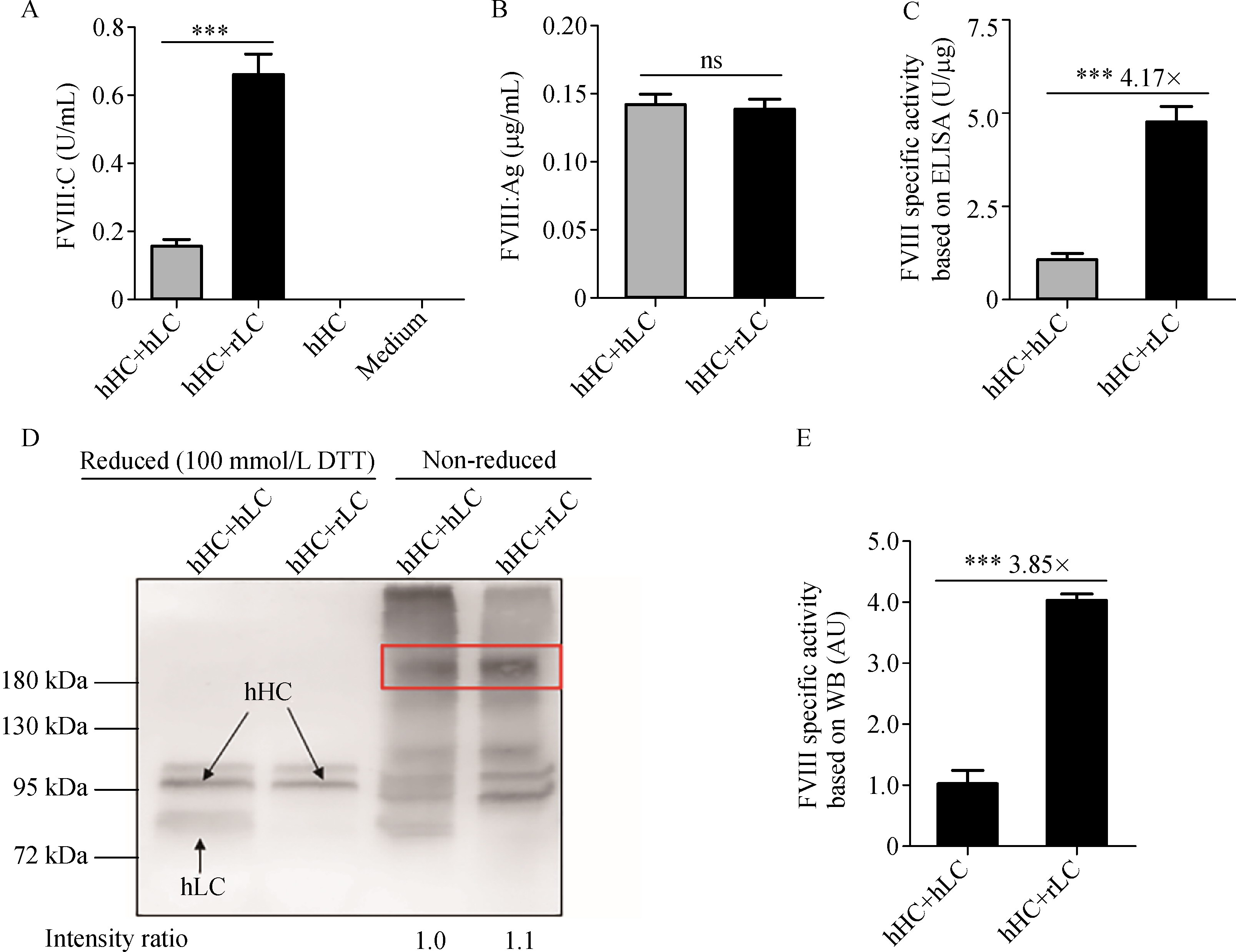
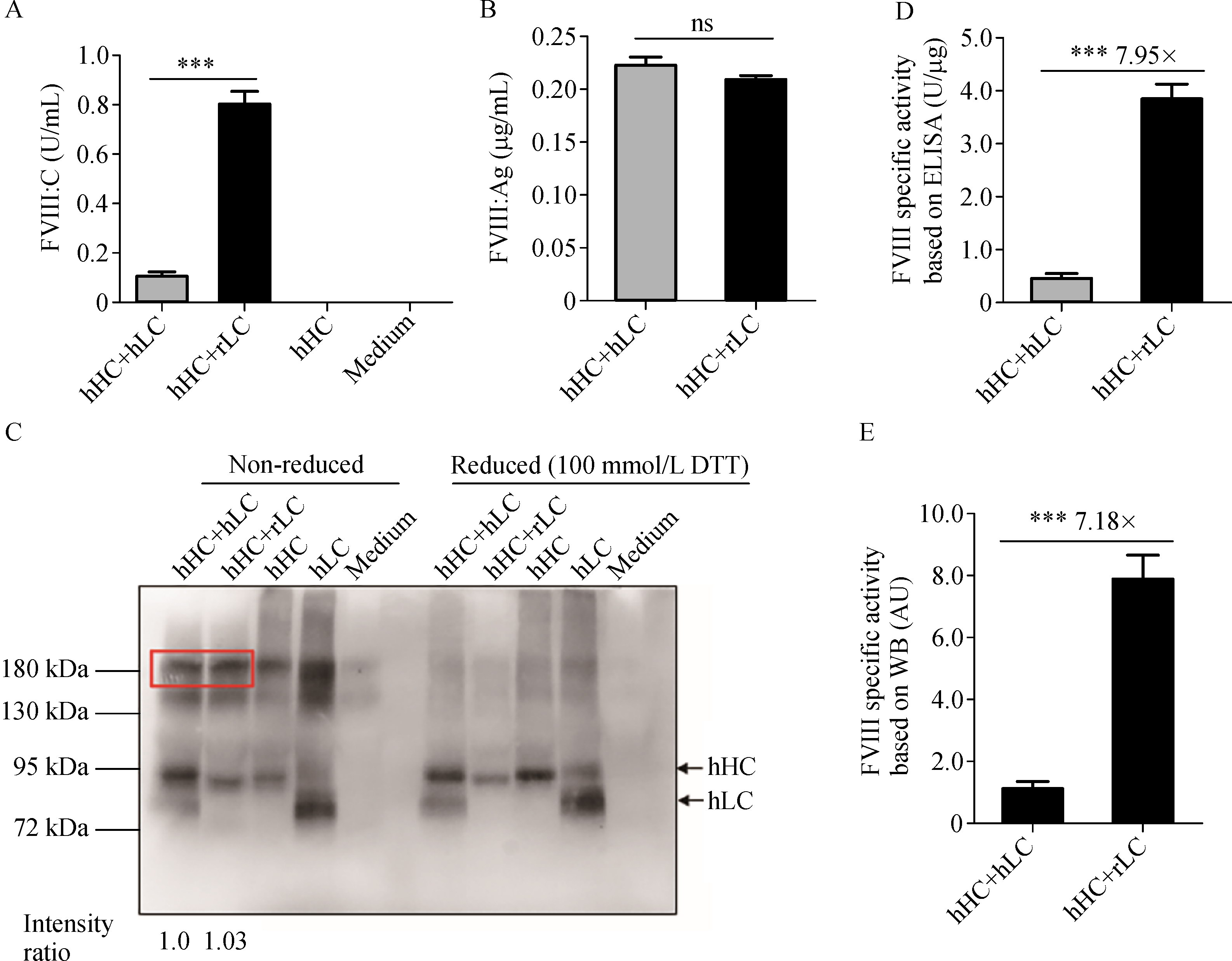
Abstract Conventional therapies for hemophilia A (HA) are prophylactic or on-demand intravenous FVIII infusions. However, they are expensive and inconvenient to perform. Thus, better strategies for HA treatment must be developed. In this study, a recombinant FVIII cDNA encoding a human/rat hybrid FVIII with an enhanced procoagulant potential for adeno-associated virus (AAV)-delivered gene therapy was developed. Plasmids containing human FVIII heavy chain (hHC), human light chain (hLC), and rat light chain (rLC) were transfected into cells and hydrodynamically injected into HA mice. Purified AAV viruses were intravenously injected into HA mice at two doses. Results showed that the hHC+ rLC protein had a higher activity than the hHC+ hLC protein at comparable expression levels. The specific activity of hHC+ rLC was about 4- to 8-fold higher than that of their counterparts. Hydrodynamic injection experiments obtained consistent results. Notably, the HA mice undergoing the AAV-delivered hHC+ rLC treatment exhibited a visibly higher activity than those treated with hHC+ hLC, and the therapeutic effects lasted for up to 40 weeks. In conclusion, the application of the hybrid FVIII (hHC+ rLC) via an AAV-delivered gene therapy substantially improved the hemorrhagic diathesis of the HA mice. These data might be of help to the development of optimized FVIII expression cassette for HA gene therapy.
|
| Keywords
hemophilia A
adeno-associated virus (AAV)
human/rat hybrid factor VIII
gene therapy
dual chain strategy
|
|
Corresponding Author(s):
Jianhua Mao,Xiaodong Xi
|
| About author: Tongcan Cui and Yizhe Hou contributed equally to this work. |
|
Just Accepted Date: 19 August 2021
Online First Date: 14 January 2022
Issue Date: 02 September 2022
|
|
| 1 |
M Makris, J Oldenburg, EP Mauser-Bunschoten, K Peerlinck, G Castaman, K; subcommittee on Factor VIII, Factor IX and Rare Bleeding Disorders. FijnvandraatThe definition, diagnosis and management of mild hemophilia A: communication from the SSC of the ISTH. J Thromb Haemost 2018; 16(12): 2530–2533
https://doi.org/10.1111/jth.14315
pmid: 30430726
|
| 2 |
PHB Bolton-Maggs, KJ Pasi. Haemophilias A and B. Lancet 2003; 361(9371): 1801–1809
https://doi.org/10.1016/S0140-6736(03)13405-8
pmid: 12781551
|
| 3 |
J Lai, C Hough, J Tarrant, D Lillicrap. Biological considerations of plasma-derived and recombinant factor VIII immunogenicity. Blood 2017; 129(24): 3147–3154
https://doi.org/10.1182/blood-2016-11-750885
pmid: 28432221
|
| 4 |
M Morfini, CAP Rapisarda. Safety of recombinant coagulation factors in treating hemophilia. Expert Opin Drug Saf 2019; 18(2): 75–85
https://doi.org/10.1080/14740338.2019.1574743
pmid: 30681006
|
| 5 |
GQ Perrin, RW Herzog, DM Markusic. Update on clinical gene therapy for hemophilia. Blood 2019; 133(5): 407–414
https://doi.org/10.1182/blood-2018-07-820720
pmid: 30559260
|
| 6 |
C Borsotti, A Follenzi. New technologies in gene therapy for inducing immune tolerance in hemophilia A. Expert Rev Clin Immunol 2018; 14(12): 1013–1019
https://doi.org/10.1080/1744666X.2018.1539667
pmid: 30345839
|
| 7 |
A Asokan, DV Schaffer, RJ Samulski. The AAV vector toolkit: poised at the clinical crossroads. Mol Ther 2012; 20(4): 699–708
https://doi.org/10.1038/mt.2011.287
pmid: 22273577
|
| 8 |
AK Zaiss, Q Liu, GP Bowen, NC Wong, JS Bartlett, DA Muruve. Differential activation of innate immune responses by adenovirus and adeno-associated virus vectors. J Virol 2002; 76(9): 4580–4590
https://doi.org/10.1128/JVI.76.9.4580-4590.2002
pmid: 11932423
|
| 9 |
AC Nathwani, EGD Tuddenham, S Rangarajan, C Rosales, J McIntosh, DC Linch, P Chowdary, A Riddell, AJ Pie, C Harrington, J O’Beirne, K Smith, J Pasi, B Glader, P Rustagi, CYC Ng, MA Kay, J Zhou, Y Spence, CL Morton, J Allay, J Coleman, S Sleep, JM Cunningham, D Srivastava, E Basner-Tschakarjan, F Mingozzi, KA High, JT Gray, UM Reiss, AW Nienhuis, AM Davidoff. Adenovirus-associated virus vector-mediated gene transfer in hemophilia B. N Engl J Med 2011; 365(25): 2357–2365
https://doi.org/10.1056/NEJMoa1108046
pmid: 22149959
|
| 10 |
S Rangarajan, L Walsh, W Lester, D Perry, B Madan, M Laffan, H Yu, C Vettermann, GF Pierce, WY Wong, KJ Pasi. AAV5-Factor VIII gene transfer in severe hemophilia A. N Engl J Med 2017; 377(26): 2519–2530
https://doi.org/10.1056/NEJMoa1708483
pmid: 29224506
|
| 11 |
AC Nathwani, AM Davidoff, EGD Tuddenham. Gene therapy for hemophilia. Hematol Oncol Clin North Am 2017; 31(5): 853–868
https://doi.org/10.1016/j.hoc.2017.06.011
pmid: 28895852
|
| 12 |
AC Nathwani, AW Nienhuis, AM Davidoff. Current status of gene therapy for hemophilia. Curr Hematol Rep 2003; 2(4): 319–327
pmid: 12901329
|
| 13 |
B Nambiar, C C Sookdeo, P Berthelette, R Jackson, S Piraino, B Burnham, S Nass, D Souza, CR O’Riordan, KA Vincent, SH Cheng, D Armentano, S Kyostio-Moore. Characteristics of minimally oversized adeno-associated virus vectors encoding human factor VIII generated using producer cell lines and triple transfection. Hum Gene Ther Methods 2017; 28(1): 23–38
https://doi.org/10.1089/hgtb.2016.124
pmid: 28166648
|
| 14 |
C Mah, R Sarkar, I Zolotukhin, M Schleissing, X Xiao, HH Kazazian, BJ Byrne. Dual vectors expressing murine factor VIII result in sustained correction of hemophilia A mice. Hum Gene Ther 2003; 14(2): 143–152
https://doi.org/10.1089/104303403321070838
pmid: 12614565
|
| 15 |
Q Wang, B Dong, J Firrman, S Roberts, AR Moore, W Cao, Y Diao, P Kapranov, R Xu, W Xiao. Efficient production of dual recombinant adeno-associated viral vectors for factor VIII delivery. Hum Gene Ther Methods 2014; 25(4): 261–268
https://doi.org/10.1089/hgtb.2014.093
pmid: 25093498
|
| 16 |
BS Doshi, VR Arruda. Gene therapy for hemophilia: what does the future hold? Ther Adv Hematol 2018; 9(9): 273–293
https://doi.org/10.1177/2040620718791933
pmid: 30210756
|
| 17 |
SA Shestopal, JJ Hao, E Karnaukhova, Y Liang, MV Ovanesov, M Lin, JH Kurasawa, TK Lee, JH Mcvey, AG Sarafanov. Expression and characterization of a codon-optimized blood coagulation factor VIII. J Thromb Haemost 2017; 15(4): 709–720
https://doi.org/10.1111/jth.13632
pmid: 28109042
|
| 18 |
J McIntosh, PJ Lenting, C Rosales, D Lee, S Rabbanian, D Raj, N Patel, EGD Tuddenham, OD Christophe, JH McVey, S Waddington, AW Nienhuis, JT Gray, P Fagone, F Mingozzi, SZ Zhou, KA High, M Cancio, CYC Ng, J Zhou, CL Morton, AM Davidoff, AC Nathwani. Therapeutic levels of FVIII following a single peripheral vein administration of rAAV vector encoding a novel human factor VIII variant. Blood 2013; 121(17): 3335–3344
https://doi.org/10.1182/blood-2012-10-462200
pmid: 23426947
|
| 19 |
GN Nguyen, LA George, JI Siner, RJ Davidson, CB Zander, XL Zheng, VR Arruda, RM Camire, DE Sabatino. Novel factor VIII variants with a modified furin cleavage site improve the efficacy of gene therapy for hemophilia A. J Thromb Haemost 2017; 15(1): 110–121
https://doi.org/10.1111/jth.13543
pmid: 27749002
|
| 20 |
MP Kosloski, KA Shetty, H Wakabayashi, PJ Fay, SV Balu-Iyer. Effects of replacement of factor VIII amino acids Asp519 and Glu665 with Val on plasma survival and efficacy in vivo. AAPS J 2014; 16(5): 1038–1045
https://doi.org/10.1208/s12248-014-9627-2
pmid: 24934295
|
| 21 |
CB Doering, JF Healey, ET Parker, RT Barrow, P Lollar. High level expression of recombinant porcine coagulation factor VIII. J Biol Chem 2002; 277(41): 38345–38349
https://doi.org/10.1074/jbc.M206959200
pmid: 12138172
|
| 22 |
CB Doering, JF Healey, ET Parker, RT Barrow, P Lollar. Identification of porcine coagulation factor VIII domains responsible for high level expression via enhanced secretion. J Biol Chem 2004; 279(8): 6546–6552
https://doi.org/10.1074/jbc.M312451200
pmid: 14660593
|
| 23 |
Q Wang, B Dong, J Firrman, W Wu, S Roberts, AR Moore, LS Liu, MPS Chin, Y Diao, J Kost, W Xiao. Evaluation of the biological differences of canine and human factor VIII in gene delivery: implications in human hemophilia treatment. Gene Ther 2016; 23(7): 597–605
https://doi.org/10.1038/gt.2016.34
pmid: 27064790
|
| 24 |
DE Sabatino, CF Freguia, R Toso, A Santos, EP Merricks, HH Kazazian, TC Nichols, RM Camire, VR Arruda. Recombinant canine B-domain-deleted FVIII exhibits high specific activity and is safe in the canine hemophilia A model. Blood 2009; 114(20): 4562–4565
https://doi.org/10.1182/blood-2009-05-220327
pmid: 19770361
|
| 25 |
P Lollar, ET Parker, PJ Fay. Coagulant properties of hybrid human/porcine factor VIII molecules. J Biol Chem 1992; 267(33): 23652–23657
https://doi.org/10.1016/S0021-9258(18)35888-5
pmid: 1429706
|
| 26 |
W Zhang, J Mao, Y Shen, G Zhang, Y Shao, Z Ruan, Y Wang, W Wu, X Wang, J Zhu, S Chen, W Xiao, X Xi. Evaluation of the activity levels of rat FVIII and human FVIII delivered by adeno-associated viral vectors both in vitro and in vivo. Blood Cells Mol Dis 2018; 73: 47–54
https://doi.org/10.1016/j.bcmd.2018.09.004
pmid: 30249384
|
| 27 |
CD Scallan, T Liu, AE Parker, SL Patarroyo-White, H Chen, H Jiang, J Vargas, D Nagy, SK Powell, JF Wright, R Sarkar, HH Kazazian, A McClelland, LB Couto. Phenotypic correction of a mouse model of hemophilia A using AAV2 vectors encoding the heavy and light chains of FVIII. Blood 2003; 102(12): 3919–3926
https://doi.org/10.1182/blood-2003-01-0222
pmid: 12893764
|
| 28 |
C Mueller, D Ratner, L Zhong, M Esteves-Sena, G Gao. Production and discovery of novel recombinant adeno-associated viral vectors. Curr Protoc Microbiol 2012; Chapter 14: s26
|
| 29 |
J Wang, J Xie, H Lu, L Chen, B Hauck, RJ Samulski, W Xiao. Existence of transient functional double-stranded DNA intermediates during recombinant AAV transduction. Proc Natl Acad Sci USA 2007; 104(32): 13104–13109
https://doi.org/10.1073/pnas.0702778104
pmid: 17664425
|
| 30 |
Y Kuang, J Wang, X Lu, S Lu, L Zhang, C Shen, J Fei, Z Wang. Generation of factor VIII gene knockout mouse by tetraploid embryo complementation technology. Chin J Med Genet (Zhonghua Yi Xue Yi Chuan Xue Za Zhi) 2010; 27(1): 1–6 (in Chinese)
pmid: 20140858
|
| 31 |
D Wang, G Zhang, J Gu, X Shao, Y Dai, J Li, X Pan, S Yao, A Xu, Y Jin, J Huang, Q Shi, J Zhu, X Xi, Z Chen, S Chen. In vivo generated hematopoietic stem cells from genome edited induced pluripotent stem cells are functional in platelet-targeted gene therapy of murine hemophilia A. Haematologica 2020; 105(4): e175–e179
https://doi.org/10.3324/haematol.2019.219089
pmid: 31296582
|
| 32 |
A Tiede. Half-life extended factor VIII for the treatment of hemophilia A. J Thromb Haemost 2015; 13(Suppl 1): S176–S179
https://doi.org/10.1111/jth.12929
pmid: 26149020
|
| 33 |
E Persson, T Foscolo, M Hansen. Reagent-specific underestimation of turoctocog alfa pegol (N8-GP) clotting activity owing to decelerated activation by thrombin. Res Pract Thromb Haemost 2019; 3(1): 114–120
https://doi.org/10.1002/rth2.12167
pmid: 30656284
|
| 34 |
AD Shapiro, MV Ragni, R Kulkarni, J Oldenberg, A Srivastava, DV Quon, KJ Pasi, H Hanabusa, I Pabinger, J Mahlangu, P Fogarty, D Lillicrap, S Kulke, J Potts, S Neelakantan, I Nestorov, S Li, JA Dumont, H Jiang, A Brennan, GF Pierce. Recombinant factor VIII Fc fusion protein: extended-interval dosing maintains low bleeding rates and correlates with von Willebrand factor levels. J Thromb Haemost 2014; 12(11): 1788–1800
https://doi.org/10.1111/jth.12723
pmid: 25196897
|
| 35 |
G Lippi, EJ Favaloro. Emicizumab (ACE910): clinical background and laboratory assessment of hemophilia A. Adv Clin Chem 2019; 88: 151–167
https://doi.org/10.1016/bs.acc.2018.10.003
pmid: 30612605
|
| 36 |
KA High, XM Anguela. Adeno-associated viral vectors for the treatment of hemophilia. Hum Mol Genet 2016; 25(R1): R36–R41
https://doi.org/10.1093/hmg/ddv475
pmid: 26614390
|
| 37 |
L Lisowski, AP Dane, K Chu, Y Zhang, SC Cunningham, EM Wilson, S Nygaard, M Grompe, IE Alexander, MA Kay. Selection and evaluation of clinically relevant AAV variants in a xenograft liver model. Nature 2014; 506(7488): 382–386
https://doi.org/10.1038/nature12875
pmid: 24390344
|
| 38 |
LA George, SK Sullivan, A Giermasz, JEJ Rasko, BJ Samelson-Jones, J Ducore, A Cuker, LM Sullivan, S Majumdar, J Teitel, CE McGuinn, MV Ragni, AY Luk, D Hui, JF Wright, Y Chen, Y Liu, K Wachtel, A Winters, S Tiefenbacher, VR Arruda, JCM van der Loo, O Zelenaia, D Takefman, ME Carr, LB Couto, XM Anguela, KA High. Hemophilia B gene therapy with a high-specific-activity factor IX variant. N Engl J Med 2017; 377(23): 2215–2227
https://doi.org/10.1056/NEJMoa1708538
pmid: 29211678
|
| 39 |
T Okuyama, RM Huber, W Bowling, R Pearline, SC Kennedy, MW Flye, KP Ponder. Liver-directed gene therapy: a retroviral vector with a complete LTR and the ApoE enhancer-α1-antitrypsin promoter dramatically increases expression of human α1-antitrypsin in vivo. Hum Gene Ther 1996; 7(5): 637–645
https://doi.org/10.1089/hum.1996.7.5-637
pmid: 8845389
|
| 40 |
P Simioni, D Tormene, G Tognin, S Gavasso, C Bulato, NP Iacobelli, JD Finn, L Spiezia, C Radu, VR Arruda. X-linked thrombophilia with a mutant factor IX (factor IX Padua). N Engl J Med 2009; 361(17): 1671–1675
https://doi.org/10.1056/NEJMoa0904377
pmid: 19846852
|
| 41 |
M Takeyama, H Wakabayashi, PJ Fay. Contribution of factor VIII light-chain residues 2007–2016 to an activated protein C-interactive site. Thromb Haemost 2013; 109(2): 187–198
https://doi.org/10.1160/TH12-08-0561
pmid: 23224054
|
| 42 |
LM O’Brien, M Mastri, PJ Fay. Regulation of factor VIIIa by human activated protein C and protein S: inactivation of cofactor in the intrinsic factor Xase. Blood 2000; 95(5): 1714–1720
https://doi.org/10.1182/blood.V95.5.1714.005k40_1714_1720
pmid: 10688829
|
| 43 |
KP Pratt, BW Shen, K Takeshima, EW Davie, K Fujikawa, BL Stoddard. Structure of the C2 domain of human factor VIII at 1.5 A resolution. Nature 1999; 402(6760): 439–442
https://doi.org/10.1038/46601
pmid: 10586887
|
| 44 |
VA Novakovic, DB Cullinan, H Wakabayashi, PJ Fay, JD Baleja, GE Gilbert. Membrane-binding properties of the Factor VIII C2 domain. Biochem J 2011; 435(1): 187–196
https://doi.org/10.1042/BJ20101797
pmid: 21210768
|
| 45 |
M Watzka, C Geisen, E Seifried, J Oldenburg. Sequence of the rat factor VIII cDNA. Thromb Haemost 2004; 91(1): 38–42
https://doi.org/10.1160/TH03-06-0336
pmid: 14691566
|
|
Viewed |
|
|
|
Full text
|
|
|
|
|
Abstract
|
|
|
|
|
Cited |
|
|
|
|
| |
Shared |
|
|
|
|
| |
Discussed |
|
|
|
|