|
|
|
DDB1- and CUL4-associated factor 8 plays a critical role in spermatogenesis |
Xiuli Zhang1, Zhizhou Xia1, Xingyu Lv2, Donghe Li1, Mingzhu Liu1, Ruihong Zhang1, Tong Ji3( ), Ping Liu1(), Ruibao Ren1() ), Ping Liu1(), Ruibao Ren1() |
1. Shanghai Institute of Hematology, State Key Laboratory for Medical Genomics, National Research Center for Translational Medicine at Shanghai, Collaborative Innovation Center of Hematology, Ruijin Hospital Affiliated to Shanghai Jiao Tong University School of Medicine, Shanghai 200025, China
2. Key Laboratory of Laparoscopic Technology of Zhejiang Province, Sir Run Run Shaw Hospital, Zhejiang University School of Medicine, Hangzhou 310058, China
3. Department of General Surgery, Sir Run Run Shaw Hospital, Zhejiang University School of Medicine, Hangzhou 310058, China |
|
|
|
|
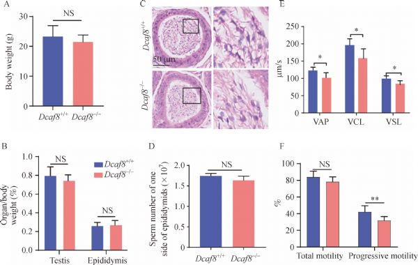
Abstract Cullin-RING E3 ubiquitin ligase (CRL)-4 is a member of the large CRL family in eukaryotes. It plays important roles in a wide range of cellular processes, organismal development, and physiological and pathological conditions. DDB1- and CUL4-associated factor 8 (DCAF8) is a WD40 repeat-containing protein, which serves as a substrate receptor for CRL4. The physiological role of DCAF8 is unknown. In this study, we constructed Dcaf8 knockout mice. Homozygous mice were viable with no noticeable abnormalities. However, the fertility of Dcaf8-deficient male mice was markedly impaired, consistent with the high expression of DCAF8 in adult mouse testis. Sperm movement characteristics, including progressive motility, path velocity, progressive velocity, and track speed, were significantly lower in Dcaf8 knockout mice than in wild-type (WT) mice. However, the total motility was similar between WT and Dcaf8 knockout sperm. More than 40% of spermatids in Dcaf8 knockout mice showed pronounced morphological abnormalities with typical bent head malformation. The acrosome and nucleus of Dcaf8 knockout sperm looked similar to those of WT sperm. In vitro tests showed that the fertilization rate of Dcaf8 knockout mice was significantly reduced. The results demonstrated that DCAF8 plays a critical role in spermatogenesis, and DCAF8 is a key component of CRL4 function in the reproductive system.
|
| Keywords
Dcaf8
male infertility
spermatogenesis
|
|
Corresponding Author(s):
Tong Ji,Ping Liu,Ruibao Ren
|
|
Just Accepted Date: 11 March 2021
Online First Date: 12 April 2021
Issue Date: 23 April 2021
|
|
| 1 |
FT Neto, PV Bach, BB Najari, PS Li, M Goldstein. Genetics of male infertility. Curr Urol Rep 2016; 17(10): 70
https://doi.org/10.1007/s11934-016-0627-x
pmid: 27502429
|
| 2 |
PN Schlegel. Evaluation of male infertility. Minerva Ginecol 2009; 61(4): 261–283
pmid: 19745794
|
| 3 |
KL O’Flynn O’Brien, AC Varghese, A Agarwal. The genetic causes of male factor infertility: a review. Fertil Steril 2010; 93(1): 1–12
https://doi.org/10.1016/j.fertnstert.2009.10.045
pmid: 20103481
|
| 4 |
C Krausz. Male infertility: pathogenesis and clinical diagnosis. Best Pract Res Clin Endocrinol Metab 2011; 25(2): 271–285
https://doi.org/10.1016/j.beem.2010.08.006
pmid: 21397198
|
| 5 |
EM Eddy. Male germ cell gene expression. Recent Prog Horm Res 2002; 57(1): 103–128
https://doi.org/10.1210/rp.57.1.103
pmid: 12017539
|
| 6 |
DM de Kretser, KL Loveland, A Meinhardt, D Simorangkir, N Wreford. Spermatogenesis. Hum Reprod 1998; 13(Suppl 1): 1–8
https://doi.org/10.1093/humrep/13.suppl_1.1
pmid: 9663765
|
| 7 |
N Schultz, FK Hamra, DL Garbers. A multitude of genes expressed solely in meiotic or postmeiotic spermatogenic cells offers a myriad of contraceptive targets. Proc Natl Acad Sci USA 2003; 100(21): 12201–12206
https://doi.org/10.1073/pnas.1635054100
pmid: 14526100
|
| 8 |
CC Hou, WX Yang. New insights to the ubiquitin-proteasome pathway (UPP) mechanism during spermatogenesis. Mol Biol Rep 2013; 40(4): 3213–3230
https://doi.org/10.1007/s11033-012-2397-y
pmid: 23268313
|
| 9 |
WM Baarends, R van der Laan, JA Grootegoed. Specific aspects of the ubiquitin system in spermatogenesis. J Endocrinol Invest 2000; 23(9): 597–604
https://doi.org/10.1007/BF03343782
pmid: 11079455
|
| 10 |
D Kopanja, N Roy, T Stoyanova, RA Hess, S Bagchi, P Raychaudhuri. Cul4A is essential for spermatogenesis and male fertility. Dev Biol 2011; 352(2): 278–287
https://doi.org/10.1016/j.ydbio.2011.01.028
pmid: 21291880
|
| 11 |
T Ma, JA Keller, X Yu. RNF8-dependent histone ubiquitination during DNA damage response and spermatogenesis. Acta Biochim Biophys Sin (Shanghai) 2011; 43(5): 339–345
https://doi.org/10.1093/abbs/gmr016
pmid: 21444325
|
| 12 |
S Wang, H Zheng, Y Esaki, F Kelly, W Yan. Cullin3 is a KLHL10-interacting protein preferentially expressed during late spermiogenesis. Biol Reprod 2006; 74(1): 102–108
https://doi.org/10.1095/biolreprod.105.045484
pmid: 16162871
|
| 13 |
LY Lu, J Wu, L Ye, GB Gavrilina, TL Saunders, X Yu. RNF8-dependent histone modifications regulate nucleosome removal during spermatogenesis. Dev Cell 2010; 18(3): 371–384
https://doi.org/10.1016/j.devcel.2010.01.010
pmid: 20153262
|
| 14 |
MD Petroski, RJ Deshaies. Function and regulation of Cullin-RING ubiquitin ligases. Nat Rev Mol Cell Biol 2005; 6(1): 9–20
https://doi.org/10.1038/nrm1547
pmid: 15688063
|
| 15 |
S Jackson, Y Xiong. CRL4s: the CUL4-RING E3 ubiquitin ligases. Trends Biochem Sci 2009; 34(11): 562–570
https://doi.org/10.1016/j.tibs.2009.07.002
pmid: 19818632
|
| 16 |
BC O’Connell, JW Harper. Ubiquitin proteasome system (UPS): what can chromatin do for you? Curr Opin Cell Biol 2007; 19(2): 206–214
https://doi.org/10.1016/j.ceb.2007.02.014
pmid: 17314036
|
| 17 |
W Zhong, H Feng, FE Santiago, ET Kipreos. CUL-4 ubiquitin ligase maintains genome stability by restraining DNA-replication licensing. Nature 2003; 423(6942): 885–889
https://doi.org/10.1038/nature01747
pmid: 12815436
|
| 18 |
K Sugasawa. The CUL4 enigma: culling DNA repair factors. Mol Cell 2009; 34(4): 403–404
https://doi.org/10.1016/j.molcel.2009.05.009
pmid: 19481520
|
| 19 |
J Cheng, J Guo, BJ North, K Tao, P Zhou, W Wei. The emerging role for Cullin 4 family of E3 ligases in tumorigenesis. Biochim Biophys Acta Rev Cancer 2019; 1871(1): 138–159
https://doi.org/10.1016/j.bbcan.2018.11.007
pmid: 30602127
|
| 20 |
H Wang, L Zhai, J Xu, HY Joo, S Jackson, H Erdjument-Bromage, P Tempst, Y Xiong, Y Zhang. Histone H3 and H4 ubiquitylation by the CUL4-DDB-ROC1 ubiquitin ligase facilitates cellular response to DNA damage. Mol Cell 2006; 22(3): 383–394
https://doi.org/10.1016/j.molcel.2006.03.035
pmid: 16678110
|
| 21 |
J Lee, P Zhou. DCAFs, the missing link of the CUL4-DDB1 ubiquitin ligase. Mol Cell 2007; 26(6): 775–780
https://doi.org/10.1016/j.molcel.2007.06.001
pmid: 17588513
|
| 22 |
S Angers, T Li, X Yi, MJ MacCoss, RT Moon, N Zheng. Molecular architecture and assembly of the DDB1-CUL4A ubiquitin ligase machinery. Nature 2006; 443(7111): 590–593
https://doi.org/10.1038/nature05175
pmid: 16964240
|
| 23 |
Y Yin, L Liu, C Yang, C Lin, GM Veith, C Wang, P Sutovsky, P Zhou, L Ma. Cell autonomous and nonautonomous function of CUL4B in mouse spermatogenesis. J Biol Chem 2016; 291(13): 6923–6935
https://doi.org/10.1074/jbc.M115.699660
pmid: 26846852
|
| 24 |
CY Lin, CY Chen, CH Yu, IS Yu, SR Lin, JT Wu, YH Lin, PL Kuo, JC Wu, SW Lin. Human X-linked intellectual disability factor CUL4B is required for post-meiotic sperm development and male fertility. Sci Rep 2016; 6(1): 20227
https://doi.org/10.1038/srep20227
pmid: 26832838
|
| 25 |
Y Yin, C Lin, ST Kim, I Roig, H Chen, L Liu, GM Veith, RU Jin, S Keeney, M Jasin, K Moley, P Zhou, L Ma. The E3 ubiquitin ligase Cullin 4A regulates meiotic progression in mouse spermatogenesis. Dev Biol 2011; 356(1): 51–62
https://doi.org/10.1016/j.ydbio.2011.05.661
pmid: 21624359
|
| 26 |
A Ali, BV Mistry, HA Ahmed, R Abdulla, HA Amer, A Prince, AM Alazami, FS Alkuraya, A Assiri. Deletion of DDB1- and CUL4-associated factor-17 (Dcaf17) gene causes spermatogenesis defects and male infertility in mice. Sci Rep 2018; 8(1): 9202
https://doi.org/10.1038/s41598-018-27379-0
pmid: 29907856
|
| 27 |
Y Wu, L Zhou, X Wang, J Lu, R Zhang, X Liang, L Wang, W Deng, YX Zeng, H Huang, T Kang. A genome-scale CRISPR-Cas9 screening method for protein stability reveals novel regulators of Cdc25A. Cell Discov 2016; 2(1): 16014
https://doi.org/10.1038/celldisc.2016.14
pmid: 27462461
|
| 28 |
M Nowak, B Suenkel, P Porras, R Migotti, F Schmidt, M Kny, X Zhu, EE Wanker, G Dittmar, J Fielitz, T Sommer. DCAF8, a novel MuRF1 interaction partner, promotes muscle atrophy. J Cell Sci 2019; 132(17): jcs233395
https://doi.org/10.1242/jcs.233395
pmid: 31391242
|
| 29 |
G Li, T Ji, J Chen, Y Fu, L Hou, Y Feng, T Zhang, T Song, J Zhao, Y Endo, H Lin, X Cai, Y Cang. CRL4DCAF8 ubiquitin ligase targets histone H3K79 and promotes H3K9 methylation in the liver. Cell Rep 2017; 18(6): 1499–1511
https://doi.org/10.1016/j.celrep.2017.01.039
pmid: 28178526
|
| 30 |
D Huang, C Liu, X Sun, X Sun, Y Qu, Y Tang, G Li, T Tong. CRL4DCAF8 and USP11 oppositely regulate the stability of myeloid leukemia factors (MLFs). Biochem Biophys Res Commun 2020; 529(2): 127–132
https://doi.org/10.1016/j.bbrc.2020.05.186
pmid: 32703400
|
| 31 |
CJ Klein, Y Wu, P Vogel, HH Goebel, C Bönnemann, K Zukosky, MV Botuyan, X Duan, S Middha, EJ Atkinson, G Mer, PJ Dyck. Ubiquitin ligase defect by DCAF8 mutation causes HMSN2 with giant axons. Neurology 2014; 82(10): 873–878
https://doi.org/10.1212/WNL.0000000000000206
pmid: 24500646
|
| 32 |
LT Gou, P Dai, JH Yang, Y Xue, YP Hu, Y Zhou, JY Kang, X Wang, H Li, MM Hua, S Zhao, SD Hu, LG Wu, HJ Shi, Y Li, XD Fu, LH Qu, ED Wang, MF Liu. Pachytene piRNAs instruct massive mRNA elimination during late spermiogenesis. Cell Res 2015; 25(2): 266
https://doi.org/10.1038/cr.2015.14
pmid: 25645811
|
| 33 |
LT Gou, JY Kang, P Dai, X Wang, F Li, S Zhao, M Zhang, MM Hua, Y Lu, Y Zhu, Z Li, H Chen, LG Wu, D Li, XD Fu, J Li, HJ Shi, MF Liu. Ubiquitination-deficient mutations in human Piwi cause male infertility by impairing histone-to-protamine exchange during spermiogenesis. Cell 2017; 169(6):1090–1104.e13
https://doi.org/10.1016/j.cell.2017.04.034
pmid: 28552346
|
| 34 |
KJ Livak, TD Schmittgen. Analysis of relative gene expression data using real-time quantitative PCR and the 2(−ΔΔC(T)) method. Methods 2001; 25(4): 402–408
https://doi.org/10.1006/meth.2001.1262
pmid: 11846609
|
| 35 |
D Shechter, HL Dormann, CD Allis, SB Hake. Extraction, purification and analysis of histones. Nat Protoc 2007; 2(6): 1445–1457
https://doi.org/10.1038/nprot.2007.202
pmid: 17545981
|
| 36 |
P Quinn, JF Kerin, GM Warnes. Improved pregnancy rate in human in vitro fertilization with the use of a medium based on the composition of human tubal fluid. Fertil Steril 1985; 44(4): 493–498
https://doi.org/10.1016/S0015-0282(16)48918-1
pmid: 3902512
|
| 37 |
Q Chen, H Peng, L Lei, Y Zhang, H Kuang, Y Cao, QX Shi, T Ma, E Duan. Aquaporin3 is a sperm water channel essential for postcopulatory sperm osmoadaptation and migration. Cell Res 2011; 21(6): 922–933
https://doi.org/10.1038/cr.2010.169
pmid: 21135872
|
| 38 |
MM Matzuk, DJ Lamb. Genetic dissection of mammalian fertility pathways. Nat Cell Biol 2002; 4(Suppl): s41–s49
https://doi.org/10.1038/ncb-nm-fertilityS41
pmid: 12479614
|
| 39 |
D Jamsai, MK O’Bryan. Mouse models in male fertility research. Asian J Androl 2011; 13(1): 139–151
https://doi.org/10.1038/aja.2010.101
pmid: 21057516
|
| 40 |
P Sutovsky. Ubiquitin-dependent proteolysis in mammalian spermatogenesis, fertilization, and sperm quality control: killing three birds with one stone. Microsc Res Tech 2003; 61(1): 88–102
https://doi.org/10.1002/jemt.10319
pmid: 12672125
|
| 41 |
M Kanatsu-Shinohara, I Onoyama, KI Nakayama, T Shinohara. Skp1-Cullin-F-box (SCF)-type ubiquitin ligase FBXW7 negatively regulates spermatogonial stem cell self-renewal. Proc Natl Acad Sci USA 2014; 111(24): 8826–8831
https://doi.org/10.1073/pnas.1401837111
pmid: 24879440
|
| 42 |
X Hou, W Zhang, Z Xiao, H Gan, X Lin, S Liao, C Han. Mining and characterization of ubiquitin E3 ligases expressed in the mouse testis. BMC Genomics 2012; 13(1): 495
https://doi.org/10.1186/1471-2164-13-495
pmid: 22992278
|
| 43 |
C Yu, YL Zhang, WW Pan, XM Li, ZW Wang, ZJ Ge, JJ Zhou, Y Cang, C Tong, QY Sun, HY Fan. CRL4 complex regulates mammalian oocyte survival and reprogramming by activation of TET proteins. Science 2013; 342(6165): 1518–1521
https://doi.org/10.1126/science.1244587
pmid: 24357321
|
| 44 |
C Yu, YW Xu, QQ Sha, HY Fan. CRL4DCAF1 is required in activated oocytes for follicle maintenance and ovulation. Mol Hum Reprod 2015; 21(2): 195–205
https://doi.org/10.1093/molehr/gau103
pmid: 25371539
|
| 45 |
C Yu, SY Ji, QQ Sha, QY Sun, HY Fan. CRL4-DCAF1 ubiquitin E3 ligase directs protein phosphatase 2A degradation to control oocyte meiotic maturation. Nat Commun 2015; 6(1): 8017
https://doi.org/10.1038/ncomms9017
pmid: 26281983
|
| 46 |
SS Suarez, AA Pacey. Sperm transport in the female reproductive tract. Hum Reprod Update 2006; 12(1): 23–37
https://doi.org/10.1093/humupd/dmi047
pmid: 16272225
|
| 47 |
KA Sutton, MK Jungnickel, HM Florman. A polycystin-1 controls postcopulatory reproductive selection in mice. Proc Natl Acad Sci USA 2008; 105(25): 8661–8666
https://doi.org/10.1073/pnas.0800603105
pmid: 18562295
|
| 48 |
J Zhang, YL Zhang, LW Zhao, JX Guo, JL Yu, SY Ji, LR Cao, SY Zhang, L Shen, XH Ou, HY Fan. Mammalian nucleolar protein DCAF13 is essential for ovarian follicle maintenance and oocyte growth by mediating rRNA processing. Cell Death Differ 2019; 26(7): 1251–1266
https://doi.org/10.1038/s41418-018-0203-7
pmid: 30283081
|
|
Viewed |
|
|
|
Full text
|
|
|
|
|
Abstract
|
|
|
|
|
Cited |
|
|
|
|
| |
Shared |
|
|
|
|
| |
Discussed |
|
|
|
|