|
|
|
Apigenin alleviates neomycin-induced oxidative damage via the Nrf2 signaling pathway in cochlear hair cells |
Gaogan Jia, Huanyu Mao, Yanping Zhang, Yusu Ni( ), Yan Chen() ), Yan Chen() |
| ENT Institute and Department of Otorhinolaryngology, Eye & ENT Hospital, Fudan University, Shanghai 200031, China; NHC Key Laboratory of Hearing Medicine (Fudan University), Shanghai 200031, China |
|
|
|
|
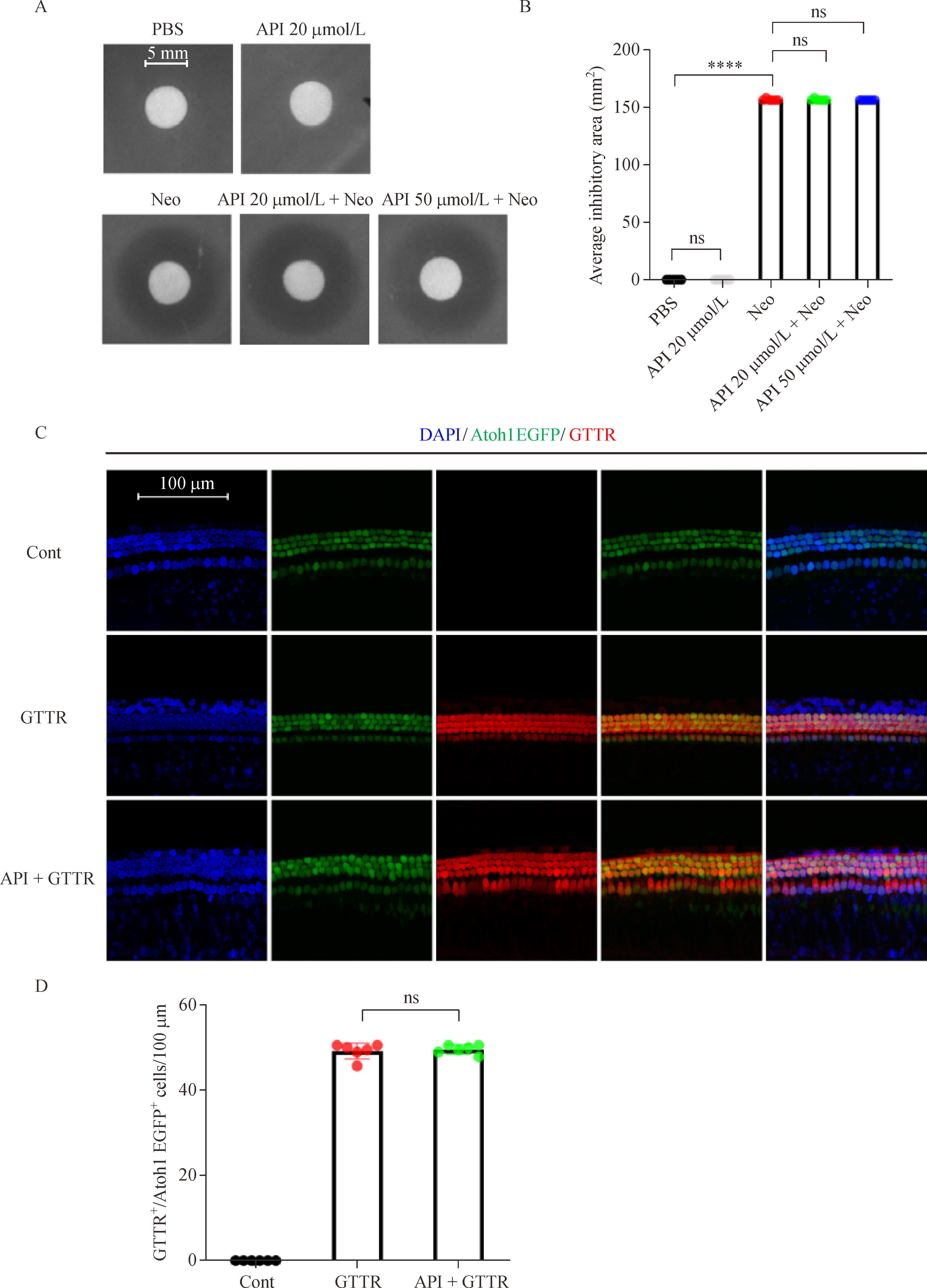
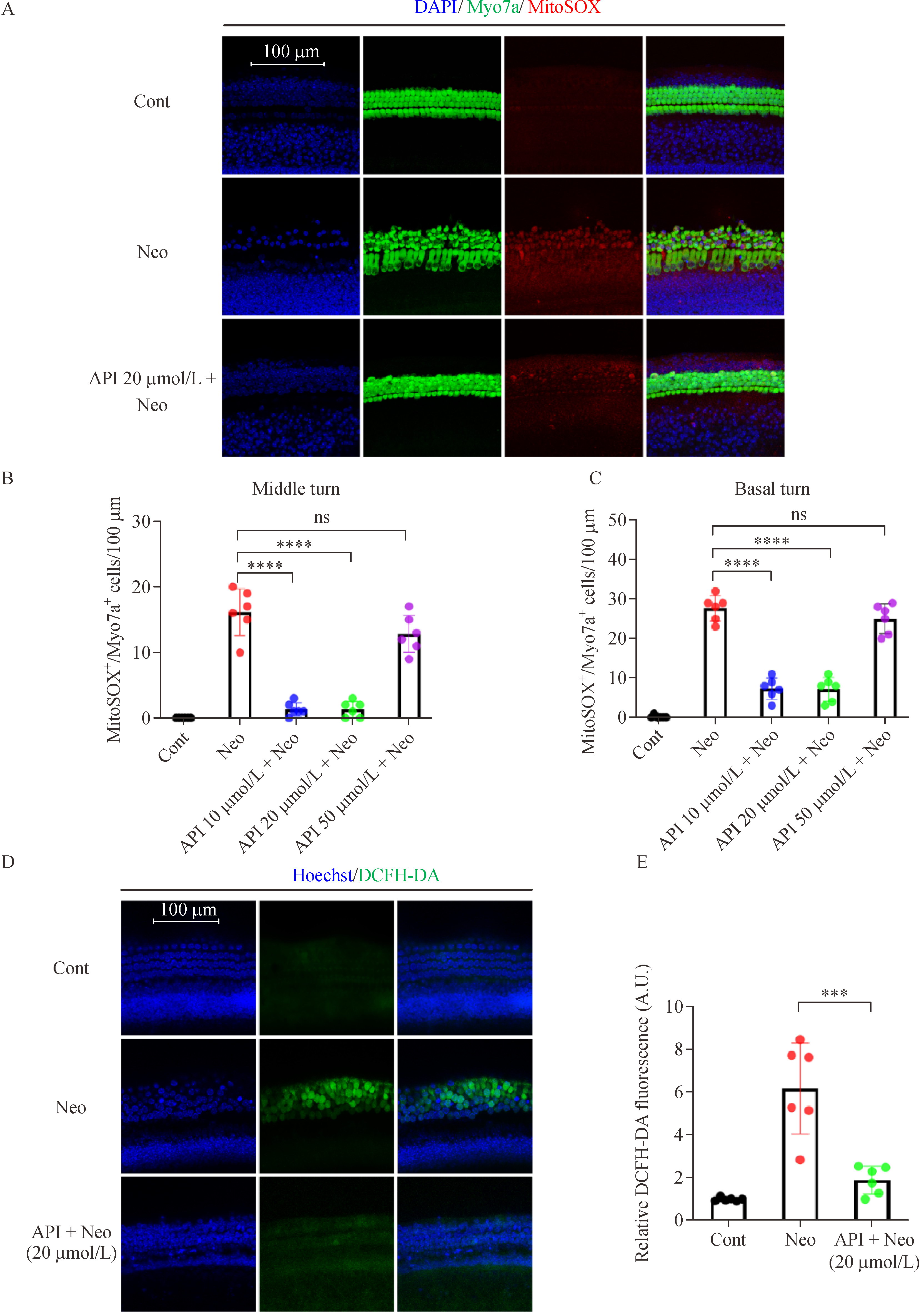
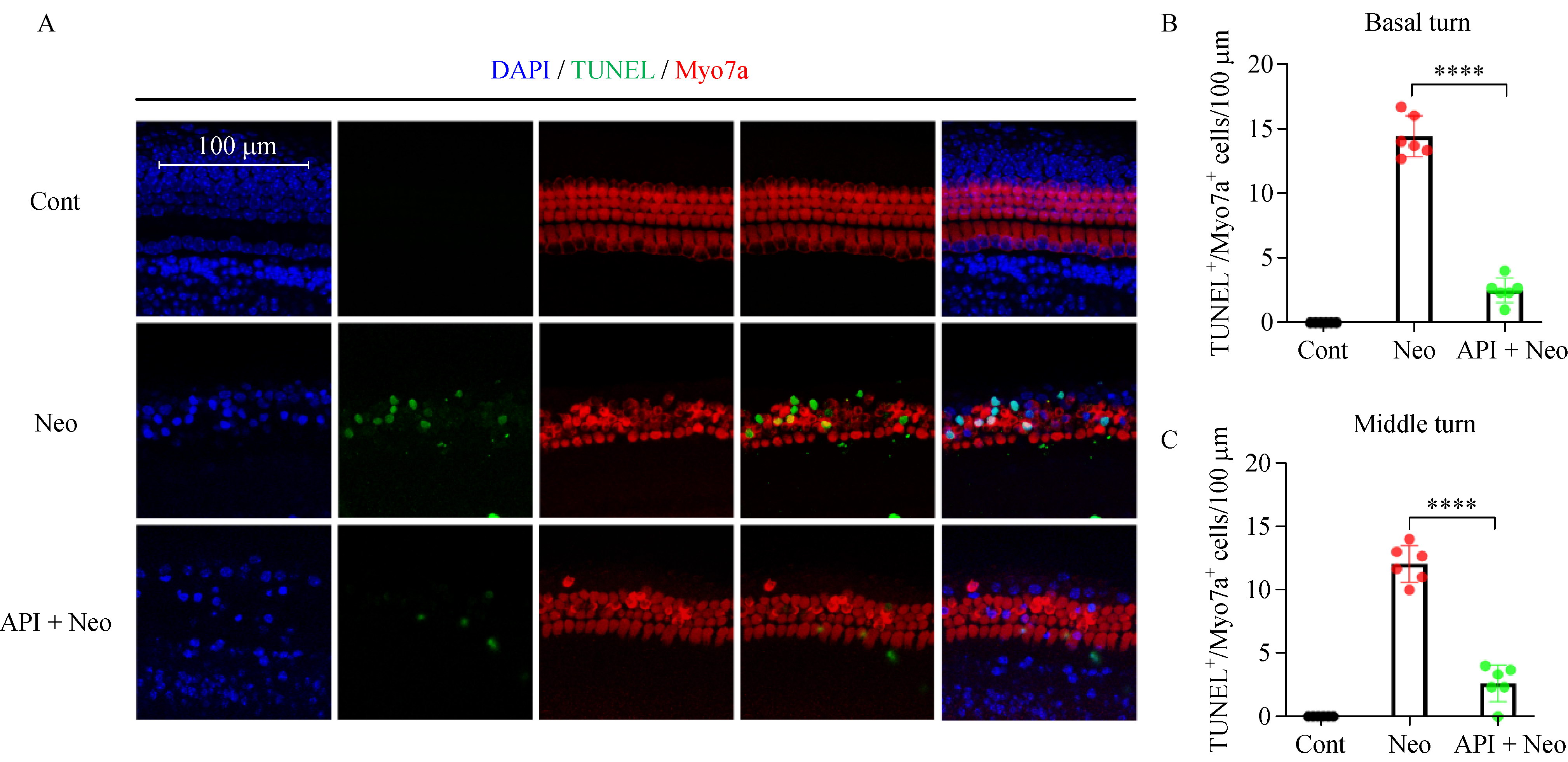
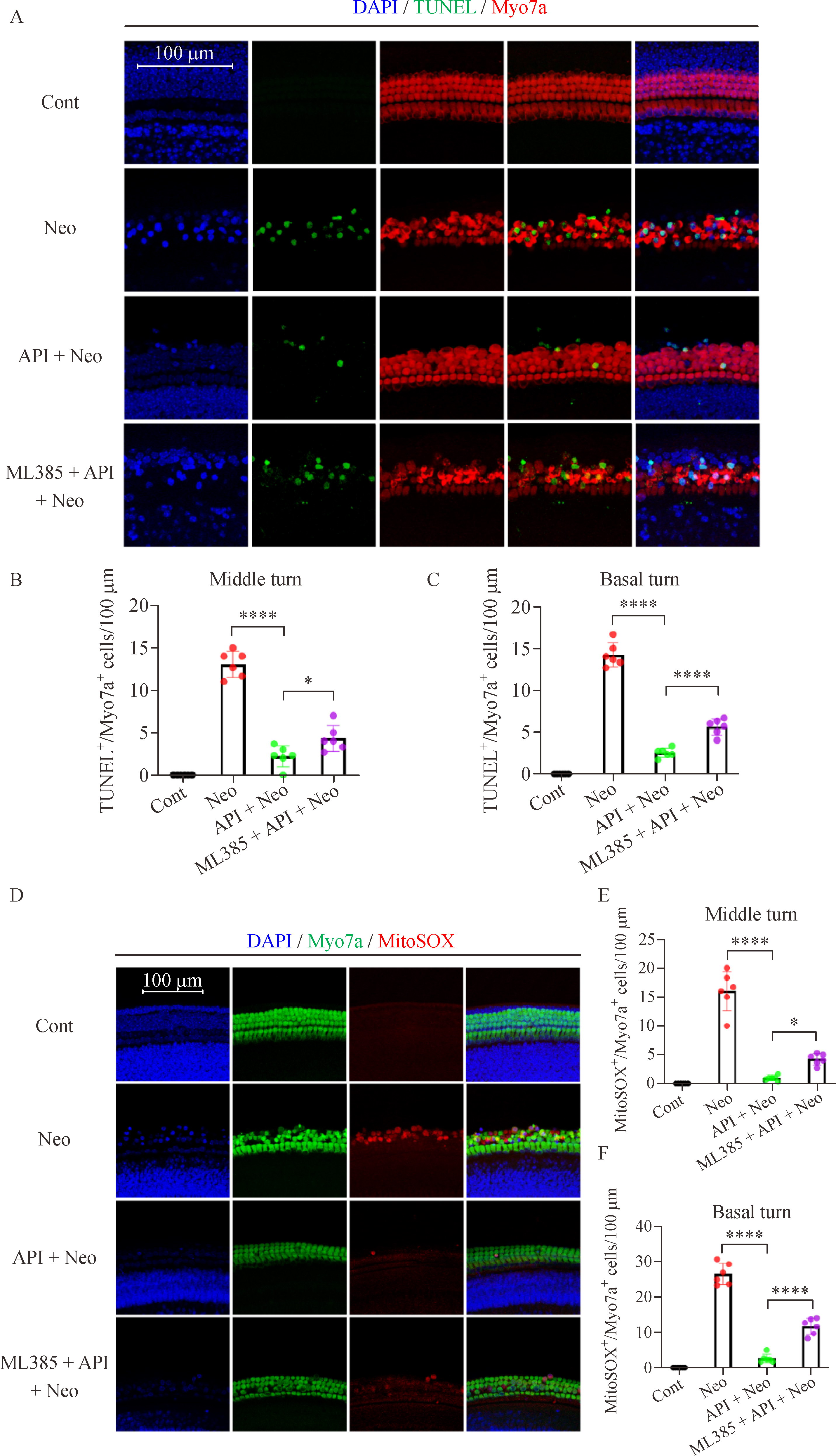
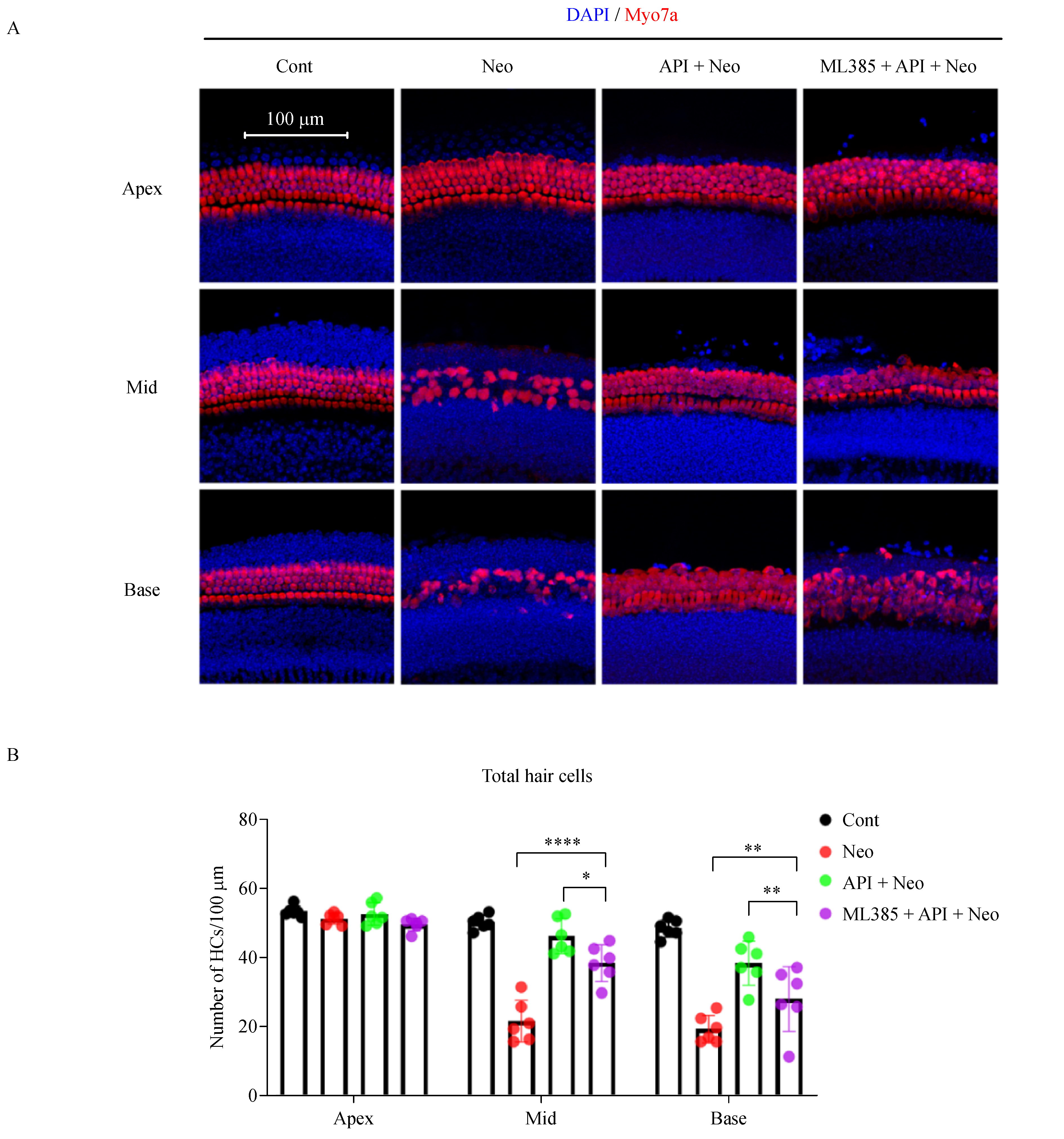
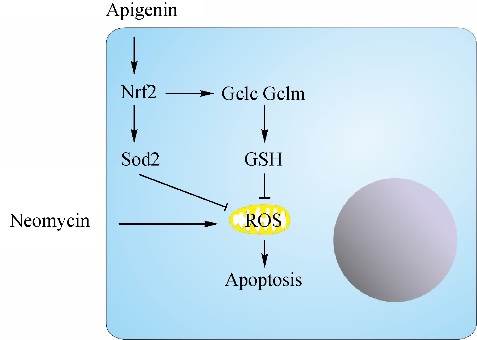
Abstract Oxidative stress plays an important role in the pathogenesis of aminoglycoside-induced hearing loss and represents a promising target for treatment. We tested the potential effect of apigenin, a natural flavonoid with anticancer, anti-inflammatory, and antioxidant activities, on neomycin-induced ototoxicity in cochlear hair cells in vitro. Results showed that apigenin significantly ameliorated the loss of hair cells and the accumulation of reactive oxygen species upon neomycin injury. Further evidence suggested that the nuclear factor erythroid 2-related factor 2 (Nrf2) signaling pathway was activated by apigenin treatment. Disruption of the Nrf2 axis abolished the effects of apigenin on the alleviation of oxidative stress and subsequent apoptosis of hair cells. This study provided evidence of the protective effect of apigenin on cochlear hair cells and its underlying mechanism.
|
| Keywords
apigenin
aminoglycosides
ototoxicity
oxidative stress
Nrf2 signaling pathway
|
|
Corresponding Author(s):
Yusu Ni,Yan Chen
|
| About author: Tongcan Cui and Yizhe Hou contributed equally to this work. |
|
Just Accepted Date: 19 August 2021
Online First Date: 16 December 2021
Issue Date: 02 September 2022
|
|
| 1 |
DJ Fink. Hearing loss in adults. N Engl J Med 2018; 378(10): 969–970
pmid: 29517216
|
| 2 |
JA Leis, JA Rutka, WL Gold. Aminoglycoside-induced ototoxicity. CMAJ 2015; 187(1): E52
https://doi.org/10.1503/cmaj.140339
pmid: 25225217
|
| 3 |
CF Dai, PS Steyger. A systemic gentamicin pathway across the stria vascularis. Hear Res 2008; 235(1–2): 114–124
https://doi.org/10.1016/j.heares.2007.10.010
pmid: 18082985
|
| 4 |
W Marcotti, LF Corns, RJ Goodyear, AK Rzadzinska, KB Avraham, KP Steel, GP Richardson, CJ Kros. The acquisition of mechano-electrical transducer current adaptation in auditory hair cells requires myosin VI. J Physiol 2016; 594(13): 3667–3681
https://doi.org/10.1113/JP272220
pmid: 27111754
|
| 5 |
A Alharazneh, L Luk, M Huth, A Monfared, PS Steyger, AG Cheng, AJ Ricci. Functional hair cell mechanotransducer channels are required for aminoglycoside ototoxicity. PLoS One 2011; 6(7): e22347
https://doi.org/10.1371/journal.pone.0022347
pmid: 21818312
|
| 6 |
Y Kawashima, GS Géléoc, K Kurima, V Labay, A Lelli, Y Asai, T Makishima, DK Wu, CC Della Santina, JR Holt, AJ Griffith. Mechanotransduction in mouse inner ear hair cells requires transmembrane channel-like genes. J Clin Invest 2011; 121(12): 4796–4809
https://doi.org/10.1172/JCI60405
pmid: 22105175
|
| 7 |
D Ruhl, TT Du, EL Wagner, JH Choi, S Li, R Reed, K Kim, M Freeman, G Hashisaki, JR Lukens, JB Shin. Necroptosis and apoptosis contribute to cisplatin and aminoglycoside ototoxicity. J Neurosci 2019; 39(15): 2951–2964
https://doi.org/10.1523/JNEUROSCI.1384-18.2019
pmid: 30733218
|
| 8 |
KN Prasad, SC Bondy. Increased oxidative stress, inflammation, and glutamate: potential preventive and therapeutic targets for hearing disorders. Mech Ageing Dev 2020; 185: 111191
https://doi.org/10.1016/j.mad.2019.111191
pmid: 31765645
|
| 9 |
E Shulman, V Belakhov, G Wei, A Kendall, EG Meyron-Holtz, D Ben-Shachar, J Schacht, T Baasov. Designer aminoglycosides that selectively inhibit cytoplasmic rather than mitochondrial ribosomes show decreased ototoxicity: a strategy for the treatment of genetic diseases. J Biol Chem 2014; 289(4): 2318–2330
https://doi.org/10.1074/jbc.M113.533588
pmid: 24302717
|
| 10 |
R Esterberg, T Linbo, SB Pickett, P Wu, HC Ou, EW Rubel, DW Raible. Mitochondrial calcium uptake underlies ROS generation during aminoglycoside-induced hair cell death. J Clin Invest 2016; 126(9): 3556–3566
https://doi.org/10.1172/JCI84939
pmid: 27500493
|
| 11 |
L Liu, Y Chen, J Qi, Y Zhang, Y He, W Ni, W Li, S Zhang, S Sun, MM Taketo, L Wang, R Chai, H Li. Wnt activation protects against neomycin-induced hair cell damage in the mouse cochlea. Cell Death Dis 2016; 7(3): e2136
https://doi.org/10.1038/cddis.2016.35
pmid: 26962686
|
| 12 |
CP Ojano-Dirain, PJ Antonelli, CG Le Prell. Mitochondria-targeted antioxidant MitoQ reduces gentamicin-induced ototoxicity. Otol Neurotol 2014; 35(3): 533–539
https://doi.org/10.1097/MAO.0000000000000192
pmid: 24518411
|
| 13 |
SA Tokgöz, E Vuralkan, ND Sonbay, M Çalişkan, C Saka, Ö Beşalti, İ Akin. Protective effects of vitamins E, B and C and L-carnitine in the prevention of cisplatin-induced ototoxicity in rats. J Laryngol Otol 2012; 126(5): 464–469
https://doi.org/10.1017/S0022215112000382
pmid: 22490890
|
| 14 |
Z He, L Guo, Y Shu, Q Fang, H Zhou, Y Liu, D Liu, L Lu, X Zhang, X Ding, D Liu, M Tang, W Kong, S Sha, H Li, X Gao, R Chai. Autophagy protects auditory hair cells against neomycin-induced damage. Autophagy 2017; 13(11): 1884–1904
https://doi.org/10.1080/15548627.2017.1359449
pmid: 28968134
|
| 15 |
Q Yang, Y Zhou, H Yin, H Li, M Zhou, G Sun, Z Cao, R Man, H Wang, J Li. PINK1 protects against gentamicin-induced sensory hair cell damage: possible relation to induction of autophagy and inhibition of p53 signal pathway. Front Mol Neurosci 2018; 11: 403
https://doi.org/10.3389/fnmol.2018.00403
pmid: 30483050
|
| 16 |
V Noack, K Pak, R Jalota, A Kurabi, AF Ryan. An antioxidant screen identifies candidates for protection of cochlear hair cells from gentamicin toxicity. Front Cell Neurosci 2017; 11: 242
https://doi.org/10.3389/fncel.2017.00242
pmid: 28867994
|
| 17 |
G Meresman, M Götte, M Laschke. Plants as source of new therapies for endometriosis: a review of preclinical and clinical studies. Hum Reprod Update 2021; 27(2): 367–392
https://doi.org/10.1093/humupd/dmaa039
pmid: 33124671
|
| 18 |
YJ Lee, KS Park, HS Nam, MK Cho, SH Lee. Apigenin causes necroptosis by inducing ROS accumulation, mitochondrial dysfunction, and ATP depletion in malignant mesothelioma cells. Korean J Physiol Pharmacol 2020; 24(6): 493–502
https://doi.org/10.4196/kjpp.2020.24.6.493
pmid: 33093271
|
| 19 |
R Ginwala, R Bhavsar, P Moore, M Bernui, N Singh, F Bearoff, M Nagarkatti, Z Khan, P Jain. Apigenin modulates dendritic cell activities and curbs inflammation via RelB inhibition in the context of neuroinflammatory diseases. J Neuroimmune Pharmacol 2021; 16(2): 403–424 doi: 10.1007/s11481-020-09933-8
pmid: 32607691
|
| 20 |
K Ren, T Jiang, HF Zhou, Y Liang, GJ Zhao. Apigenin retards atherogenesis by promoting ABCA1-mediated cholesterol efflux and suppressing inflammation. Cell Physiol Biochem 2018; 47(5): 2170–2184
https://doi.org/10.1159/000491528
pmid: 29975943
|
| 21 |
I Bougioukas, V Didilis, A Emmert, AF Jebran, R Waldmann-Beushausen, T Stojanovic, FA Schoendube, BC Danner. Apigenin reduces NF-κB and subsequent cytokine production as protective effect in a rodent animal model of lung ischemia-reperfusion injury. J Invest Surg 2018; 31(2): 96–106 doi:10.1080/08941939.2017.1296512
pmid: 28340319
|
| 22 |
Y Ogura, M Kitada, J Xu, I Monno, D Koya. CD38 inhibition by apigenin ameliorates mitochondrial oxidative stress through restoration of the intracellular NAD+/NADH ratio and Sirt3 activity in renal tubular cells in diabetic rats. Aging (Albany NY) 2020; 12(12): 11325–11336
https://doi.org/10.18632/aging.103410
pmid: 32507768
|
| 23 |
B Salehi, A Venditti, M Sharifi-Rad, D Kręgiel, J Sharifi-Rad, A Durazzo, M Lucarini, A Santini, EB Souto, E Novellino, H Antolak, E Azzini, WN Setzer, N Martins. The therapeutic potential of apigenin. Int J Mol Sci 2019; 20(6): 1305
https://doi.org/10.3390/ijms20061305
pmid: 30875872
|
| 24 |
T Tateya, S Sakamoto, F Ishidate, T Hirashima, I Imayoshi, R Kageyama. Three-dimensional live imaging of Atoh1 reveals the dynamics of hair cell induction and organization in the developing cochlea. Development 2019; 146(21): dev177881
https://doi.org/10.1242/dev.177881
pmid: 31676552
|
| 25 |
X Qian, Z He, Y Wang, B Chen, A Hetrick, C Dai, F Chi, H Li, D Ren. Hair cell uptake of gentamicin in the developing mouse utricle. J Cell Physiol 2021; 236(7): 5235–5252
pmid: 33368220
|
| 26 |
M Zallocchi, S Hati, Z Xu, W Hausman, H Liu, DZ He, J Zuo. Characterization of quinoxaline derivatives for protection against iatrogenically induced hearing loss. JCI Insight 2021; 6(5): 141561
https://doi.org/10.1172/jci.insight.141561
pmid: 33476306
|
| 27 |
LL Cunningham, AG Cheng, EW Rubel. Caspase activation in hair cells of the mouse utricle exposed to neomycin. J Neurosci 2002; 22(19): 8532–8540
https://doi.org/10.1523/JNEUROSCI.22-19-08532.2002
pmid: 12351727
|
| 28 |
Z Zhong, X Fu, H Li, J Chen, M Wang, S Gao, L Zhang, C Cheng, Y Zhang, P Li, S Zhang, X Qian, Y Shu, R Chai, X Gao. Citicoline protects auditory hair cells against neomycin-induced damage. Front Cell Dev Biol 2020; 8: 712
https://doi.org/10.3389/fcell.2020.00712
pmid: 32984303
|
| 29 |
X Xu, M Li, W Chen, H Yu, Y Yang, L Hang. Apigenin attenuates oxidative injury in ARPE-19 cells thorough activation of Nrf2 pathway. Oxid Med Cell Longev 2016; 2016: 4378461
https://doi.org/10.1155/2016/4378461
pmid: 27656262
|
| 30 |
Y Zhang, Y Yang, H Yu, M Li, L Hang, X Xu. Apigenin protects mouse retina against oxidative damage by regulating the Nrf2 pathway and autophagy. Oxid Med Cell Longev 2020; 2020: 9420704
pmid: 32509154
|
| 31 |
W Xu, T Zhao, H Xiao. The implication of oxidative stress and AMPK-Nrf2 antioxidative signaling in pneumonia pathogenesis. Front Endocrinol (Lausanne) 2020; 11: 400
https://doi.org/10.3389/fendo.2020.00400
pmid: 32625169
|
| 32 |
U Müller, PG Barr-Gillespie. New treatment options for hearing loss. Nat Rev Drug Discov 2015; 14(5): 346–365
https://doi.org/10.1038/nrd4533
pmid: 25792261
|
| 33 |
HG Rizk, JA Lee, YF Liu, L Endriukaitis, JL Isaac, WM Bullington. Drug-induced ototoxicity: a comprehensive review and reference guide. Pharmacotherapy 2020; 40(12): 1265–1275
https://doi.org/10.1002/phar.2478
pmid: 33080070
|
| 34 |
JN Cobley. Mechanisms of mitochondrial ROS production in assisted reproduction: the known, the unknown, and the intriguing. Antioxidants 2020; 9(10): 933
https://doi.org/10.3390/antiox9100933
pmid: 33003362
|
| 35 |
L Wang, Z Ai, T Khoyratty, K Zec, HL Eames, E van Grinsven, A Hudak, S Morris, D Ahern, C Monaco, EB Eruslanov, R Luqmani, IA Udalova. ROS-producing immature neutrophils in giant cell arteritis are linked to vascular pathologies. JCI Insight 2020; 5(20): e139163
https://doi.org/10.1172/jci.insight.139163
pmid: 32960815
|
| 36 |
CT Madreiter-Sokolowski, C Thomas, M Ristow. Interrelation between ROS and Ca2+ in aging and age-related diseases. Redox Biol 2020; 36: 101678
https://doi.org/10.1016/j.redox.2020.101678
pmid: 32810740
|
| 37 |
W Chen, D Li. Reactive oxygen species (ROS)-responsive nanomedicine for solving ischemia-reperfusion injury. Front Chem 2020; 8: 732
https://doi.org/10.3389/fchem.2020.00732
pmid: 32974285
|
| 38 |
S Banerjee, S Ghosh, A Mandal, N Ghosh, PC Sil. ROS-associated immune response and metabolism: a mechanistic approach with implication of various diseases. Arch Toxicol 2020; 94(7): 2293–2317
https://doi.org/10.1007/s00204-020-02801-7
pmid: 32524152
|
| 39 |
T Tsubata. Involvement of reactive oxygen species (ROS) in BCR signaling as a second messenger. Adv Exp Med Biol 2020; 1254: 37–46
https://doi.org/10.1007/978-981-15-3532-1_3
pmid: 32323267
|
| 40 |
H Sies, DP Jones. Reactive oxygen species (ROS) as pleiotropic physiological signalling agents. Nat Rev Mol Cell Biol 2020; 21(7): 363–383
https://doi.org/10.1038/s41580-020-0230-3
pmid: 32231263
|
| 41 |
RM Kluck, E Bossy-Wetzel, DR Green, DD Newmeyer. The release of cytochrome c from mitochondria: a primary site for Bcl-2 regulation of apoptosis. Science 1997; 275(5303): 1132–1136
https://doi.org/10.1126/science.275.5303.1132
pmid: 9027315
|
| 42 |
SJ Chong, IC Low, S Pervaiz. Mitochondrial ROS and involvement of Bcl-2 as a mitochondrial ROS regulator. Mitochondrion 2014; 19(Pt A): 39–48
https://doi.org/10.1016/j.mito.2014.06.002
pmid: 24954615
|
| 43 |
L Wang, Q Duan, T Wang, M Ahmed, N Zhang, Y Li, L Li, X Yao. Mitochondrial respiratory chain inhibitors involved in ROS production induced by acute high concentrations of iodide and the effects of SOD as a protective factor. Oxid Med Cell Longev 2015; 2015: 217670
https://doi.org/10.1155/2015/217670
pmid: 26294939
|
| 44 |
P Zhang, T Li, X Wu, EC Nice, C Huang, Y Zhang. Oxidative stress and diabetes: antioxidative strategies. Front Med 2020; 14(5): 583–600
https://doi.org/10.1007/s11684-019-0729-1
pmid: 32248333
|
| 45 |
M Yang, ZH Jiang, CG Li, YJ Zhu, Z Li, YZ Tang, CL Ni. Apigenin prevents metabolic syndrome in high-fructose diet-fed mice by Keap1-Nrf2 pathway. Biomed Pharmacother 2018; 105: 1283–1290
https://doi.org/10.1016/j.biopha.2018.06.108
pmid: 30021365
|
| 46 |
M Galicia-Moreno, S Lucano-Landeros, HC Monroy-Ramirez, J Silva-Gomez, J Gutierrez-Cuevas, A Santos, J Armendariz-Borunda. Roles of Nrf2 in liver diseases: molecular, pharmacological, and epigenetic aspects. Antioxidants 2020; 9(10): 980
https://doi.org/10.3390/antiox9100980
pmid: 33066023
|
| 47 |
A Owusu-Ansah, SH Choi, A Petrosiute, JJ Letterio, AY Huang. Triterpenoid inducers of Nrf2 signaling as potential therapeutic agents in sickle cell disease: a review. Front Med 2015; 9(1): 46–56
https://doi.org/10.1007/s11684-015-0375-1
pmid: 25511620
|
| 48 |
D Moretti, S Tambone, M Cerretani, P Fezzardi, A Missineo, L Sherman, I Munoz-Sajuan, S Harper, C Dominquez, R Pacifici, L Tomei, L Park, A Bresciani. NRF2 activation by reversible KEAP1 binding induces the antioxidant response in primary neurons and astrocytes of a Huntington’s disease mouse model. Free Radic Biol Med 2021; 162: 243–254
pmid: 33096251
|
| 49 |
A Cuadrado, AI Rojo, G Wells, JD Hayes, SP Cousin, WL Rumsey, OC Attucks, S Franklin, AL Levonen, TW Kensler, AT Dinkova-Kostova. Therapeutic targeting of the NRF2 and KEAP1 partnership in chronic diseases. Nat Rev Drug Discov 2019; 18(4): 295–317
https://doi.org/10.1038/s41573-018-0008-x
pmid: 30610225
|
| 50 |
GS Drummond, J Baum, M Greenberg, D Lewis, NG Abraham. HO-1 overexpression and underexpression: clinical implications. Arch Biochem Biophys 2019; 673: 108073
https://doi.org/10.1016/j.abb.2019.108073
pmid: 31425676
|
| 51 |
Y Honkura, H Matsuo, S Murakami, M Sakiyama, K Mizutari, A Shiotani, M Yamamoto, I Morita, N Shinomiya, T Kawase, Y Katori, H Motohashi. NRF2 is a key target for prevention of noise-induced hearing loss by reducing oxidative damage of cochlea. Sci Rep 2016; 6(1): 19329
https://doi.org/10.1038/srep19329
pmid: 26776972
|
| 52 |
W Zhang, H Xiong, J Pang, Z Su, L Lai, H Lin, B Jian, W He, H Yang, Y Zheng. Nrf2 activation protects auditory hair cells from cisplatin-induced ototoxicity independent on mitochondrial ROS production. Toxicol Lett 2020; 331: 1–10
https://doi.org/10.1016/j.toxlet.2020.04.005
pmid: 32428544
|
| 53 |
Y Zhang, D Chen, L Zhao, W Li, Y Ni, Y Chen, H Li. Nfatc4 deficiency attenuates ototoxicity by suppressing Tnf-mediated hair cell apoptosis in the mouse cochlea. Front Immunol 2019; 10: 1660
https://doi.org/10.3389/fimmu.2019.01660
pmid: 31379853
|
| 54 |
CH Huang, PL Kuo, YL Hsu, TT Chang, HI Tseng, YT Chu, CH Kuo, HN Chen, CH Hung. The natural flavonoid apigenin suppresses Th1- and Th2-related chemokine production by human monocyte THP-1 cells through mitogen-activated protein kinase pathways. J Med Food 2010; 13(2): 391–398
https://doi.org/10.1089/jmf.2009.1229
pmid: 20170340
|
| 55 |
C Nicholas, S Batra, MA Vargo, OH Voss, MA Gavrilin, MD Wewers, DC Guttridge, E Grotewold, AI Doseff. Apigenin blocks lipopolysaccharide-induced lethality in vivo and proinflammatory cytokines expression by inactivating NF-κB through the suppression of p65 phosphorylation. J Immunol 2007; 179(10): 7121–7127
https://doi.org/10.4049/jimmunol.179.10.7121
pmid: 17982104
|
| 56 |
F Li, F Lang, H Zhang, L Xu, Y Wang, C Zhai, E Hao. Apigenin alleviates endotoxin-induced myocardial toxicity by modulating inflammation, oxidative stress, and autophagy. Oxid Med Cell Longev 2017; 2017: 2302896
https://doi.org/10.1155/2017/2302896
pmid: 28828145
|
| 57 |
E de Font-Réaulx Rojas, G Dorazco-Barragan. Clinical stabilisation in neurodegenerative diseases: clinical study in phase II. Rev Neurol 2010; 50(9): 520–528 (in Spanish)
pmid: 20443170
|
| 58 |
R Shoara, MH Hashempur, A Ashraf, A Salehi, S Dehshahri, Z Habibagahi. Efficacy and safety of topical Matricaria chamomilla L. (chamomile) oil for knee osteoarthritis: a randomized controlled clinical trial. Complement Ther Clin Pract 2015; 21(3): 181–187
https://doi.org/10.1016/j.ctcp.2015.06.003
pmid: 26256137
|
| 59 |
JG Qiu, L Wang, WJ Liu, JF Wang, EJ Zhao, FM Zhou, XB Ji, LH Wang, ZK Xia, W Wang, MC Lin, LZ Liu, YX Huang, BH Jiang. Apigenin inhibits IL-6 transcription and suppresses esophageal carcinogenesis. Front Pharmacol 2019; 10: 1002
https://doi.org/10.3389/fphar.2019.01002
pmid: 31572184
|
| 60 |
M Granato, MS Gilardini Montani, R Santarelli, G D’Orazi, A Faggioni, M Cirone. Apigenin, by activating p53 and inhibiting STAT3, modulates the balance between pro-apoptotic and pro-survival pathways to induce PEL cell death. J Exp Clin Cancer Res 2017; 36(1): 167
https://doi.org/10.1186/s13046-017-0632-z
pmid: 29179721
|
| 61 |
D Tang, K Chen, L Huang, J Li. Pharmacokinetic properties and drug interactions of apigenin, a natural flavone. Expert Opin Drug Metab Toxicol 2017; 13(3): 323–330
https://doi.org/10.1080/17425255.2017.1251903
pmid: 27766890
|
| 62 |
Z Sang, K Wang, J Shi, X Cheng, G Zhu, R Wei, Q Ma, L Yu, Y Zhao, Z Tan, W Liu. Apigenin-rivastigmine hybrids as multi-target-directed liagnds for the treatment of Alzheimer’s disease. Eur J Med Chem 2020; 187: 111958
https://doi.org/10.1016/j.ejmech.2019.111958
pmid: 31865014
|
| 63 |
Y Huang, X Zhao, Y Zu, L Wang, Y Deng, M Wu, H Wang. Enhanced solubility and bioavailability of apigenin via preparation of solid dispersions of mesoporous silica nanoparticles. Iran J Pharm Res 2019; 18(1): 168–182
pmid: 31089353
|
|
Viewed |
|
|
|
Full text
|
|
|
|
|
Abstract
|
|
|
|
|
Cited |
|
|
|
|
| |
Shared |
|
|
|
|
| |
Discussed |
|
|
|
|