|
|
|
In vivo skin imaging prototypes “made in Latvia” |
Janis SPIGULIS( ) ) |
| Biophotonics Laboratory, Institute of Atomic Physics and Spectroscopy, University of Latvia, Riga, LV-1586, Latvia |
|
|
|
|
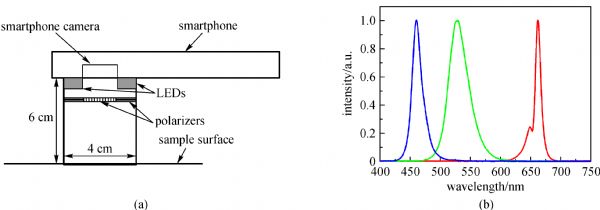
Abstract This paper briefly reviews the operational principles and designs of portable in vivo skin imaging prototypes developed at the Biophotonics Laboratory of the Institute of Atomic Physics and Spectroscopy, University of Latvia. Four types of imaging devices are presented. Multi-spectral imagers ensure distant mapping of specific skin parameters (e.g., distribution of skin chromophores). Autofluorescence photobleaching rate imagers show potential for skin tumor assessment and margin delineation. Photoplethysmography video-imagers remotely detect cutaneous blood pulsations and provide real-time information on the human cardiovascular state. Multimodal skin imagers perform the above-mentioned functions by acquiring several spectral and video images using the same image sensor.
|
| Keywords
multispectral skin imaging
autofluorescence photobleaching
remote photoplethysmography
|
|
Corresponding Author(s):
Janis SPIGULIS
|
|
Just Accepted Date: 04 July 2017
Online First Date: 21 August 2017
Issue Date: 26 September 2017
|
|
| 1 |
Spigulis J. Biophotonic technologies for noninvasive assessment of skin condition and blood microcirculation. Latvian Journal of Physics and Technical Sciences 2012, 49(5): 63–80
|
| 2 |
( accessed on 12.03.2017)
|
| 3 |
Jakovels D, Spigulis J, Rogule L. RGB mapping of hemoglobin distribution in skin. Proceedings of the Society for Photo-Instrumentation Engineers, 2011, 8087: 80872B
https://doi.org/10.1117/12.889665
|
| 4 |
Jakovels D, Kuzmina I, Berzina A, Valeine L, Spigulis J. Noncontact monitoring of vascular lesion phototherapy efficiency by RGB multispectral imaging. Journal of Biomedical Optics, 2013, 18(12): 126019
https://doi.org/10.1117/1.JBO.18.12.126019
pmid: 24362928
|
| 5 |
Jakovels D, Spigulis J. 2-D mapping of skin chromophores in the spectral range 500−700 nm. Journal of Biophotonics, 2010, 3(3): 125–129
https://doi.org/10.1002/jbio.200910069
pmid: 19894217
|
| 6 |
Jakovels D, Spigulis J. RGB imaging device for mapping and monitoring of hemoglobin distribution in skin. Lithuanian Journal of Physics, 2012, 52(1): 50–54
https://doi.org/10.3952/physics.v52i1.2267
|
| 7 |
Philips Vital Signs Camera. ( accessed on 12.03.2017)
|
| 8 |
The best heart disease iPhone & Android Apps of the year. ( accessed on 12.03.2017)
|
| 9 |
Skinvision. ( accessed on 12.03.2017)
|
| 10 |
Spigulis J, Lacis M, Kuzmina I, Lihacovs A, Upmalis V, Rupenheits Z. Method and device for smartphone mapping of tissue compounds. WO 2017/012675 A1, 2017
|
| 11 |
Kuzmina I, Lacis M, Spigulis J, Berzina A, Valeine L. Study of smartphone suitability for mapping of skin chromophores. Journal of Biomedical Optics, 2015, 20(9): 090503
https://doi.org/10.1117/1.JBO.20.9.090503
pmid: 26405818
|
| 12 |
( accessed on 12.03.2017)
|
| 13 |
( accessed on 12.03.2017)
|
| 14 |
Diebele I, Kuzmina I, Lihachev A, Kapostinsh J, Derjabo A, Valeine L, Spigulis J. Clinical evaluation of melanomas and common nevi by spectral imaging. Biomedical Optics Express, 2012, 3(3): 467–472
https://doi.org/10.1364/BOE.3.000467
pmid: 22435095
|
| 15 |
Bekina A, Diebele I, Rubins U, Zaharans J, Derjabo A, Spigulis J. Multispectral assessment of skin malformations by modified video-microscope. Latvian Journal of Physics and Technical Sciences, 2012, 49(5): 4–8
|
| 16 |
Bekina A, Rubins U, Lihacova I, Zaharans J, Spigulis J. Skin chromophore mapping by means of a modified video-microscope for skin malformation diagnosis. Proceedings of the Society for Photo-Instrumentation Engineers, 2013, 8856: 88562G
https://doi.org/10.1117/12.2023960
|
| 17 |
Rubins U, Zaharans J, Lihacova I, Spigulis J. Multispectral video-microscope modified for skin diagnostics. Latvian Journal of Physics and Technical Sciences, 2014, 51(5): 65–70
|
| 18 |
Spigulis J, Elste L. Method and device for imaging of spectral reflectance at several wavelength bands. WO2013135311 (A1), 2012
|
| 19 |
Spigulis J, Jakovels D, Rubins U. Multi-spectral skin imaging by a consumer photo-camera. Proceedings of the Society for Photo-Instrumentation Engineers, 2010, 7557: 75570M
https://doi.org/10.1117/12.845492
|
| 20 |
Spigulis J, Oshina I. Snapshot RGB mapping of skin melanin and hemoglobin. Journal of Biomedical Optics, 2015, 20(5): 050503
https://doi.org/10.1117/1.JBO.20.5.050503
pmid: 25992844
|
| 21 |
Spigulis J, Oshina I, Berzina A, Bykov A. Smartphone snapshot mapping of skin chromophores under triple-wavelength laser illumination. Journal of Biomedical Optics, 2017, 22(9): 091508
https://doi.org/10.1117/1.JBO.22.9.091508
pmid: 28253387
|
| 22 |
Prahl S. Tabulated molar extinction coefficient for hemoglobin in water. ( accessed 30 November 2016)
|
| 23 |
Sarna T, Swartz H M. The physical properties of melanin. ( accessed 30 November 2016)
|
| 24 |
Spigulis J, Elste L. Single-snapshot RGB multispectral imaging at fixed wavelengths: proof of concept. Proceedings of the Society for Photo-Instrumentation Engineers, 2014, 8937: 89370L
https://doi.org/10.1117/12.2039442
|
| 25 |
Spigulis J, Oshina I. Method and device for chromophore mapping under illumination by several spectral lines. LV patent 15106 B, 2016
|
| 26 |
Rubins U, Kviesis-Kipge E, Spigulis J. Device for obtaining speckle-free images at illumination by scattered laser beams. LV patent application P-17–17, 2017
|
| 27 |
Oshina I, Spigulis J, Rubins U, Kviesis-Kipge E, Lauberts K. Express RGB mapping of three to five skin chromophores. OSA Technical Digests, 2017 (ECBO Proceedings, Munich, in press)
|
| 28 |
Lihachev A, Lesins Jh D, Jakovels J, Spigulis. Low power cw-laser signatures on human skin. Quantum Electronics, 2011, 40(12): 1077–1080
https://doi.org/10.1070/QE2010v040n12ABEH014470
|
| 29 |
Stratonnikov A A, Polikarpov V S, Loschenov V B. Photobleaching of endogenous fluorochroms in tissues in vivo during laser irradiation. Proceedings of the Society for Photo-Instrumentation Engineers, 2001, 4241: 13–24
https://doi.org/10.1117/12.431555
|
| 30 |
Lesinsh J, Lihachev A, Rudys R, Bagdonas S, Spigulis J. Skin autofluorescence photobleaching and photo-memory. Proceedings of the Society for Photo-Instrumentation Engineers, 2011, 8092: 80920N
https://doi.org/10.1117/12.889861
|
| 31 |
Spigulis J, Lihachev A, Erts R. Imaging of laser-excited tissue autofluorescence bleaching rates. Applied Optics, 2009, 48(10): D163–D168
https://doi.org/10.1364/AO.48.00D163
pmid: 19340105
|
| 32 |
Lihachev A, Derjabo A, Ferulova I, Lange M, Lihacova I, Spigulis J. Autofluorescence imaging of basal cell carcinoma by smartphone RGB camera. Journal of Biomedical Optics, 2015, 20(12): 120502
https://doi.org/10.1117/1.JBO.20.12.120502
pmid: 26662298
|
| 33 |
Allen J. Photoplethysmography and its application in clinical physiological measurement. Physiological Measurement, 2007, 28(3): R1–R39
https://doi.org/10.1088/0967-3334/28/3/R01
pmid: 17322588
|
| 34 |
Spigulis J. Optical noninvasive monitoring of skin blood pulsations. Applied Optics, 2005, 44(10): 1850–1857
https://doi.org/10.1364/AO.44.001850
pmid: 15813522
|
| 35 |
Rubins U, Upmalis V, Rubenis O, Jakovels D, Spigulis J. Real-time photoplethysmography imaging system. Proceedings of IFMBE, 2011, 34: 183–186
|
| 36 |
Rubins U, Spigulis J, Miscuks A. Photoplethysmography imaging algorithm for continuous monitoring of regional anesthesia. In: Proceedings of the 14th ACM/IEEE Symposium on Embedded Systems for Real-Time Multimedia, ESTIMedia'16. 2016: 67–71
|
| 37 |
Rubins U, Spigulis J, Miscuks A. Application of color magnification technique for revealing skin microcircuration changes under regional anaesthetic input. Proceedings of the Society for Photo-Instrumentation Engineers, 2013, 9032: 903203
https://doi.org/10.1117/12.2044574
|
| 38 |
Spigulis J, Gailite L, Lihachev A, Erts R. Simultaneous recording of skin blood pulsations at different vascular depths by multiwavelength photoplethysmography. Applied Optics, 2007, 46(10): 1754–1759
https://doi.org/10.1364/AO.46.001754
pmid: 17356618
|
| 39 |
Marcinkevics Z, Rubins U, Zaharans J, Miscuks A, Urtane E, Ozolina-Moll L. Imaging photoplethysmography for clinical assessment of cutaneous microcirculation at two different depths. Journal of Biomedical Optics, 2016, 21(3): 035005
https://doi.org/10.1117/1.JBO.21.3.035005
pmid: 27027825
|
| 40 |
Spigulis J, Garancis V, Rubins U, Zaharans E, Zaharans J, Elste L. A device for multimodal imaging of skin. Proceedings of the Society for Photo-Instrumentation Engineers, 2013, 8574: 85740J
https://doi.org/10.1117/12.2003510
|
| 41 |
Spigulis J, Rubins U, Kviesis-Kipge E, Rubenis O. SkImager: a concept device for in-vivo skin assessment by multimodal imaging. Proceedings of the Estonian Academy of Sciences, 2014, 63(3): 213–220
https://doi.org/10.3176/proc.2014.3.02
|
| 42 |
Embedded linux on board computer decsription, ( accessed on 12.03.2017)
|
| 43 |
Industrial USB cameras description, ( accessed on 12.03.2017)
|
| 44 |
Bliznuks D, Jakovels D, Saknite I, Spigulis J. Mobile platform for online processing of multimodal skin optical images: using online Matlab server for processing remission, fluorescence and laser speckle images, obtained by using novel handheld device. In: Proceedings of BioPhotonics 2015 (Florence). 2015: 7304024
|
| 45 |
Jakovels D, Saknite I, Bliznuks D, Spigulis J, Kadikis R. Benign-atypical nevi discrimination using diffuse reflectance and fluorescence multispectral imaging system. In: Proceedings of BioPhotonics 2015 (Florence). 2015: 7304026
|
|
Viewed |
|
|
|
Full text
|
|
|
|
|
Abstract
|
|
|
|
|
Cited |
|
|
|
|
| |
Shared |
|
|
|
|
| |
Discussed |
|
|
|
|