|
|
|
Multifunctional modification of Fe3O4 nanoparticles for diagnosis and treatment of diseases: A review |
Miao QIN1, Mengjie XU1, Lulu NIU3, Yizhu CHENG1, Xiaolian NIU1, Jinlong KONG1, Xiumei ZHANG1, Yan WEI1,2, Di HUANG1,2( ) ) |
1. Department of Biomedical Engineering, Research Center for Nano-biomaterials & Regenerative Medicine, College of Biomedical Engineering, Taiyuan University of Technology, Taiyuan 030024, China
2. Shanxi Key Laboratory of Material Strength & Structural Impact, Institute of Biomedical Engineering, Taiyuan University of Technology, Taiyuan 030024, China
3. Wellead Medical Co. Ltd., Guangzhou 511434, China |
|
|
|
|
Abstract With the rapid improvements in nanomaterials and imaging technology, great progresses have been made in diagnosis and treatment of diseases during the past decades. Fe3O4 magnetic nanoparticles (MNPs) with good biocompatibility and superparamagnetic property are usually used as contrast agent for diagnosis of diseases in magnetic resonance imaging (MRI). Currently, the combination of multiple imaging technologies has been considered as new tendency in diagnosis and treatment of diseases, which could enhance the accuracy and reliability of disease diagnosis and provide new strategies for disease treatment. Therefore, novel contrast agents used for multifunctional imaging are urgently needed. Fe3O4 MNPs are believed to be a potential candidate for construction of multifunctional platform in diagnosis and treatment of diseases. In recent years, there are a plethora of studies concerning the construction of multifunctional platform presented based on Fe3O4 MNPs. In this review, we introduce fabrication methods and modification strategies of Fe3O4 MNPs, expecting great improvements for diagnosis and treatment of diseases in the future.
|
| Keywords
Fe3O4 MNPs
preparation methods
modification strategy
multifunctional platform
|
|
Corresponding Author(s):
Di HUANG
|
|
Online First Date: 07 February 2021
Issue Date: 11 March 2021
|
|
| 1 |
N Lewinski, V Colvin, R Drezek. Cytotoxicity of nanoparticles. Small, 2008, 4(1): 26–49
https://doi.org/10.1002/smll.200700595
pmid: 18165959
|
| 2 |
P Formoso, R Muzzalupo, L Tavano, et al.. Nanotechnology for the environment and medicine. Mini-Reviews in Medicinal Chemistry, 2016, 16(8): 668–675
https://doi.org/10.2174/1389557515666150709105129
pmid: 26955878
|
| 3 |
N Hoshyar, S Gray, H B Han, et al.. The effect of nanoparticle size on in vivo pharmacokinetics and cellular interaction. Nanomedicine, 2016, 11(6): 673–692
https://doi.org/10.2217/nnm.16.5
pmid: 27003448
|
| 4 |
O C Farokhzad, R Langer. Impact of nanotechnology on drug delivery. ACS Nano, 2009, 3(1): 16–20
https://doi.org/10.1021/nn900002m
pmid: 19206243
|
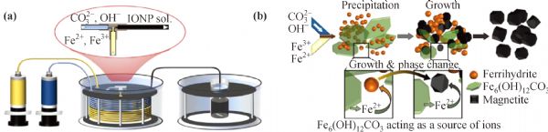
| 5 |
S C Baetke, T Lammers, F Kiessling. Applications of nanoparticles for diagnosis and therapy of cancer. The British Journal of Radiology, 2015, 88(1054): 20150207
https://doi.org/10.1259/bjr.20150207
pmid: 25969868
|
| 6 |
J Gupta. Nanotechnology applications in medicine and dentistry. Journal of Investigative and Clinical Dentistry, 2011, 2(2): 81–88
https://doi.org/10.1111/j.2041-1626.2011.00046.x
pmid: 25426600
|
| 7 |
A Zhang, C M Lieber. Nano-bioelectronics. Chemical Reviews, 2016, 116(1): 215–257
https://doi.org/10.1021/acs.chemrev.5b00608
pmid: 26691648
|
| 8 |
M R Angle, B Cui, N A Melosh. Nanotechnology and neurophysiology. Current Opinion in Neurobiology, 2015, 32: 132–140
https://doi.org/10.1016/j.conb.2015.03.014
pmid: 25889532
|
| 9 |
A Noy. Bionanoelectronics. Advanced Materials, 2011, 23(7): 807–820
https://doi.org/10.1002/adma.201003751
pmid: 21328478
|
| 10 |
F D Guerra, M F Attia, D C Whitehead, et al.. Nanotechnology for environmental remediation: Materials and applications. Molecules, 2018, 23(7): 1760
https://doi.org/10.3390/molecules23071760
pmid: 30021974
|
| 11 |
X Hu, J Xu, C Wu, et al.. Ethylenediamine grafted to graphene oxide@Fe3O4 for chromium(VI) decontamination: Performance, modelling, and fractional factorial design. PLoS One, 2017, 12(10): e0187166
https://doi.org/10.1371/journal.pone.0187166
pmid: 29084287
|
| 12 |
S Ma, S Zhan, Y Jia, et al.. Superior antibacterial activity of Fe3O4–TiO2 nanosheets under solar light. ACS Applied Materials & Interfaces, 2015, 7(39): 21875–21883
https://doi.org/10.1021/acsami.5b06264
pmid: 26372171
|
| 13 |
R Sadeghi, R J Rodriguez, Y Yao, et al.. Advances in nanotechnology as they pertain to food and agriculture: Benefits and risks. Annual Review of Food Science and Technology, 2017, 8(1): 467–492
https://doi.org/10.1146/annurev-food-041715-033338
pmid: 28125343
|
| 14 |
I Iavicoli, V Leso, D H Beezhold, et al.. Nanotechnology in agriculture: Opportunities, toxicological implications, and occupational risks. Toxicology and Applied Pharmacology, 2017, 329: 96–111
https://doi.org/10.1016/j.taap.2017.05.025
pmid: 28554660
|
| 15 |
G Das, J K Patra, S Paramithiotis, et al.. The sustainability challenge of food and environmental nanotechnology: Current status and imminent perceptions. International Journal of Environmental Research and Public Health, 2019, 16(23): 4848
https://doi.org/10.3390/ijerph16234848
pmid: 31810271
|
| 16 |
M Rossi, D Passeri, A Sinibaldi, et al.. Nanotechnology for food packaging and food quality assessment. Advances in Food and Nutrition Research, 2017, 82: 149–204
https://doi.org/10.1016/bs.afnr.2017.01.002
pmid: 28427532
|
| 17 |
M Wei, W D Le. The role of nanomaterials in autophagy. Advances in Experimental Medicine and Biology, 2019, 1206: 273–286
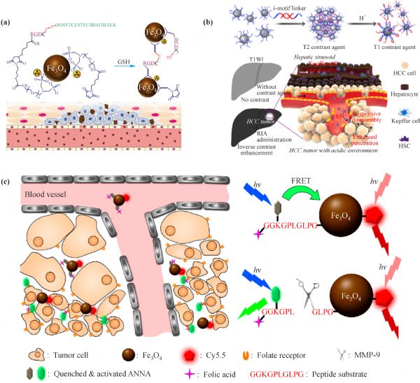
https://doi.org/10.1007/978-981-15-0602-4_14
pmid: 31776991
|
| 18 |
R Mohammadinejad, M A Moosavi, S Tavakol, et al.. Necrotic, apoptotic and autophagic cell fates triggered by nanoparticles. Autophagy, 2019, 15(1): 4–33
https://doi.org/10.1080/15548627.2018.1509171
pmid: 30160607
|
| 19 |
K El-Boubbou. Magnetic iron oxide nanoparticles as drug carriers: Preparation, conjugation and delivery. Nanomedicine, 2018, 13(8): 929–952
https://doi.org/10.2217/nnm-2017-0320
pmid: 29546817
|
| 20 |
C Song, W Sun, Y Xiao, et al.. Ultrasmall iron oxide nanoparticles: Synthesis, surface modification, assembly, and biomedical applications. Drug Discovery Today, 2019, 24(3): 835–844
https://doi.org/10.1016/j.drudis.2019.01.001
pmid: 30639557
|
| 21 |
A K Gupta, M Gupta. Synthesis and surface engineering of iron oxide nanoparticles for biomedical applications. Biomaterials, 2005, 26(18): 3995–4021
https://doi.org/10.1016/j.biomaterials.2004.10.012
pmid: 15626447
|
| 22 |
Y Yuan, Z Ding, J Qian, et al.. Casp3/7-instructed intracellular aggregation of Fe3O4 nanoparticles enhances T2 MR imaging of tumor apoptosis. Nano Letters, 2016, 16(4): 2686–2691
https://doi.org/10.1021/acs.nanolett.6b00331
pmid: 27031226
|
| 23 |
Y Chen, Q Zhou, X Li, et al.. Ultrasmall paramagnetic iron oxide nanoprobe targeting epidermal growth factor receptor for in vivo magnetic resonance imaging of hepatocellular carcinoma. Bioconjugate Chemistry, 2017, 28(11): 2794–2803
https://doi.org/10.1021/acs.bioconjchem.7b00501
pmid: 28972742
|
| 24 |
J Huang, L Wang, X Zhong, et al.. Facile non-hydrothermal synthesis of oligosaccharides coated sub-5 nm magnetic iron oxide nanoparticles with dual MRI contrast enhancement effect. Journal of Materials Chemistry B: Materials for Biology and Medicine, 2014, 2(33): 5344–5351
https://doi.org/10.1039/C4TB00811A
pmid: 25181490
|
| 25 |
P Martinkova, M Brtnicky, J Kynicky, et al.. Iron oxide nanoparticles: Innovative tool in cancer diagnosis and therapy. Advanced Healthcare Materials, 2018, 7(5): 1700932
https://doi.org/10.1002/adhm.201700932
pmid: 29205944
|
| 26 |
R Qiao, Q Jia, J Zeng, et al.. Magnetic iron oxide nanoparticles and their applications in magnetic resonance imaging. Acta Biophysica Sinica, 2011, 27(4): 272–288 (in Chinese)
https://doi.org/10.3724/SP.J.1260.2011.00272
|
| 27 |
R Das, N Rinaldi-Montes, J Alonso, et al.. Boosted hyperthermia therapy by combined ac magnetic and photothermal exposures in Ag/Fe3O4 nanoflowers. ACS Applied Materials & Interfaces, 2016, 8(38): 25162–25169
https://doi.org/10.1021/acsami.6b09942
pmid: 27589410
|
| 28 |
O S Nielsen, M Horsman, J Overgaard. A future for hyperthermia in cancer treatment? European Journal of Cancer, 2001, 37(13): 1587–1589
https://doi.org/10.1016/S0959-8049(01)00193-9
pmid: 11527682
|
| 29 |
T Vangijzegem, D Stanicki, S Laurent. Magnetic iron oxide nanoparticles for drug delivery: Applications and characteristics. Expert Opinion on Drug Delivery, 2019, 16(1): 69–78
https://doi.org/10.1080/17425247.2019.1554647
pmid: 30496697
|
| 30 |
R Tietze, J Zaloga, H Unterweger, et al.. Magnetic nanoparticle-based drug delivery for cancer therapy. Biochemical and Biophysical Research Communications, 2015, 468(3): 463–470
https://doi.org/10.1016/j.bbrc.2015.08.022
pmid: 26271592
|
| 31 |
X Hu, M Ma, M Zeng, et al.. Supercritical carbon dioxide anchored Fe3O4 nanoparticles on graphene foam and lithium battery performance. ACS Applied Materials & Interfaces, 2014, 6(24): 22527–22533
https://doi.org/10.1021/am5066255
pmid: 25438281
|
| 32 |
V Harnchana, S Chaiyachad, S Pimanpang, et al.. Hierarchical Fe3O4-reduced graphene oxide nanocomposite grown on NaCl crystals for triiodide reduction in dye-sensitized solar cells. Scientific Reports, 2019, 9: 1494
https://doi.org/10.1038/s41598-018-38050-z
pmid: 30728432
|
| 33 |
T Niemiec, M Dudek, N Dziekan, et al.. The method of coating Fe3O4 with carbon nanoparticles to modify biological properties of oxide measured in vitro. Journal of AOAC International, 2017, 100(4): 905–915
https://doi.org/10.5740/jaoacint.17-0165
pmid: 28623660
|
| 34 |
S S Chen, H Xu, H J Xu, et al.. A facile ultrasonication assisted method for Fe3O4@SiO2–Ag nanospheres with excellent antibacterial activity. Dalton Transactions, 2015, 44(19): 9140–9148
https://doi.org/10.1039/C5DT00977D
pmid: 25901793
|
| 35 |
Z Jiao, Y Zhang, H Fan. Ultrasonic-microwave method in preparation of polypyrrole-coated magnetic particles for vitamin D extraction in milk. Journal of Chromatography A, 2016, 1457: 7–13
https://doi.org/10.1016/j.chroma.2016.06.041
pmid: 27371018
|
| 36 |
H Montaseri, S Alipour, M A Vakilinezhad. Development, evaluation and optimization of superparamagnetite nanoparticles prepared by co-precipitation method. Research in Pharmaceutical Sciences, 2017, 12(4): 274–282
https://doi.org/10.4103/1735-5362.212044
pmid: 28855938
|
| 37 |
J Park, K An, Y Hwang, et al.. Ultra-large-scale syntheses of monodisperse nanocrystals. Nature Materials, 2004, 3(12): 891–895
https://doi.org/10.1038/nmat1251
pmid: 15568032
|
| 38 |
X Yu, G Cheng, M D Zhou, et al.. On-demand one-step synthesis of monodisperse functional polymeric microspheres with droplet microfluidics. Langmuir, 2015, 31(13): 3982–3992
https://doi.org/10.1021/acs.langmuir.5b00617
pmid: 25782525
|
| 39 |
P Li, L Li, Y Zhao, et al.. Selective binding and magnetic separation of histidine-tagged proteins using Fe3O4/Cu-apatite nanoparticles. Journal of Inorganic Biochemistry, 2016, 156: 49–54
https://doi.org/10.1016/j.jinorgbio.2015.12.017
pmid: 26773852
|
| 40 |
A H Lu, E L Salabas, F Schüth. Magnetic nanoparticles: Synthesis, protection, functionalization, and application. Angewandte Chemie International Edition in English, 2007, 46(8): 1222–1244
https://doi.org/10.1002/anie.200602866
pmid: 17278160
|
| 41 |
D Ling, N Lee, T Hyeon. Chemical synthesis and assembly of uniformly sized iron oxide nanoparticles for medical applications. Accounts of Chemical Research, 2015, 48(5): 1276– 1285
https://doi.org/10.1021/acs.accounts.5b00038
pmid: 25922976
|
| 42 |
J Park, N R Kadasala, S A Abouelmagd, et al.. Polymer–iron oxide composite nanoparticles for EPR-independent drug delivery. Biomaterials, 2016, 101: 285–295
https://doi.org/10.1016/j.biomaterials.2016.06.007
pmid: 27310916
|
| 43 |
W J Syu, C C Huang, J K Hsiao, et al.. Co-precipitation synthesis of near-infrared iron oxide nanocrystals on magnetically targeted imaging and photothermal cancer therapy via photoablative protein denature. Nanotheranostics, 2019, 3(3): 236–254
https://doi.org/10.7150/ntno.24124
pmid: 31263656
|
| 44 |
L Gan, Z Lu, D Cao, et al.. Effects of cetyltrimethylammonium bromide on the morphology of green synthesized Fe3O4 nanoparticles used to remove phosphate. Materials Science and Engineering C, 2018, 82: 41–45
https://doi.org/10.1016/j.msec.2017.08.073
pmid: 29025673
|
| 45 |
H Wang, X Zhao, W Meng, et al.. Cetyltrimethylammonium bromide-coated Fe3O4 magnetic nanoparticles for analysis of 15 trace polycyclic aromatic hydrocarbons in aquatic environments by ultraperformance, liquid chromatography with fluorescence detection. Analytical Chemistry, 2015, 87(15): 7667–7675
https://doi.org/10.1021/acs.analchem.5b01077
pmid: 26153060
|
| 46 |
H Nosrati, M Salehiabar, H K Manjili, et al.. Preparation of magnetic albumin nanoparticles via a simple and one-pot desolvation and co-precipitation method for medical and pharmaceutical applications. International Journal of Biological Macromolecules, 2018, 108: 909–915
https://doi.org/10.1016/j.ijbiomac.2017.10.180
pmid: 29101048
|
| 47 |
M Anbarasu, M Anandan, E Chinnasamy, et al.. Synthesis and characterization of polyethylene glycol (PEG) coated Fe3O4 nanoparticles by chemical co-precipitation method for biomedical applications. Spectrochimica Acta Part A: Molecular Spectroscopy, 2015, 135: 536–539
https://doi.org/10.1016/j.saa.2014.07.059
pmid: 25123943
|
| 48 |
C Li. A targeted approach to cancer imaging and therapy. Nature Materials, 2014, 13(2): 110–115
https://doi.org/10.1038/nmat3877
pmid: 24452345
|
| 49 |
A P LaGrow, M O Besenhard, A Hodzic, et al.. Unravelling the growth mechanism of the co-precipitation of iron oxide nanoparticles with the aid of synchrotron X-ray diffraction in solution. Nanoscale, 2019, 11(14): 6620–6628
https://doi.org/10.1039/C9NR00531E
pmid: 30896010
|
| 50 |
Y W Jun, Y M Huh, J S Choi, et al.. Nanoscale size effect of magnetic nanocrystals and their utilization for cancer diagnosis via magnetic resonance imaging. Journal of the American Chemical Society, 2005, 127(16): 5732–5733
https://doi.org/10.1021/ja0422155
pmid: 15839639
|
| 51 |
R Hufschmid, H Arami, R M Ferguson, et al.. Synthesis of phase-pure and monodisperse iron oxide nanoparticles by thermal decomposition. Nanoscale, 2015, 7(25): 11142–11154
https://doi.org/10.1039/C5NR01651G
pmid: 26059262
|
| 52 |
H Guo, Y Zhang, W Liang, et al.. An inorganic magnetic fluorescent nanoprobe with favorable biocompatibility for dual-modality bioimaging and drug delivery. Journal of Inorganic Biochemistry, 2019, 192: 72–81
https://doi.org/10.1016/j.jinorgbio.2018.12.002
pmid: 30612028
|
| 53 |
F B Effenberger, R A Couto, P K Kiyohara, et al.. Economically attractive route for the preparation of high quality magnetic nanoparticles by the thermal decomposition of iron(III) acetylacetonate. Nanotechnology, 2017, 28(11): 115603
https://doi.org/10.1088/1361-6528/aa5ab0
pmid: 28192283
|
| 54 |
V Patsula, L Kosinová, M Lovrić, et al.. Superparamagnetic Fe3O4 nanoparticles: synthesis by thermal decomposition of iron(III) glucuronate and application in magnetic resonance imaging. ACS Applied Materials & Interfaces, 2016, 8(11): 7238–7247
https://doi.org/10.1021/acsami.5b12720
pmid: 26928653
|
| 55 |
I A Barbosa, P C de Sousa Filho, D L da Silva, et al.. Metalloporphyrins immobilized in Fe3O4@SiO2 mesoporous submicrospheres: Reusable biomimetic catalysts for hydrocarbon oxidation. Journal of Colloid and Interface Science, 2016, 469: 296–309
https://doi.org/10.1016/j.jcis.2016.01.059
pmid: 26897566
|
| 56 |
S Mumtaz, S Wang, S Z Hussain, et al.. Dopamine coated Fe3O4 nanoparticles as enzyme mimics for the sensitive detection of bacteria. Chemical Communications, 2017, 53(91): 12306–12308
https://doi.org/10.1039/C7CC07149C
pmid: 29094116
|
| 57 |
Y Liu, D L Purich, C Wu, et al.. Ionic functionalization of hydrophobic colloidal nanoparticles to form ionic nanoparticles with enzyme like properties. Journal of the American Chemical Society, 2015, 137(47): 14952–14958
https://doi.org/10.1021/jacs.5b08533
pmid: 26562739
|
| 58 |
X Wang, J Zhuang, Q Peng, et al.. A general strategy for nanocrystal synthesis. Nature, 2005, 437(7055): 121–124
https://doi.org/10.1038/nature03968
pmid: 16136139
|
| 59 |
H Cai, X An, J Cui, et al.. Facile hydrothermal synthesis and surface functionalization of polyethyleneimine-coated iron oxide nanoparticles for biomedical applications. ACS Applied Materials & Interfaces, 2013, 5(5): 1722–1731
https://doi.org/10.1021/am302883m
pmid: 23388099
|
| 60 |
R P Bagwe, J R Kanicky, B J Palla, et al.. Improved drug delivery using microemulsions: rationale, recent progress, and new horizons. Critical Reviews in Therapeutic Drug Carrier Systems, 2001, 18(1): 77–140
pmid: 11326744
|
| 61 |
F Bai, D Wang, Z Huo, et al.. A versatile bottom-up assembly approach to colloidal spheres from nanocrystals. Angewandte Chemie International Edition, 2007, 46(35): 6650–6653
https://doi.org/10.1002/anie.200701355
pmid: 17661295
|
| 62 |
Z L Liu, X Wang, K Yao, et al.. Synthesis of magnetite nanoparticles in W/O microemulsion. Journal of Materials Science, 2004, 39(7): 2633–2636
https://doi.org/10.1023/B:JMSC.0000020046.68106.22
|
| 63 |
L Zhuang, W Zhang, Y Zhao, et al.. Preparation and characterization of Fe3O4 particles with novel nanosheets morphology and magnetochromatic property by a modified solvothermal method. Scientific Reports, 2015, 5(1): 9320
https://doi.org/10.1038/srep09320
pmid: 25799320
|
| 64 |
T A Lastovina, A P Budnyk, E A Kudryavtsev, et al.. Solvothermal synthesis of Sm3+-doped Fe3O4 nanoparticles. Materials Science and Engineering C, 2017, 80: 110–116
https://doi.org/10.1016/j.msec.2017.05.087
pmid: 28866144
|
| 65 |
L Gao, Y Tang, C Wang, et al.. Highly-efficient amphiphilic magnetic nanocomposites based on a simple sol–gel modification for adsorption of phthalate esters. Journal of Colloid and Interface Science, 2019, 552: 142–152
https://doi.org/10.1016/j.jcis.2019.05.031
pmid: 31121431
|
| 66 |
Y Zeng, R Hao, B Xing, et al.. One-pot synthesis of Fe3O4 nanoprisms with controlled electrochemical properties. Chemical Communications, 2010, 46(22): 3920–3922
https://doi.org/10.1039/c0cc00246a
pmid: 20428516
|
| 67 |
E Carenza, V Barceló, A Morancho, et al.. Rapid synthesis of water-dispersible superparamagnetic iron oxide nanoparticles by a microwave-assisted route for safe labeling of endothelial progenitor cells. Acta Biomaterialia, 2014, 10(8): 3775–3785
https://doi.org/10.1016/j.actbio.2014.04.010
pmid: 24755438
|
| 68 |
D Shan, S Deng, T Zhao, et al.. Preparation of ultrafine magnetic biochar and activated carbon for pharmaceutical adsorption and subsequent degradation by ball milling. Journal of Hazardous Materials, 2016, 305: 156–163
https://doi.org/10.1016/j.jhazmat.2015.11.047
pmid: 26685062
|
| 69 |
M Chang, Y J Chang, P Y Chao, et al.. Exosome purification based on PEG-coated Fe3O4 nanoparticles. PLoS One, 2018, 13(6): e0199438
https://doi.org/10.1371/journal.pone.0199438
pmid: 29933408
|
| 70 |
N Shahabadi, M Falsafi, K Mansouri. Improving antiproliferative effect of the anticancer drug cytarabine on human promyelocytic leukemia cells by coating on Fe3O4@SiO2 nanoparticles. Colloids and Surfaces B: Biointerfaces, 2016, 141: 213–222
https://doi.org/10.1016/j.colsurfb.2016.01.054
pmid: 26852105
|
| 71 |
C Achilli, S Grandi, G F Guidetti, et al.. Fe3O4@SiO2 core–shell nanoparticles for biomedical purposes: Adverse effects on blood cells. Biomaterials Science, 2016, 4(10): 1417–1421
https://doi.org/10.1039/C6BM00374E
pmid: 27517098
|
| 72 |
Y Wei, G Yin, C Ma, et al.. Synthesis and cellular compatibility of biomineralized Fe3O4 nanoparticles in tumor cells targeting peptides. Colloids and Surfaces B: Biointerfaces, 2013, 107: 180–188
https://doi.org/10.1016/j.colsurfb.2013.01.058
pmid: 23500729
|
| 73 |
G Wang, W Gao, X Zhang, et al.. Au nanocage functionalized with ultra-small Fe3O4 nanoparticles for targeting T1–T2 dual MRI and CT imaging of tumor. Scientific Reports, 2016, 6: 28258
https://doi.org/10.1038/srep28258
pmid: 27312564
|
| 74 |
M Felber, R Alberto. 99mTc radiolabelling of Fe3O4–Au core–shell and Au–Fe3O4 dumbbell-like nanoparticles. Nanoscale, 2015, 7(15): 6653–6660
https://doi.org/10.1039/C5NR00269A
pmid: 25797603
|
| 75 |
M Gou, S Li, L Zhang, et al.. Facile one-pot synthesis of carbon/calcium phosphate/Fe3O4 composite nanoparticles for simultaneous imaging and pH/NIR-responsive drug delivery. Chemical Communications, 2016, 52(74): 11068–11071
https://doi.org/10.1039/C6CC05515J
pmid: 27561158
|
| 76 |
S Shen, B Ding, S Zhang, et al.. Near-infrared light-responsive nanoparticles with thermosensitive yolk–shell structure for multimodal imaging and chemo-photothermal therapy of tumor. Nanomedicine, 2017, 13(5): 1607–1616
https://doi.org/10.1016/j.nano.2017.02.014
pmid: 28285157
|
| 77 |
S Nayak, L A Lyon. Soft nanotechnology with soft nanoparticles. Angewandte Chemie International Edition, 2005, 44(47): 7686–7708
https://doi.org/10.1002/anie.200501321
pmid: 16283684
|
| 78 |
Z Tang, X Zhao, T Zhao, et al.. Magnetic nanoparticles interaction with humic acid: In the presence of surfactants. Environmental Science & Technology, 2016, 50(16): 8640– 8648
https://doi.org/10.1021/acs.est.6b01749
pmid: 27404337
|
| 79 |
B Dutta, N G Shetake, B K Barick, et al.. pH sensitive surfactant-stabilized Fe3O4 magnetic nanocarriers for dual drug delivery. Colloids and Surfaces B: Biointerfaces, 2018, 162: 163–171
https://doi.org/10.1016/j.colsurfb.2017.11.054
pmid: 29190467
|
| 80 |
C Justin, A V Samrot, D P Sruthi, et al.. Preparation, characterization and utilization of core/shell super paramagnetic iron oxide nanoparticles for curcumin delivery. PLoS One, 2018, 13(7): e0200440
https://doi.org/10.1371/journal.pone.0200440
pmid: 30021002
|
| 81 |
H K Can, S Kavlak, S ParviziKhosroshahi, et al.. Preparation, characterization and dynamical mechanical properties of dextran-coated iron oxide nanoparticles (DIONPs). Artificial Cells Nanomedicine and Biotechnology, 2018, 46(2): 421–431
https://doi.org/10.1080/21691401.2017.1315428
pmid: 28423951
|
| 82 |
M Barrow, A Taylor, J C Carrión, et al.. Co-precipitation of DEAE-dextran coated SPIONs: How synthesis conditions affect particle properties, stem cell labelling and MR contrast. Contrast Media & Molecular Imaging, 2016, 11(5): 362–370
https://doi.org/10.1002/cmmi.1700
pmid: 27358113
|
| 83 |
D Wu, L Zhu, Y Li, et al.. Superparamagnetic chitosan nanocomplexes for colorectal tumor-targeted delivery of irinotecan. International Journal of Pharmaceutics, 2020, 584: 119394
https://doi.org/10.1016/j.ijpharm.2020.119394
pmid: 32407940
|
| 84 |
P I P Soares, D Machado, C Laia, et al.. Thermal and magnetic properties of chitosan–iron oxide nanoparticles. Carbohydrate Polymers, 2016, 149: 382–390
https://doi.org/10.1016/j.carbpol.2016.04.123
pmid: 27261762
|
| 85 |
X Zhang, Y Wang, S Yang. Simultaneous removal of Co(II) and 1-naphthol by core–shell structured Fe3O4@cyclodextrin magnetic nanoparticles. Carbohydrate Polymers, 2014, 114: 521–529
https://doi.org/10.1016/j.carbpol.2014.08.072
pmid: 25263922
|
| 86 |
J Xie, J Huang, X Li, et al.. Iron oxide nanoparticle platform for biomedical applications. Current Medicinal Chemistry, 2009, 16(10): 1278–1294
https://doi.org/10.2174/092986709787846604
pmid: 19355885
|
| 87 |
F Wang, X Li, W Li, et al.. Dextran coated Fe3O4 nanoparticles as a near-infrared laser-driven photothermal agent for efficient ablation of cancer cells in vitro and in vivo. Materials Science and Engineering C, 2018, 90: 46–56
https://doi.org/10.1016/j.msec.2018.04.030
pmid: 29853113
|
| 88 |
F Assa, H Jafarizadeh-Malmiri, H Ajamein, et al.. Chitosan magnetic nanoparticles for drug delivery systems. Critical Reviews in Biotechnology, 2017, 37(4): 492–509
https://doi.org/10.1080/07388551.2016.1185389
pmid: 27248312
|
| 89 |
H Xing, S Zhang, W Bu, et al.. Ultrasmall NaGdF4 nanodots for efficient MR angiography and atherosclerotic plaque imaging. Advanced Materials, 2014, 26(23): 3867–3872
https://doi.org/10.1002/adma.201305222
pmid: 24677351
|
| 90 |
A K Gupta, S Wells. Surface-modified superparamagnetic nanoparticles for drug delivery: Preparation, characterization, and cytotoxicity studies. IEEE Transactions on Nanobioscience, 2004, 3(1): 66–73
https://doi.org/10.1109/TNB.2003.820277
pmid: 15382647
|
| 91 |
G Yang, W Ma, B Zhang, et al.. The labeling of stem cells by superparamagnetic iron oxide nanoparticles modified with PEG/PVP or PEG/PEI. Materials Science and Engineering C, 2016, 62: 384–390
https://doi.org/10.1016/j.msec.2016.01.090
pmid: 26952437
|
| 92 |
Y Chen, F Zhang, Q Wang, et al.. The synthesis of LA-Fe3O4@PDA-PEG-DOX for photothermal therapy-chemotherapy. Dalton Transactions, 2018, 47(7): 2435–2443
https://doi.org/10.1039/C7DT04080F
pmid: 29379913
|
| 93 |
L Wang, M Wang, B Zhou, et al.. PEGylated reduced-graphene oxide hybridized with Fe3O4 nanoparticles for cancer photothermal-immunotherapy. Journal of Materials Chemistry B: Materials for Biology and Medicine, 2019, 7(46): 7406–7414
https://doi.org/10.1039/C9TB00630C
pmid: 31710067
|
| 94 |
C Yang, W Guo, L Cui, et al.. pH-responsive magnetic core–shell nanocomposites for drug delivery. Langmuir, 2014, 30(32): 9819–9827
https://doi.org/10.1021/la501833u
pmid: 25073728
|
| 95 |
H Wei, N Insin, J Lee, et al.. Compact zwitterion-coated iron oxide nanoparticles for biological applications. Nano Letters, 2012, 12(1): 22–25
https://doi.org/10.1021/nl202721q
pmid: 22185195
|
| 96 |
Y Liu, T Chen, C Wu, et al.. Facile surface functionalization of hydrophobic magnetic nanoparticles. Journal of the American Chemical Society, 2014, 136(36): 12552–12555
https://doi.org/10.1021/ja5060324
pmid: 25140614
|
| 97 |
J J Wang, K F Lei, F Han. Tumor microenvironment: Recent advances in various cancer treatments. European Review for Medical and Pharmacological Sciences, 2018, 22(12): 3855–3864
pmid: 29949179
|
| 98 |
M A Zaimy, N Saffarzadeh, A Mohammadi, et al.. New methods in the diagnosis of cancer and gene therapy of cancer based on nanoparticles. Cancer Gene Therapy, 2017, 24(6): 233–243
https://doi.org/10.1038/cgt.2017.16
pmid: 28574057
|
| 99 |
L Li, F Gao, W Jiang, et al.. Folic acid-conjugated superparamagnetic iron oxide nanoparticles for tumor-targeting MR imaging. Drug Delivery, 2016, 23(5): 1726–1733
https://doi.org/10.3109/10717544.2015.1006404 PMID:25715808
|
| 100 |
H Choi, S R Choi, R Zhou, et al.. Iron oxide nanoparticles as magnetic resonance contrast agent for tumor imaging via folate receptor-targeted delivery. Academic Radiology, 2004, 11(9): 996–1004
https://doi.org/10.1016/j.acra.2004.04.018
pmid: 15350580
|
| 101 |
J Yang, Y Luo, Y Xu, et al.. Conjugation of iron oxide nanoparticles with RGD-modified dendrimers for targeted tumor MR imaging. ACS Applied Materials & Interfaces, 2015, 7(9): 5420–5428
https://doi.org/10.1021/am508983n
pmid: 25695661
|
| 102 |
Y Luo, J Yang, Y Yan, et al.. RGD-functionalized ultrasmall iron oxide nanoparticles for targeted T1-weighted MR imaging of gliomas. Nanoscale, 2015, 7(34): 14538–14546
https://doi.org/10.1039/C5NR04003E
pmid: 26260703
|
| 103 |
H Zhang, J Li, Y Hu, et al.. Folic acid-targeted iron oxide nanoparticles as contrast agents for magnetic resonance imaging of human ovarian cancer. Journal of Ovarian Research, 2016, 9(1): 19
https://doi.org/10.1186/s13048-016-0230-2
pmid: 27025582
|
| 104 |
Y Cui, C Zhang, R Luo, et al.. Noninvasive monitoring of early antiangiogenic therapy response in human nasopharyngeal carcinoma xenograft model using MRI with RGD-conjugated ultrasmall superparamagnetic iron oxide nanoparticles. International Journal of Nanomedicine, 2016, 11: 5671–5682
https://doi.org/10.2147/IJN.S115357
pmid: 27895477
|
| 105 |
E Hirata, E Sahai. Tumor microenvironment and differential responses to therapy. Cold Spring Harbor Perspectives in Medicine, 2017, 7(7): a026781
https://doi.org/10.1101/cshperspect.a026781
pmid: 28213438
|
| 106 |
T Wu, Y Dai. Tumor microenvironment and therapeutic response. Cancer Letters, 2017, 387: 61–68
https://doi.org/10.1016/j.canlet.2016.01.043
pmid: 26845449
|
| 107 |
H Li, Y Zhao, Y Jia, et al.. Covalently assembled dopamine nanoparticle as an intrinsic photosensitizer and pH-responsive nanocarrier for potential application in anticancer therapy. Chemical Communications, 2019, 55(100): 15057–15060
https://doi.org/10.1039/C9CC08294H
pmid: 31777882
|
| 108 |
E Li, Y Yang, G Hao, et al.. Multifunctional magnetic mesoporous silica nanoagents for in vivo enzyme-responsive drug delivery and MR imaging. Nanotheranostics, 2018, 2(3): 233–242
https://doi.org/10.7150/ntno.25565
pmid: 29868348
|
| 109 |
T Yang, D Niu, J Chen, et al.. Biodegradable organosilica magnetic micelles for magnetically targeted MRI and GSH-triggered tumor chemotherapy. Biomaterials Science, 2019, 7(7): 2951–2960
https://doi.org/10.1039/C9BM00342H
pmid: 31099352
|
| 110 |
Z Gao, Y Hou, J Zeng, et al.. Tumor microenvironment-triggered aggregation of antiphagocytosis 99mTc-labeled Fe3O4 nanoprobes for enhanced tumor imaging in vivo. Advanced Materials, 2017, 29(24): 1701095
https://doi.org/10.1002/adma.201701095
pmid: 28402594
|
| 111 |
J Lu, J Sun, F Li, et al.. Highly sensitive diagnosis of small hepatocellular carcinoma using pH-responsive iron oxide nanocluster assemblies. Journal of the American Chemical Society, 2018, 140(32): 10071–10074
https://doi.org/10.1021/jacs.8b04169
pmid: 30059219
|
| 112 |
T Ma, Y Hou, J Zeng, et al.. Dual-ratiometric target-triggered fluorescent probe for simultaneous quantitative visualization of tumor microenvironment protease activity and pH in vivo. Journal of the American Chemical Society, 2018, 140(1): 211–218
https://doi.org/10.1021/jacs.7b08900
pmid: 29237264
|
| 113 |
M Qin, Y Peng, M Xu, et al.. Uniform Fe3O4/Gd2O3-DHCA nanocubes for dual-mode magnetic resonance imaging. Beilstein Journal of Nanotechnology, 2020, 11: 1000–1009
https://doi.org/10.3762/bjnano.11.84
pmid: 32704462
|
| 114 |
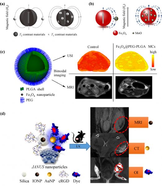
Z Zhou, D Huang, J Bao, et al.. A synergistically enhanced T1–T2 dual-modal contrast agent. Advanced Materials, 2012, 24(46): 6223–6228
https://doi.org/10.1002/adma.201203169
pmid: 22972529
|
| 115 |
C Lu, P Dong, L Pi, et al.. Hydroxyl-PEG-phosphonic acid-stabilized superparamagnetic manganese oxide-doped iron oxide nanoparticles with synergistic effects for dual-mode MR imaging. Langmuir, 2019, 35(29): 9474–9482
https://doi.org/10.1021/acs.langmuir.9b00736
pmid: 31241339
|
| 116 |
S Xu, F Yang, X Zhou, et al.. Uniform PEGylated PLGA microcapsules with embedded Fe3O4 nanoparticles for US/MR dual-modality imaging. ACS Applied Materials & Interfaces, 2015, 7(36): 20460–20468
https://doi.org/10.1021/acsami.5b06594
pmid: 26327472
|
| 117 |
X Cui, D Mathe, N Kovács, et al.. Synthesis, characterization, and application of core–shell Co0.16Fe2.84O4@NaYF4(Yb, Er) and Fe3O4@NaYF4(Yb, Tm) nanoparticle as trimodal (MRI, PET/SPECT, and optical) imaging agents. Bioconjugate Chemistry, 2016, 27(2): 319–328
https://doi.org/10.1021/acs.bioconjchem.5b00338
pmid: 26172432
|
| 118 |
A Sánchez, K Ovejero Paredes, J Ruiz-Cabello, et al.. Hybrid decorated core@shell Janus nanoparticles as a flexible platform for targeted multimodal molecular bioimaging of cancer. ACS Applied Materials & Interfaces, 2018, 10(37): 31032–31043
https://doi.org/10.1021/acsami.8b10452
pmid: 30141615
|
| 119 |
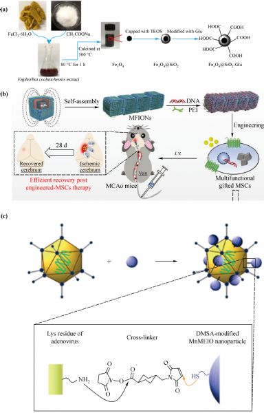
W, Cai M, Guo X Weng, et al.. Modified green synthesis of Fe3O4@SiO2 nanoparticles for pH responsive drug release. Materials Science and Engineering C, 2020, 112: 110900
https://doi.org/10.1016/j.msec.2020.110900
|
| 120 |
T Y Zhang, F Y Li, Q H Xu, et al.. Ferrimagnetic nanochains-based mesenchymal stem cell engineering for highly efficient post-stroke recovery. Advanced Functional Materials, 2019, 29(24): 1900603
https://doi.org/10.1002/adfm.201900603
|
| 121 |
Y M Huh, E S Lee, J H Lee, et al.. Hybrid nanoparticles for magnetic resonance imaging of target-specific viral gene delivery. Advanced Materials, 2007, 19(20): 3109–3112
https://doi.org/10.1002/adma.200701952
|
| 122 |
O K Arriortua, M Insausti, L Lezama, et al.. RGD-functionalized Fe3O4 nanoparticles for magnetic hyperthermia. Colloids and Surfaces B: Biointerfaces, 2018, 165: 315–324
https://doi.org/10.1016/j.colsurfb.2018.02.031
pmid: 29501962
|
| 123 |
C Xu, Y Zheng, W Gao, et al.. Magnetic hyperthermia ablation of tumors using injectable Fe3O4/calcium phosphate cement. ACS Applied Materials & Interfaces, 2015, 7(25): 13866–13875
https://doi.org/10.1021/acsami.5b02077
pmid: 26065316
|
| 124 |
L Li, S Fu, C Chen, et al.. Microenvironment-driven bioelimination of magnetoplasmonic nanoassemblies and their multimodal imaging-guided tumor photothermal therapy. ACS Nano, 2016, 10(7): 7094–7105
https://doi.org/10.1021/acsnano.6b03238
pmid: 27309678
|
| 125 |
Y Liu, Z Yang, X Huang, et al.. Glutathione-responsive self-assembled magnetic gold nanowreath for enhanced tumor imaging and imaging-guided photothermal therapy. ACS Nano, 2018, 12(8): 8129–8137
https://doi.org/10.1021/acsnano.8b02980
pmid: 30001110
|
|
Viewed |
|
|
|
Full text
|
|
|
|
|
Abstract
|
|
|
|
|
Cited |
|
|
|
|
| |
Shared |
|
|
|
|
| |
Discussed |
|
|
|
|