|
|
|
Functional ferritin nanoparticles for biomedical applications |
Zhantong Wang1,2, Haiyan Gao1, Yang Zhang1, Gang Liu1( ), Gang Niu2, Xiaoyuan Chen2() ), Gang Niu2, Xiaoyuan Chen2() |
1. State Key Laboratory of Molecular Vaccinology and Molecular Diagnostics & Center for Molecular Imaging and Translational Medicine, School of Public Health, Xiamen University, Xiamen 361102, China
2. Laboratory of Molecular Imaging and Nanomedicine (LOMIN), National Institute of Biomedical Imaging and Bioengineering (NIBIB), National Institutes of Health, Bethesda, MD 20892, USA |
|
|
|
|
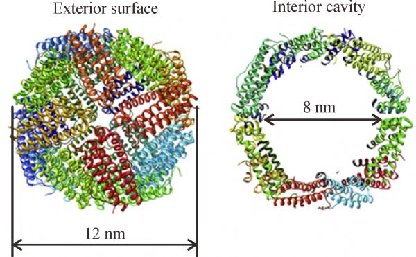
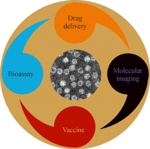
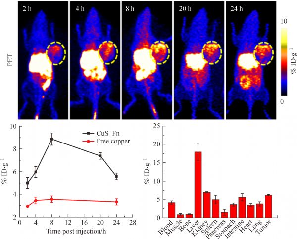
Abstract Ferritin, a major iron storage protein with a hollow interior cavity, has been reported recently to play many important roles in biomedical and bioengineering applications. Owing to the unique architecture and surface properties, ferritin nanoparticles offer favorable characteristics and can be either genetically or chemically modified to impart functionalities to their surfaces, and therapeutics or probes can be encapsulated in their interiors by controlled and reversible assembly/disassembly. There has been an outburst of interest regarding the employment of functional ferritin nanoparticles in nanomedicine. This review will highlight the recent advances in ferritin nanoparticles for drug delivery, bioassay, and molecular imaging with a particular focus on their biomedical applications.
|
| Keywords
nanomedicine
ferritin
drug delivery
bioassay
molecular imaging
|
|
Corresponding Author(s):
Gang Liu,Xiaoyuan Chen
|
|
Just Accepted Date: 05 January 2017
Online First Date: 15 February 2017
Issue Date: 06 November 2017
|
|
| 1 |
Worwood M, Cook J D. Serum ferritin. Critical Reviews in Clinical Laboratory Sciences, 1979, 10(2): 171–204
https://doi.org/10.3109/10408367909147133
|
| 2 |
Meldrum F C, Heywood B R, Mann S. Magnetoferritin: In vitro synthesis of a novel magnetic protein. Science, 1992, 257(5069): 522–523
https://doi.org/10.1126/science.1636086
|
| 3 |
Zeth K, Hoiczyk E, Okuda M. Ferroxidase-mediated iron oxide biomineralization: Novel pathways to multifunctional nanoparticles. Trends in Biochemical Sciences, 2016, 41(2): 190–203
https://doi.org/10.1016/j.tibs.2015.11.011
|
| 4 |
Chasteen N D, Harrison P M. Mineralization in ferritin: An efficient means of iron storage. Journal of Structural Biology, 1999, 126(3): 182–194
https://doi.org/10.1006/jsbi.1999.4118
|
| 5 |
Uchida M, Kang S, Reichhardt C, Harlen K, Douglas T. The ferritin superfamily: Supramolecular templates for materials synthesis. Biochimica et Biophysica Acta, 2010, 1800(8): 834–845
https://doi.org/10.1016/j.bbagen.2009.12.005
|
| 6 |
Bulte J W, Douglas T, Mann S, Frankel R B, Moskowitz B M, Brooks R A, Baumgarner C D, Vymazal J, Strub M P, Frank J A. Magnetoferritin: Characterization of a novel superparamagnetic MR contrast agent. Journal of Magnetic Resonance Imaging, 1994, 4(3): 497–505
https://doi.org/10.1002/jmri.1880040343
|
| 7 |
Uchida M, Flenniken M L, Allen M, Willits D A, Crowley B E, Brumfield S, Willis A F, Jackiw L, Jutila M, Young M J, Douglas T. Targeting of cancer cells with ferrimagnetic ferritin cage nanoparticles. Journal of the American Chemical Society, 2006, 128(51): 16626–16633
https://doi.org/10.1021/ja0655690
|
| 8 |
Okuda M, Iwahori K, Yamashita I, Yoshimura H. Fabrication of nickel and chromium nanoparticles using the protein cage of apoferritin. Biotechnology and Bioengineering, 2003, 84(2): 187–194
https://doi.org/10.1002/bit.10748
|
| 9 |
Galvez N, Sanchez P, Dominguez-Vera J M. Preparation of Cu and CuFe prussian blue derivative nanoparticles using the apoferritin cavity as nanoreactor. Dalton Transactions (Cambridge, England), 2005, 15(15): 2492–2494
https://doi.org/10.1039/b506290j
|
| 10 |
Jeong G H, Yamazaki A, Suzuki S, Yoshimura H, Kobayashi Y, Homma Y. Cobalt-filled apoferritin for suspended single-walled carbon nanotube growth with narrow diameter distribution. Journal of the American Chemical Society, 2005, 127(23): 8238–8239
https://doi.org/10.1021/ja0505144
|
| 11 |
Fan R, Chew S W, Cheong V V, Orner B P. Fabrication of gold nanoparticles inside unmodified horse spleen apoferritin. Small, 2010, 6(14): 1483–1487
https://doi.org/10.1002/smll.201000457
|
| 12 |
Zhen Z, Tang W, Chen H, Lin X, Todd T, Wang G, Cowger T, Chen X, Xie J. RGD-modified apoferritin nanoparticles for efficient drug delivery to tumors. ACS Nano, 2013, 7(6): 4830–4837
https://doi.org/10.1021/nn305791q
|
| 13 |
Zhen Z, Tang W, Guo C, Chen H, Lin X, Liu G, Fei B, Chen X, Xu B, Xie J. Ferritin nanocages to encapsulate and deliver photosensitizers for efficient photodynamic therapy against cancer. ACS Nano, 2013, 7(8): 6988–6996
https://doi.org/10.1021/nn402199g
|
| 14 |
Tian Y, Yan X, Saha M L, Niu Z, Stang P J. Hierarchical self-assembly of responsive organoplatinum(ii) metallacycle-TMV complexes with turn-on fluorescence. Journal of the American Chemical Society, 2016, 138(37): 12033–12036
https://doi.org/10.1021/jacs.6b07402
|
| 15 |
Harrison P M, Arosio P. The ferritins: Molecular properties, iron storage function and cellular regulation. Biochimica et Biophysica Acta, 1996, 1275(3): 161–203
https://doi.org/10.1016/0005-2728(96)00022-9
|
| 16 |
Lin X, Xie J, Niu G, Zhang F, Gao H, Yang M, Quan Q, Aronova M A, Zhang G, Lee S, et al. Chimeric ferritin nanocages for multiple function loading and multimodal imaging. Nano Letters, 2011, 11(2): 814–819
https://doi.org/10.1021/nl104141g
|
| 17 |
Yamashita I, Iwahori K, Kumagai S. Ferritin in the field of nanodevices. Biochimica et Biophysica Acta, 2010, 1800(8): 846–857
https://doi.org/10.1016/j.bbagen.2010.03.005
|
| 18 |
Jolley C C, Uchida M, Reichhardt C, Harrington R, Kang S, Klem M T, Parise J B, Douglas T. Size and crystallinity in protein-templated inorganic nanoparticles. Chemistry of Materials, 2010, 22(16): 4612–4618
https://doi.org/10.1021/cm100657w
|
| 19 |
Zhang L, Swift J, Butts C A, Yerubandi V, Dmochowski I J. Structure and activity of apoferritin-stabilized gold nanoparticles. Journal of Inorganic Biochemistry, 2007, 101(11-12): 1719–1729
https://doi.org/10.1016/j.jinorgbio.2007.07.023
|
| 20 |
Rother M, Nussbaumer M G, Renggli K, Bruns N. Protein cages and synthetic polymers: A fruitful symbiosis for drug delivery applications, bionanotechnology and materials science. Chemical Society Reviews, 2016, 45(22): 6213–6249
https://doi.org/10.1039/C6CS00177G
|
| 21 |
Ghirlando R, Mutskova R, Schwartz C. Enrichment and characterization of ferritin for nanomaterial applications. Nanotechnology, 2016, 27(4): 045102
https://doi.org/10.1088/0957-4484/27/4/045102
|
| 22 |
Konijn A, Meyron-Holtz E, Levy R, Ben-Bassat H, Matzner Y. Specific binding of placental acidic isoferritin to cells of the T-cell line HD-MAR. FEBS Letters, 1990, 263(2): 229–232
https://doi.org/10.1016/0014-5793(90)81380-7
|
| 23 |
Bretscher M S, Thomson J N. Distribution of ferritin receptors and coated pits on giant Hela cells. EMBO Journal, 1983, 2(4): 599–603
|
| 24 |
Lei Y, Hamada Y, Li J, Cong L, Wang N, Li Y, Zheng W, Jiang X. Targeted tumor delivery and controlled release of neuronal drugs with ferritin nanoparticles to regulate pancreatic cancer progression. Journal of Controlled Release, 2016, 232: 131–142
https://doi.org/10.1016/j.jconrel.2016.03.023
|
| 25 |
Zhao Y, Liang M, Li X, Fan K, Xiao J, Li Y, Shi H, Wang F, Choi H S, Cheng D, et al. Bioengineered magnetoferritin nanoprobes for single-dose nuclear-magnetic resonance tumor imaging. ACS Nano, 2016, 10(4): 4184–4191
https://doi.org/10.1021/acsnano.5b07408
|
| 26 |
Adams P C, Powell L W, Halliday J W. Isolation of a human hepatic ferritin receptor. Hepatology (Baltimore, MD.), 1988, 8(4): 719–721
https://doi.org/10.1002/hep.1840080402
|
| 27 |
Chen X. Multimodality imaging of tumor integrin alphavbeta3 expression. Mini-Reviews in Medicinal Chemistry, 2006, 6(2): 227–233
https://doi.org/10.2174/138955706775475975
|
| 28 |
Liu Y, Wang Z, Zhang H, Lang L, Ma Y, He Q, Lu N, Huang P, Song J, Liu Z, et al. A photothermally responsive nanoprobe for bioimaging based on edman degradation. Nanoscale, 2016, 8(20): 10553–10557
https://doi.org/10.1039/C6NR01400C
|
| 29 |
Kitagawa T, Kosuge H, Uchida M, Dua M M, Iida Y, Dalman R L, Douglas T, McConnell M V. Rgd-conjugated human ferritin nanoparticles for imaging vascular inflammation and angiogenesis in experimental carotid and aortic disease. Molecular Imaging & Biology, 2012, 14(3): 315–324
https://doi.org/10.1007/s11307-011-0495-1
|
| 30 |
Choi H S, Nasr K, Alyabyev S, Feith D, Lee J H, Kim S H, Ashitate Y, Hyun H, Patonay G, Strekowski L, et al. Synthesis and in vivo fate of zwitterionic near—infrared fluorophores. Angewandte Chemie International Edition, 2011, 50(28): 6258–6263
https://doi.org/10.1002/anie.201102459
|
| 31 |
Agostinis P, Berg K, Cengel K A, Foster T H, Girotti A W, Gollnick S O, Hahn S M, Hamblin M R, Juzeniene A, Kessel D, et al. Photodynamic therapy of cancer: An update. CA: a Cancer Journal for Clinicians, 2011, 61(4): 250–281
https://doi.org/10.3322/caac.20114
|
| 32 |
Ochsner M. Photophysical and photobiological processes in the photodynamic therapy of tumours. Journal of Photochemistry and Photobiology. B, Biology, 1997, 39(1): 1–18
https://doi.org/10.1016/S1011-1344(96)07428-3
|
| 33 |
Brown S B, Brown E A, Walker I. The present and future role of photodynamic therapy in cancer treatment. Lancet Oncology, 2004, 5(8): 497–508
https://doi.org/10.1016/S1470-2045(04)01529-3
|
| 34 |
Cairnduff F, Stringer M R, Hudson E J, Ash D V, Brown S B. Superficial photodynamic therapy with topical 5-aminolaevulinic acid for superficial primary and secondary skin cancer. British Journal of Cancer, 1994, 69(3): 605–608
https://doi.org/10.1038/bjc.1994.112
|
| 35 |
Falvo E, Tremante E, Fraioli R, Leonetti C, Zamparelli C, Boffi A, Morea V, Ceci P, Giacomini P. Antibody-drug conjugates: Targeting melanoma with cisplatin encapsulated in protein-cage nanoparticles based on human ferritin. Nanoscale, 2013, 5(24): 12278–12285
https://doi.org/10.1039/c3nr04268e
|
| 36 |
MacKie R. Melanoma prevention and early detection. British Medical Bulletin, 1995, 51(3): 570–583
|
| 37 |
Siegel R, Naishadham D, Jemal A. Cancer statistics, 2012. CA: a Cancer Journal for Clinicians, 2012, 62(1): 10–29
https://doi.org/10.3322/caac.20138
|
| 38 |
Greenlee R T, Murray T, Bolden S, Wingo P A. Cancer statistics, 2000. CA: a Cancer Journal for Clinicians, 2000, 50(1): 7–33
https://doi.org/10.3322/canjclin.50.1.7
|
| 39 |
Rigel D S, Carucci J A. Malignant melanoma: Prevention, early detection, and treatment in the 21st century. CA: a Cancer Journal for Clinicians, 2000, 50(4): 215–236
https://doi.org/10.3322/canjclin.50.4.215
|
| 40 |
Morgenstern D A, Asher R A, Fawcett J W. Chondroitin sulphate proteoglycans in the CNS injury response. Progress in Brain Research, 2002, 137: 313–332
https://doi.org/10.1016/S0079-6123(02)37024-9
|
| 41 |
Eisenmann K M, McCarthy J B, Simpson M A, Keely P J, Guan J L, Tachibana K, Lim L, Manser E, Furcht L T, Iida J. Melanoma chondroitin sulphate proteoglycan regulates cell spreading through Cdc42, Ack-1 and p130cas. Nature Cell Biology, 1999, 1(8): 507–513
https://doi.org/10.1038/70302
|
| 42 |
Thon N, Haas C A, Rauch U, Merten T, Fässler R, Frotscher M, Deller T. The chondroitin sulphate proteoglycan brevican is upregulated by astrocytes after entorhinal cortex lesions in adult rats. European Journal of Neuroscience, 2000, 12(7): 2547–2558
https://doi.org/10.1046/j.1460-9568.2000.00109.x
|
| 43 |
Levine J, Nishiyama A. The NG2 chondroitin sulfate proteoglycan: A multifunctional proteoglycan associated with immature cells. Perspectives on Developmental Neurobiology, 1996, 3(4): 245–259
|
| 44 |
Oohira A, Matsui F, Watanabe E, Kushima Y, Maeda N. Developmentally regulated expression of a brain specific species of chondroitin sulfate proteoglycan, neurocan, identified with a monoclonal antibody LG2 in the rat cerebrum. Neuroscience, 1994, 60(1): 145–157
https://doi.org/10.1016/0306-4522(94)90210-0
|
| 45 |
Levine J M, Stallcup W B. Plasticity of developing cerebellar cells in vitro studied with antibodies against the NG2 antigen. Journal of Neuroscience, 1987, 7(9): 2721–2731
|
| 46 |
Maeda H, Wu J, Sawa T, Matsumura Y, Hori K. Tumor vascular permeability and the EPR effect in macromolecular therapeutics: A review. Journal of Controlled Release, 2000, 65(1): 271–284
https://doi.org/10.1016/S0168-3659(99)00248-5
|
| 47 |
Mamo T, Poland G A. Nanovaccinology: The next generation of vaccines meets 21st century materials science and engineering. Vaccine, 2012, 30(47): 6609–6611
https://doi.org/10.1016/j.vaccine.2012.08.023
|
| 48 |
des Rieux A, Fievez V, Garinot M, Schneider Y J, Préat V. Nanoparticles as potential oral delivery systems of proteins and vaccines: A mechanistic approach. Journal of Controlled Release, 2006, 116(1): 1–27
https://doi.org/10.1016/j.jconrel.2006.08.013
|
| 49 |
Singh M, Chakrapani A, O’Hagan D. Nanoparticles and microparticles as vaccine-delivery systems. Expert Review of Vaccines, 2007, 6(5): 797–808
https://doi.org/10.1586/14760584.6.5.797
|
| 50 |
Oyewumi M O, Kumar A, Cui Z. Nano-microparticles as immune adjuvants: Correlating particle sizes and the resultant immune responses. Expert Review of Vaccines, 2010, 9(9): 1095–1107
https://doi.org/10.1586/erv.10.89
|
| 51 |
Zhao L, Seth A, Wibowo N, Zhao C X, Mitter N, Yu C, Middelberg A P. Nanoparticle vaccines. Vaccine, 2014, 32(3): 327–337
https://doi.org/10.1016/j.vaccine.2013.11.069
|
| 52 |
Zhao K, Chen G, Shi X, Gao T, Li W, Zhao Y, Zhang F, Wu J, Cui X, Wang Y F. Preparation and efficacy of a live newcastle disease virus vaccine encapsulated in chitosan nanoparticles. PLoS One, 2012, 7(12): e53314
https://doi.org/10.1371/journal.pone.0053314
|
| 53 |
Borges O, Cordeiro-da-Silva A, Tavares J, Santarém N, de Sousa A, Borchard G, Junginger H E. Immune response by nasal delivery of hepatitis B surface antigen and codelivery of a CpG ODN in alginate coated chitosan nanoparticles. European Journal of Pharmaceutics and Biopharmaceutics, 2008, 69(2): 405–416
https://doi.org/10.1016/j.ejpb.2008.01.019
|
| 54 |
Nochi T, Yuki Y, Takahashi H, Sawada S, Mejima M, Kohda T, Harada N, Kong I G, Sato A, Kataoka N, et al. Nanogel antigenic protein-delivery system for adjuvant-free intranasal vaccines. Nature Materials, 2010, 9(7): 572–578
https://doi.org/10.1038/nmat2784
|
| 55 |
Stone J W, Thornburg N J, Blum D L, Kuhn S J, Wright D W, Crowe J E Jr. Gold nanorod vaccine for respiratory syncytial virus. Nanotechnology, 2013, 24(29): 295102
https://doi.org/10.1088/0957-4484/24/29/295102
|
| 56 |
Wang T, Zou M, Jiang H, Ji Z, Gao P, Cheng G. Synthesis of a novel kind of carbon nanoparticle with large mesopores and macropores and its application as an oral vaccine adjuvant. European Journal of Pharmaceutical Sciences, 2011, 44(5): 653–659
https://doi.org/10.1016/j.ejps.2011.10.012
|
| 57 |
Glück R, Moser C, Metcalfe I C. Influenza virosomes as an efficient system for adjuvanted vaccine delivery. Expert Opinion on Biological Therapy, 2004, 4(7): 1139–1145
https://doi.org/10.1517/14712598.4.7.1139
|
| 58 |
Zhu F C, Zhang J, Zhang X F, Zhou C, Wang Z Z, Huang S J, Wang H, Yang C L, Jiang H M, Cai J P, et al. Efficacy and safety of a recombinant hepatitise vaccine in healthy adults: A large-scale, randomised, double-blind placebo-controlled, phase 3 trial. Lancet, 2010, 376(9744): 895–902
https://doi.org/10.1016/S0140-6736(10)61030-6
|
| 59 |
Sliepen K, Ozorowski G, Burger J A, van Montfort T, Stunnenberg M, LaBranche C, Montefiori D C, Moore J P, Ward A B, Sanders R W. Presenting native-like HIV-1 envelope trimers on ferritin nanoparticles improves their immunogenicity. Retrovirology, 2015, 12(82): 15–21
|
| 60 |
Champion C I, Kickhoefer V A, Liu G, Moniz R J, Freed A S, Bergmann L L, Vaccari D, Raval-Fernandes S, Chan A M, Rome L H, Kelly K A. A vault nanoparticle vaccine induces protective mucosal immunity. PLoS One, 2009, 4(4): e5409
https://doi.org/10.1371/journal.pone.0005409
|
| 61 |
Kanekiyo M, Wei C J, Yassine H M, McTamney P M, Boyington J C, Whittle J R, Rao S S, Kong W P, Wang L, Nabel G J. Self-assembling influenza nanoparticle vaccines elicit broadly neutralizing H1N1 antibodies. Nature, 2013, 499(7456): 102–106
https://doi.org/10.1038/nature12202
|
| 62 |
Cho K J, Shin H J, Lee J H, Kim K J, Park S S, Lee Y, Lee C, Park S S, Kim K H. The crystal structure of ferritin from helicobacter pylori reveals unusual conformational changes for iron uptake. Journal of Molecular Biology, 2009, 390(1): 83–98
https://doi.org/10.1016/j.jmb.2009.04.078
|
| 63 |
Steinman R M. Decisions about dendritic cells: Past, present, and future. Annual Review of Immunology, 2012, 30(1): 1–22
https://doi.org/10.1146/annurev-immunol-100311-102839
|
| 64 |
Gilboa E. DC-based cancer vaccines. Journal of Clinical Investigation, 2007, 117(5): 1195–1203
https://doi.org/10.1172/JCI31205
|
| 65 |
Aarntzen E, Figdor C, Adema G, Punt C, De Vries I. Dendritic cell vaccination and immune monitoring. Cancer Immunology, Immunotherapy, 2008, 57(10): 1559–1568
https://doi.org/10.1007/s00262-008-0553-y
|
| 66 |
Han J A, Kang Y J, Shin C, Ra J S, Shin H H, Hong S Y, Do Y, Kang S. Ferritin protein cage nanoparticles as versatile antigen delivery nanoplatforms for dendritic cell (DC)-based vaccine development. Nanomedicine; Nanotechnology, Biology, and Medicine, 2013, 10(3): 561–569
https://doi.org/10.1016/j.nano.2013.11.003
|
| 67 |
Shimonkevitz R, Colon S, Kappler J W, Marrack P, Grey H M. Antigen recognition by H-2-restricted T cells. II. A tryptic ovalbumin peptide that substitutes for processed antigen. Journal of Immunology (Baltimore, MD: 1950), 1984, 133(4): 2067–2074
|
| 68 |
Liu D, Wang Z, Jin A, Huang X, Sun X, Wang F, Yan Q, Ge S, Xia N, Niu G, Liu G, Hight Walker A R, Chen X. Acetylcholinesterase—catalyzed hydrolysis allows ultrasensitive detection of pathogens with the naked eye. Angewandte Chemie International Edition in English, 2013, 52(52): 14065–14069
https://doi.org/10.1002/anie.201307952
|
| 69 |
Lee S H, Lee H, Park J S, Choi H, Han K Y, Seo H S, Ahn K Y, Han S S, Cho Y, Lee K H, et al. A novel approach to ultrasensitive diagnosis using supramolecular protein nanoparticles. FASEB Journal, 2007, 21(7): 1324–1334
https://doi.org/10.1096/fj.06-7303com
|
| 70 |
Abbaspour A, Noori A. Electrochemical detection of individual single nucleotide polymorphisms using monobase-modified apoferritin-encapsulated nanoparticles. Biosensors & Bioelectronics, 2012, 37(1): 11–18
https://doi.org/10.1016/j.bios.2012.04.017
|
| 71 |
Tang Z, Wu H, Zhang Y, Li Z, Lin Y. Enzyme-mimic activity of ferric nano-core residing in ferritin and its biosensing applications. Analytical Chemistry, 2011, 83(22): 8611–8616
https://doi.org/10.1021/ac202049q
|
| 72 |
Men D, Zhang T T, Hou L W, Zhou J, Zhang Z P, Shi Y Y, Zhang J L, Cui Z Q, Deng J Y, Wang D B, et al. Self-assembly of ferritin nanoparticles into an enzyme nanocomposite with tunable size for ultrasensitive immunoassay. ACS Nano, 2015, 9(11): 10852–10860
https://doi.org/10.1021/acsnano.5b03607
|
| 73 |
Liu G, Wang J, Wu H, Lin Y. Versatile apoferritin nanoparticle labels for assay of protein. Analytical Chemistry, 2006, 78(21): 7417–7423
https://doi.org/10.1021/ac060653j
|
| 74 |
Liu G, Wu H, Wang J, Lin Y. Apoferritin-templated synthesis of metal phosphate nanoparticle labels for electrochemical immunoassay. Small, 2006, 2(10): 1139–1143
https://doi.org/10.1002/smll.200600206
|
| 75 |
Beutler B, Cerami A. Cachectin and tumour necrosis factor as two sides of the same biological coin. Nature, 1986, 320(6063): 584–588
https://doi.org/10.1038/320584a0
|
| 76 |
Scuderi P, Lam K, Ryan K, Petersen E, Sterling K, Finley P, Ray C G, Slymen D, Salmon S. Raised serum levels of tumour necrosis factor in parasitic infections. Lancet, 1986, 328(8520): 1364–1365
https://doi.org/10.1016/S0140-6736(86)92007-6
|
| 77 |
Yu F, Li G, Qu B, Cao W. Electrochemical detection of DNA hybridization based on signal DNA probe modified with Au and apoferritin nanoparticles. Biosensors & Bioelectronics, 2010, 26(3): 1114–1117
https://doi.org/10.1016/j.bios.2010.08.018
|
| 78 |
Li L, Fang C J, Ryan J C, Niemi E C, Lebrón J A, Björkman P J, Arase H, Torti F M, Torti S V, Nakamura M C, et al. Binding and uptake of H-ferritin are mediated by human transferrin receptor-1. Proceedings of the National Academy of Sciences of the United States of America, 2010, 107(8): 3505–3510
https://doi.org/10.1073/pnas.0913192107
|
| 79 |
Fan K, Cao C, Pan Y, Lu D, Yang D, Feng J, Song L, Liang M, Yan X. Magnetoferritin nanoparticles for targeting and visualizing tumour tissues. Nature Nanotechnology, 2012, 7(7): 459–464
https://doi.org/10.1038/nnano.2012.90
|
| 80 |
Lee E J, Ahn K Y, Lee J H, Park J S, Song J A, Sim S J, Lee E B, Cha Y J, Lee J. A novel bioassay platform using ferritin-based nanoprobe hydrogel. Advanced Materials, 2012, 24(35): 4739–4744
https://doi.org/10.1002/adma.201200728
|
| 81 |
Zhao J, Liu M, Zhang Y, Li H, Lin Y, Yao S. Apoferritin protein nanoparticles dually-labeled with aptamer and HRP as a sensing probe for thrombin detection. Analytica Chimica Acta, 2012, 1(759): 53–60
|
| 82 |
John A, Tuszynski G. The role of matrix metalloproteinases in tumor angiogenesis and tumor metastasis. Pathology Oncology Research, 2001, 7(1): 14–23
https://doi.org/10.1007/BF03032599
|
| 83 |
Stetler-Stevenson W G, Aznavoorian S, Liotta L A. Tumor cell interactions with the extracellular matrix during invasion and metastasis. Annual Review of Cell Biology, 1993, 9(1): 541–573
https://doi.org/10.1146/annurev.cb.09.110193.002545
|
| 84 |
Malemud C J. Matrix metalloproteinases (MMPs) in health and disease: An overview. Frontiers in Bioscience: A Journal and Virtual Library, 2006, 11: 1696
|
| 85 |
Lin X, Xie J, Zhu L, Lee S, Niu G, Ma Y, Kim K, Chen X. Hybrid ferritin nanoparticles as activatable probes for tumor imaging. Angewandte Chemie International Edition in English, 2011, 50(7): 1569–1572
https://doi.org/10.1002/anie.201006757
|
| 86 |
Zhu L, Ma Y, Kiesewetter D O, Wang Y, Lang L, Lee S, Niu G, Chen X. Rational design of matrix metalloproteinase-13 activatable probes for enhanced specificity. ACS Chemical Biology, 2013, 9(2): 510–516
https://doi.org/10.1021/cb400698s
|
| 87 |
Zhu L, Xie J, Swierczewska M, Zhang F, Quan Q, Ma Y, Fang X, Kim K, Lee S, Chen X. Real-time video imaging of protease expression in vivo. Theranostics, 2011, 1: 18–27
https://doi.org/10.7150/thno/v01p0018
|
| 88 |
Wang J, Zhang L, Chen M, Gao S, Zhu L. Activatable ferritin nanocomplex for real-time monitoring of caspase-3 activation during photodynamic therapy. ACS Applied Materials & Interfaces, 2015, 7(41): 23248–23256
https://doi.org/10.1021/acsami.5b07316
|
| 89 |
Choi S H, Na H B, Park Y I, An K, Kwon S G, Jang Y, Park M H, Moon J, Son J S, Song I C, et al. Simple and generalized synthesis of oxide-metal heterostructured nanoparticles and their applications in multimodal biomedical probes. Journal of the American Chemical Society, 2008, 130(46): 15573–15580
https://doi.org/10.1021/ja805311x
|
| 90 |
Wang H, Cao F, De A, Cao Y, Contag C, Gambhir S S, Wu J C, Chen X. Trafficking mesenchymal stem cell engraftment and differentiation in tumor-bearing mice by bioluminescence imaging. Stem Cells (Dayton, OH), 2009, 27(7): 1548–1558
https://doi.org/10.1002/stem.81
|
| 91 |
Xu C, Yuan Z, Kohler N, Kim J, Chung M A, Sun S. Fept nanoparticles as an Fe reservoir for controlled Fe release and tumor inhibition. Journal of the American Chemical Society, 2009, 131(42): 15346–15351
https://doi.org/10.1021/ja905938a
|
| 92 |
Bhirde A, Xie J, Swierczewska M, Chen X. Nanoparticles for cell labeling. Nanoscale, 2011, 3(1): 142–153
https://doi.org/10.1039/C0NR00493F
|
| 93 |
Terashima M, Uchida M, Kosuge H, Tsao P S, Young M J, Conolly S M, Douglas T, McConnell M V. Human ferritin cages for imaging vascular macrophages. Biomaterials, 2011, 32(5): 1430–1437
https://doi.org/10.1016/j.biomaterials.2010.09.029
|
| 94 |
Mills P H, Ahrens E T. Theoretical MRI contrast model for exogenous T2 agents. Magnetic Resonance in Medicine, 2007, 57(2): 442–447
https://doi.org/10.1002/mrm.21145
|
| 95 |
Charlton J R, Pearl V M, Denotti A R, Lee J B, Swaminathan S, Scindia Y M, Charlton N P, Baldelomar E J, Beeman S C, Bennett K M. Biocompatibility of ferritin-based nanoparticles as targeted mri contrast agents. Nanomedicine; Nanotechnology, Biology, and Medicine, 2016, 12(6): 1735–1745
https://doi.org/10.1016/j.nano.2016.03.007
|
| 96 |
Domínguez-Vera J M, Fernandez B, Galvez N. Native and synthetic ferritins for nanobiomedical applications: Recent advances and new perspectives. Future Medicinal Chemistry, 2010, 2(4): 609–618
https://doi.org/10.4155/fmc.09.171
|
| 97 |
Maraloiu V A, Appaix F, Broisat A, Le Guellec D, Teodorescu V S, Ghezzi C, van der Sanden B, Blanchin M G. Multiscale investigation of uspio nanoparticles in atherosclerotic plaques and their catabolism and storage in vivo. Nanomedicine; Nanotechnology, Biology, and Medicine, 2016, 12(1): 191–200
https://doi.org/10.1016/j.nano.2015.08.005
|
| 98 |
Xie H, Cheng Y C, Kokeny P, Liu S, Hsieh C Y, Haacke E M, Palihawadana Arachchige M, Lawes G. A quantitative study of susceptibility and additional frequency shift of three common materials in MRI. Magnetic Resonance in Medicine, 2016, 76(4): 1263–1269
https://doi.org/10.1002/mrm.26035
|
| 99 |
Choi S H, Cho H R, Kim H S, Kim Y H, Kang K W, Kim H, Moon W K. Imaging and quantification of metastatic melanoma cells in lymph nodes with a ferritin MR reporter in living mice. NMR in Biomedicine, 2012, 25(5): 737–745
https://doi.org/10.1002/nbm.1788
|
| 100 |
Fan K, Gao L, Yan X. Human ferritin for tumor detection and therapy. Wiley Interdisciplinary Reviews. Nanomedicine and Nanobiotechnology, 2013, 5(4): 287–298
https://doi.org/10.1002/wnan.1221
|
| 101 |
Schenck J F, Zimmerman E A. High-field magnetic resonance imaging of brain iron: Birth of a biomarker? NMR in Biomedicine, 2004, 17(7): 433–445
https://doi.org/10.1002/nbm.922
|
| 102 |
Christoforidis A, Haritandi A, Tsitouridis I, Tsatra I, Tsantali H, Karyda S, Dimitriadis A S, Athanassiou-Metaxa M. Correlative study of iron accumulation in liver, myocardium, and pituitary assessed with MRI in young thalassemic patients. Journal of Pediatric Hematology/Oncology, 2006, 28(5): 311–315
https://doi.org/10.1097/01.mph.0000212915.22265.3b
|
| 103 |
Bartzokis G, Cummings J L, Markham C H, Marmarelis P Z, Treciokas L J, Tishler T A, Marder S R, Mintz J. MRI evaluation of brain iron in earlier-and later-onset Parkinson’s disease and normal subjects. Magnetic Resonance Imaging, 1999, 17(2): 213–222
https://doi.org/10.1016/S0730-725X(98)00155-6
|
| 104 |
Bartzokis G, Tishler T. MRI evaluation of basal ganglia ferritin iron and neurotoxicity in Alzheimer’s and Huntingon’s disease. Cellular and Molecular Biology, 2000, 46(4): 821–833
|
| 105 |
Bennett K M, Zhou H, Sumner J P, Dodd S J, Bouraoud N, Doi K, Star R A, Koretsky A P. MRI of the basement membrane using charged nanoparticles as contrast agents. Magnetic Resonance in Medicine, 2008, 60(3): 564–574
https://doi.org/10.1002/mrm.21684
|
| 106 |
Kim J W, Choi S H, Lillehei P T, Chu S H, King G C, Watt G D. Cobalt oxide hollow nanoparticles derived by bio-templating. Chemical Communications, 2005, (32): 4101–4103
https://doi.org/10.1039/b505097a
|
| 107 |
Deng Q Y, Yang B, Wang J F, Whiteley C G, Wang X N. Biological synthesis of platinum nanoparticles with apoferritin. Biotechnology Letters, 2009, 31(10): 1505–1509
https://doi.org/10.1007/s10529-009-0040-3
|
| 108 |
Sun C, Yang H, Yuan Y, Tian X, Wang L, Guo Y, Xu L, Lei J, Gao N, Anderson G J, et al. Controlling assembly of paired gold clusters within apoferritin nanoreactor for in vivo kidney targeting and biomedical imaging. Journal of the American Chemical Society, 2011, 133(22): 8617–8624
https://doi.org/10.1021/ja200746p
|
| 109 |
Uchida M, Terashima M, Cunningham C H, Suzuki Y, Willits D A, Willis A F, Yang P C, Tsao P S, McConnell M V, Young M J, et al. A human ferritin iron oxide nano-composite magnetic resonance contrast agent. Magnetic Resonance in Medicine, 2008, 60(5): 1073–1081
https://doi.org/10.1002/mrm.21761
|
| 110 |
Jezierska A, Motyl T. Matrix metalloproteinase-2 involvement in breast cancer progression: A mini-review. Medical Science Monitor, 2009, 15(2): RA32–40
|
| 111 |
Ravanti L, Kähäri V. Matrix metalloproteinases in wound repair. International Journal of Molecular Medicine, 2000, 6(4): 391–798
|
| 112 |
Matsumura S, Aoki I, Saga T, Shiba K. A tumor-environment-responsive nanocarrier that evolves its surface properties upon sensing matrix metalloproteinase-2 and initiates agglomeration to enhance t(2) relaxivity for magnetic resonance imaging. Molecular Pharmaceutics, 2011, 8(5): 1970–1974
https://doi.org/10.1021/mp2001999
|
| 113 |
Makino A, Harada H, Okada T, Kimura H, Amano H, Saji H, Hiraoka M, Kimura S. Effective encapsulation of a new cationic gadolinium chelate into apoferritin and its evaluation as an MRI contrast agent. Nanomedicine; Nanotechnology, Biology, and Medicine, 2011, 7(5): 638–646
https://doi.org/10.1016/j.nano.2011.01.015
|
| 114 |
Sanchez P, Valero E, Galvez N, Dominguez-Vera J M, Marinone M, Poletti G, Corti M, Lascialfari A. MRI relaxation properties of water-soluble apoferritin-encapsulated gadolinium oxide-hydroxide nanoparticles. Dalton Transactions (Cambridge, England), 2009, (5): 800–804
https://doi.org/10.1039/B809645G
|
| 115 |
Lee S, Chen X. Dual-modality probes for in vivo molecular imaging. Molecular Imaging, 2009, 8(2): 87–100
|
| 116 |
Cai W, Chen X. Multimodality molecular imaging of tumor angiogenesis. Journal of Nuclear Medicine, 2008, 49(Suppl 2): 113S–128S
https://doi.org/10.2967/jnumed.107.045922
|
| 117 |
Cai W, Niu G, Chen X. Multimodality imaging of the HER-kinase axis in cancer. European Journal of Nuclear Medicine and Molecular Imaging, 2008, 35(1): 186–208
https://doi.org/10.1007/s00259-007-0560-9
|
| 118 |
Wang Z, Huang P, Jacobson O, Wang Z, Liu Y, Lin L, Lin J, Lu N, Zhang H, Tian R, et al. Biomineralization-inspired synthesis of copper sulfide-ferritin nanocages as cancer theranostics. ACS Nano, 2016, 10(3): 3453–3460
https://doi.org/10.1021/acsnano.5b07521
|
| 119 |
Xu G, Zhao L, He Z. Performance of whole-body pet/ct for the detection of distant malignancies in various cancers: A systematic review and meta-analysis. Journal of Nuclear Medicine, 2012, 53(12): 1847–1854
https://doi.org/10.2967/jnumed.112.105049
|
| 120 |
Ford E C, Herman J, Yorke E, Wahl R L. 18F-FDG PET/CT for image-guided and intensity-modulated radiotherapy. Journal of Nuclear Medicine, 2009, 50(10): 1655–1665
https://doi.org/10.2967/jnumed.108.055780
|
| 121 |
Vach W, Hoilund-Carlsen P F, Gerke O, Weber W A. Generating evidence for clinical benefit of PET/CT in diagnosing cancer patients. Journal of Nuclear Medicine, 2011, 52(Suppl 2): 77S–85S
https://doi.org/10.2967/jnumed.110.085704
|
| 122 |
Cai W, Sam Gambhir S, Chen X. Multimodality tumor imaging targeting integrin alphavbeta3. BioTechniques, 2005, 39(6 Suppl): S14–S25
https://doi.org/10.2144/000112091
|
| 123 |
Vikram D S, Zweier J L, Kuppusamy P. Methods for noninvasive imaging of tissue hypoxia. Antioxidants & Redox Signaling, 2007, 9(10): 1745–1756
|
| 124 |
Huang P, Lin J, Li W, Rong P, Wang Z, Wang S, Wang X, Sun X, Aronova M, Niu G, et al. Biodegradable gold nanovesicles with an ultrastrong plasmonic coupling effect for photoacoustic imaging and photothermal therapy. Angewandte Chemie International Edition, 2013, 52(52): 13958–13964
https://doi.org/10.1002/anie.201308986
|
| 125 |
Yang M, Fan Q, Zhang R, Cheng K, Yan J, Pan D, Ma X, Lu A, Cheng Z. Dragon fruit-like biocage as an iron trapping nanoplatform for high efficiency targeted cancer multimodality imaging. Biomaterials, 2015, 69: 30–37
https://doi.org/10.1016/j.biomaterials.2015.08.001
|
| 126 |
Vannucci L, Falvo E, Failla C M, Carbo M, Fornara M, Canese R, Cecchetti S, Rajsiglova L, Stakheev D, Krizan J, et al. In vivo targeting of cutaneous melanoma using an melanoma stimulating hormone-engineered human protein cage with fluorophore and magnetic resonance imaging tracers. Journal of Biomedical Nanotechnology, 2015, 11(1): 81–92
https://doi.org/10.1166/jbn.2015.1946
|
| 127 |
Liang M, Fan K, Zhou M, Duan D, Zheng J, Yang D, Feng J, Yan X. H-ferritin-nanocaged doxorubicin nanoparticles specifically target and kill tumors with a single-dose injection. Proceedings of the National Academy of Sciences of the United States of America, 2014, 111(41): 14900–14905
https://doi.org/10.1073/pnas.1407808111
|
|
Viewed |
|
|
|
Full text
|
|
|
|
|
Abstract
|
|
|
|
|
Cited |
|
|
|
|
| |
Shared |
|
|
|
|
| |
Discussed |
|
|
|
|