|
|
|
Anodization of titanium alloys for orthopedic applications |
Merve İzmir1, Batur Ercan1,2( ) ) |
1. Department of Metallurgical and Materials Engineering, Middle East Technical University, 06800 Ankara, Turkey
2. Biomedical Engineering Program, Middle East Technical University, 066800 Ankara, Turkey |
|
|
|
|
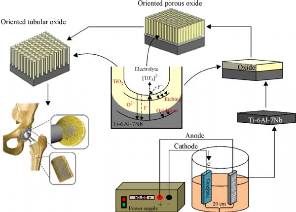
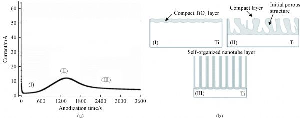
Abstract In recent years, nanostructured oxide films on titanium alloy surfaces have gained significant interest due to their electrical, catalytic and biological properties. In literature, there is variety of different approaches to fabricate nanostructured oxide films. Among these methods, anodization technique, which allows fine-tuning of oxide film thickness, feature size, topography and chemistry, is one of the most popular approaches to fabricate nanostructured oxide films on titanium alloys, and it has been widely investigated for orthopedic applications. Briefly, anodization is the growth of a controlled oxide film on a metallic component attached to the anode of an electrochemical cell. This review provides an overview of the anodization technique to grow nanostructured oxide films on titanium and titanium alloys and summarizes the interactions between anodized titanium alloy surfaces with cells in terms of cellular adhesion, proliferation and differentiation. It will start with summarizing the mechanism of nanofeatured oxide fabrication on titanium alloys and then switch its focus on the latest findings for anodization of titanium alloys, including the use of fluoride free electrolytes and anodization of 3D titanium foams. The review will also highlight areas requiring further research to successfully translate anodized titanium alloys to clinics for orthopedic applications.
|
| Keywords
titanium alloys
anodization
biocompatibility
orthopedics
|
|
Corresponding Author(s):
Batur Ercan
|
|
Just Accepted Date: 10 July 2018
Online First Date: 03 December 2018
Issue Date: 25 February 2019
|
|
| 1 |
BLi, T Webster. Orthopedic Biomaterials: Advances and Applications. Cham: Springer International Publishing, 2017, 31–32
|
| 2 |
S MKurtz, K L Ong, J Schmier, FMowat, KSaleh, EDybvik, JKärrholm, GGarellick, L IHavelin, OFurnes, HMalchau, ELau. Future clinical and economic impact of revision total hip and knee arthroplasty. Journal of Bone and Joint Surgery (American), 2007, 89(Suppl 3): 144–151
pmid: 17908880
|
| 3 |
D AEtzioni, J H Liu, M A Maggard, C Y Ko. The aging population and its impact on the surgery workforce. Annals of Surgery, 2003, 238(2): 170–177
https://doi.org/10.1097/01.SLA.0000081085.98792.3d
pmid: 12894008
|
| 4 |
R DRamiah, A M Ashmore, E Whitley, G CBannister. Ten-year life expectancy after primary total hip replacement. Journal of Bone and Joint Surgery. British Volume, 2007, 89(10): 1299–1302
https://doi.org/10.1302/0301-620X.89B10.18735
pmid: 17957067
|
| 5 |
VSansone, D Pagani, MMelato. The effects on bone cells of metal ions released from orthopaedic implants: A review. Clinical Cases in Mineral and Bone Metabolism, 2013, 10(1): 34–40
pmid: 23858309
|
| 6 |
C DEtkin, B D Springer. The American joint replacement registry—the first 5 years. Arthroplasty Today, 2017, 3(2): 67–69
https://doi.org/10.1016/j.artd.2017.02.002
pmid: 28695176
|
| 7 |
VZwilling, E Darque-Ceretti, ABoutry-Forveille, DDavid, M YPerrin, MAucouturier. Structure and physicochemistry of anodic oxide films on titanium and TA6V alloy. Surface and Interface Analysis, 1999, 27(7): 629–637
https://doi.org/:10.1002/(SICI)1096-9918(199907)27:7<629::AID-SIA551>3.0.CO;2-0
|
| 8 |
MLong, H J Rack. Titanium alloys in total joint replacement—a materials science perspective. Biomaterials, 1998, 19(18): 1621–1639
https://doi.org/10.1016/S0142-9612(97)00146-4
pmid: 9839998
|
| 9 |
EBütev, Z Esen, ŞBor. Characterization of Ti6Al7Nb alloy foams surface treated in aqueous NaOH and CaCl2 solutions. Journal of the Mechanical Behavior of Biomedical Materials, 2016, 60: 127–138
https://doi.org/10.1016/j.jmbbm.2015.12.040
pmid: 26807769
|
| 10 |
KPrasad, O Bazaka, MChua, MRochford, LFedrick, JSpoor, RSymes, MTieppo, CCollins, ACao, et al. Metallic biomaterials: Current challenges and opportunities. Materials, 2017, 10(8): 884
https://doi.org/10.3390/ma10080884
pmid: 28773240
|
| 11 |
MÖzcan, C Hämmerle. Titanium as a reconstruction and implant material in dentistry: Advantages and pitfalls. Materials, 2012, 5(9): 1528–1545
https://doi.org/10.3390/ma5091528
|
| 12 |
S DRogers, D W Howie, S E Graves, M J Pearcy, D R Haynes. In vitro human monocyte response to wear particles of titanium alloy containing vanadium or niobium. Journal of Bone and Joint Surgery, 1997, 79(2): 311–315
https://doi.org/10.1302/0301-620X.79B2.7192
pmid: 9119864
|
| 13 |
K CPopat, L Leoni, C AGrimes, T ADesai. Influence of engineered titania nanotubular surfaces on bone cells. Biomaterials, 2007, 28(21): 3188–3197
https://doi.org/10.1016/j.biomaterials.2007.03.020
pmid: 17449092
|
| 14 |
L CPalmer, C J Newcomb, S R Kaltz, E D Spoerke, S I Stupp. Biomimetic systems for hydroxyapatite mineralization inspired by bone and enamel. Chemical Reviews, 2008, 108(11): 4754–4783
https://doi.org/10.1021/cr8004422
pmid: 19006400
|
| 15 |
S JLiliensiek, PNealey, C JMurphy. Characterization of endothelial basement membrane nanotopography in rhesus macaque as a guide for vessel tissue engineering. Tissue Engineering. Part A, 2009, 15(9): 2643–2651
https://doi.org/10.1089/ten.tea.2008.0284
pmid: 19207042
|
| 16 |
ARafieerad, E Zalnezhad, ABushroa, AHamouda, MSarraf, BNasiri-Tabrizi. Self-organized TiO2 nanotube layer on Ti-6Al-7Nb for biomedical application. Surface and Coatings Technology, 2015, 265: 24–31
https://doi.org/10.1016/j.surfcoat.2015.01.067
|
| 17 |
J MMacak, H Tsuchiya, LTaveira, AGhicov, PSchmuki. Self-organized nanotubular oxide layers on Ti-6Al-7Nb and Ti-6Al-4V formed by anodization in NH4F solutions. Journal of Biomedical Materials Research Part A, 2005, 75(4): 928–933
https://doi.org/10.1002/jbm.a.30501
pmid: 16138327
|
| 18 |
SMahshid, A Dolati, MGoodarzi, MAskari, AGhahramaninezhad. Self-organized titanium oxide nanotubes prepared in phosphate electrolytes: Effect of voltage and fluorine concentration. ECS Transactions, 2010, 28(7): 67–74
|
| 19 |
AAkhlag, E U Haq, W Akhtar, MArshad, ZAhmad. Synthesis and characterization of titania nanotubes by anodizing of titanium in fluoride containing electrolytes. Applied Nanoscience, 2017, 7(8): 701–710
https://doi.org/10.1007/s13204-017-0608-5
|
| 20 |
KIndira, U K Mudali, T Nishimura, N.Rajendran A review on TiO2 nanotubes: Influence of anodization parameters, formation mechanism, properties, corrosion behavior, and biomedical applications. Journal of Bio- and Tribo-Corrosion, 2015, 1(28): 7–14
|
| 21 |
JMacak, H Tsuchiya, AGhicov, KYasuda, RHahn, S Bauer, PSchmuki. TiO2 nanotubes: Self-organized electrochemical formation, properties and applications. Current Opinion in Solid State and Materials Science, 2007, 11(1–2): 3–18
https://doi.org/10.1016/j.cossms.2007.08.004
|
| 22 |
PRoy, S Berger, PSchmuki. TiO2 nanotubes: Synthesis and applications. Angewandte Chemie International Edition, 2011, 50(13): 2904–2939
https://doi.org/10.1002/anie.201001374
pmid: 21394857
|
| 23 |
HOmidvar, S Goodarzi, ASeif, A RAzadmehr. Influence of anodization parameters on the morphology of TiO2 nanotube arrays. Superlattices and Microstructures, 2011, 50(1): 26–39
https://doi.org/10.1016/j.spmi.2011.04.006
|
| 24 |
A LEscada, R Z Nakazato, A P Claro. Influence of anodization parameters in the TiO2 nanotubes formation on Ti-7.5Mo alloy surface for biomedical application. Materials Research, 2017, 20(5): 1282–1290
https://doi.org/10.1590/1980-5373-mr-2016-0520
|
| 25 |
DRegonini, C Bowen, AJaroenworaluck, RStevens. A review of growth mechanism, structure and crystallinity of anodized TiO2 nanotubes. Materials Science and Engineering: R: Reports, 2013, 74(12): 377–406
https://doi.org/10.1016/j.mser.2013.10.001
|
| 26 |
XFeng, J M Macak, P Schmuki. Robust self-organization of oxide nanotubes over a wide pH range. Chemistry of Materials, 2007, 19(7): 1534–1536
https://doi.org/10.1021/cm063042g
|
| 27 |
SSreekantan, Z Lockman, RHazan, MTasbihi, L KTong, A RMohamed. Influence of electrolyte pH on TiO2 nanotube formation by Ti anodization. Journal of Alloys and Compounds, 2009, 485(1–2): 478–483
https://doi.org/10.1016/j.jallcom.2009.05.152
|
| 28 |
JChen, J Lin, XChen. Self-assembled TiO2 nanotube arrays with U-shaped profile by controlling anodization temperature. Journal of Nanomaterials, 2010, 1: 38
|
| 29 |
DGong, C A Grimes, O K Varghese, W Hu, R SSingh, ZChen, E C Dickey. Titanium oxide nanotube arrays prepared by anodic oxidation. Journal of Materials Research, 2001, 16(12): 3331–3334
https://doi.org/10.1557/JMR.2001.0457
|
| 30 |
MKulkarni, A Mazare, PSchmuki, AIglic. Influence of anodization parameters on morphology of TiO2 nanostructured surfaces. Advanced Materials Letters, 2016, 7(1): 23–28
https://doi.org/10.5185/amlett.2016.6156
|
| 31 |
MGöttlicher, M Rohnke, AKunz, JThomas, R AHenning, TLeichtweiß, JJanek. Anodization of titanium in radio frequency oxygen discharge. Microstructure, kinetics and transport mechanism. Solid State Ionics, 2016, 290: 130–139
https://doi.org/10.1016/j.ssi.2016.04.013
|
| 32 |
MGöttlicher, M Rohnke, AHelth, TLeichtweiß, TGemming, AGebert, JEckert, JJanek. Controlled surface modification of Ti-40Nb implant alloy by electrochemically assisted inductively coupled RF plasma oxidation. Acta Biomaterialia, 2013, 9(11): 9201–9210
https://doi.org/10.1016/j.actbio.2013.07.015
pmid: 23891813
|
| 33 |
VZwilling, E Darque-Ceretti, ABoutry-Forveille, DDavid, M YPerrin, MAucouturier. Structure and physicochemistry of anodic oxide films on titanium and TA6V alloy. Surface and Interface Analysis, 1999, 27(7): 629–637
https://doi.org/10.1002/(SICI)1096-9918(199907)27:7<629::AID-SIA551>3.0.CO;2-0
|
| 34 |
RBeranek, H Hildebrand, PSchmuki. Self-organized porous titanium oxide prepared in H2SO4/HF electrolytes. Electrochemical and Solid-State Letters, 2003, 6(3): 12–14
https://doi.org/10.1149/1.1545192
|
| 35 |
QCai, M Paulose, O KVarghese, C AGrimes. The effect of electrolyte composition on the fabrication of self-organized titanium oxide nanotube arrays by anodic oxidation. Journal of Materials Research, 2005, 20(01): 230–236
https://doi.org/10.1557/JMR.2005.0020
|
| 36 |
J MMacak, M Zlamal, JKrysa, PSchmuki. Self-organized TiO2 nanotube layers as highly efficient photocatalysts. Small, 2007, 3(2): 300–304
https://doi.org/10.1002/smll.200600426
pmid: 17230591
|
| 37 |
AMazare, M Dilea, DIonita, ITitorencu, VTrusca, EVasile. Changing bioperformance of TiO2 amorphous nanotubes as an effect of inducing crystallinity. Bioelectrochemistry, 2012, 87: 124–131
https://doi.org/10.1016/j.bioelechem.2012.01.002
pmid: 22341625
|
| 38 |
AKaczmarek, T Klekiel, EKrasicka-Cydzik. Fluoride concentration effect on the anodic growth of self-aligned oxide nanotube array on Ti6Al7Nb alloy. Surface and Interface Analysis, 2010, 42(6–7): 510–514
https://doi.org/10.1002/sia.3303
|
| 39 |
I SPark, H J Oh, T S Bae. Bioactivity and generation of anodized nanotubular TiO2 layer of Ti-6Al-4V alloy in glycerol solution. Thin Solid Films, 2013, 548: 292–298
https://doi.org/10.1016/j.tsf.2013.08.096
|
| 40 |
LMohan, C Anandan, NRajendran. Electrochemical behavior and effect of heat treatment on morphology, crystalline structure of self-organized TiO2 nanotube arrays on Ti-6Al-7Nb for biomedical applications. Materials Science and Engineering C, 2015, 50: 394–401
https://doi.org/10.1016/j.msec.2015.02.013
pmid: 25746285
|
| 41 |
HJha, R Hahn, PSchmuki. Ultrafast oxide nanotube formation on TiNb, TiZr and TiTa alloys by rapid breakdown anodization. Electrochimica Acta, 2010, 55(28): 8883–8887
|
| 41 |
A LEscada, R Z Nakazato, A P Claro. Influence of anodization parameters in the TiO2 nanotubes formation on Ti-7.5Mo alloy surface for biomedical application. Materials Research, 2017, 20(5): 1282–1290
https://doi.org/10.1590/1980-5373-mr-2016-0520
|
| 42 |
P CChen, S J Hsieh, C C Chen, J Zou. The microstructure and capacitance characterizations of anodic titanium based alloy oxide nanotube. Journal of Nanomaterials, 2013, 2013: 157494
|
| 43 |
WKim, H Choe, W ABrantley. Nanostructured surface changes of Ti-35Ta-xZr alloys with changes in anodization factors. Thin Solid Films, 2011, 519(15): 4663–4667
https://doi.org/10.1016/j.tsf.2011.01.013
|
| 44 |
AOssowska, S Sobieszczyk, MSupernak, AZielinski. Morphology and properties of nanotubular oxide layer on the Ti-13Zr-13Nb alloy. Surface and Coatings Technology, 2014, 258(15): 1239–1248
https://doi.org/10.1016/j.surfcoat.2014.06.054
|
| 45 |
TSaito, T Furuta, J HHwang, SKuramoto, KNishino, NSuzuki, RChen, A Yamada, KIto, YSeno, et al. Multifunctional alloys obtained via a dislocation-free plastic deformation mechanism. Science, 2003, 300(5618): 464–467
https://doi.org/10.1126/science.1081957
pmid: 12702870
|
| 46 |
Y HChiu, T H Lai, C Y Chen, P Y Hsieh, K Ozasa, MNiinomi, KOkada, T MChang, NMatsushita, MSone, et al. Fully depleted Ti-Nb-Ta-Zr-O nanotubes: Interfacial charge dynamics and solar hydrogen production. ACS Applied Materials & Interfaces, 2018, 10(27): 22997–23008
https://doi.org/10.1021/acsami.8b00727
pmid: 29664283
|
| 47 |
CRichter, Z Wu, EPanaitescu, RWilley, LMenon. Ultra-high-aspect-ratio titania nanotubes. Advanced Materials, 2007, 19(7): 946–948
https://doi.org/10.1002/adma.200602389
|
| 48 |
Y LCheong, F K Yam, S W Ng, Z Hassan, S SNg, I MLow. Fabrication of titanium dioxide nanotubes in fluoride-free electrolyte via rapid breakdown anodization. Journal of Porous Materials, 2015, 22(6): 1437–1444
https://doi.org/10.1007/s10934-015-0024-8
|
| 49 |
N KAllam, K Shankar, C AGrimes. Photoelectrochemical and water photoelectrolysis properties of ordered TiO2 nanotubes fabricated by Ti anodization in fluoride-free HCl electrolytes. Journal of Materials Chemistry, 2008, 18(20): 2341–2348
https://doi.org/10.1039/b718580d
|
| 50 |
SKim, M Seong, JChoi. Rapid breakdown anodization for the preparation of titania nanotubes in halogen-free acids. Journal of the Electrochemical Society, 2015, 162(6): 205–208
https://doi.org/10.1149/2.0291506jes
|
| 51 |
ZBi, M P Paranthaman, P A Menchhofer, R R Dehoff, C A Bridges, M Chi, SDai. Self-organized amorphous TiO2 nanotube arrays on porous Ti foam for rechargeable lithium and sodium ion batteries. Journal of Power Sources, 2013, 222: 461–466
https://doi.org/10.1016/j.jpowsour.2012.09.019
|
| 52 |
J SKang, H Choi, JKim, HPark, J Y Kim, J W Choi, S H Yu, K J Lee, Y S Kang, S H Park, et al. Solar cells: Multidimensional anodized titanium foam photoelectrode for efficient utilization of photons in mesoscopic solar cells. Small, 2017, 13(34): 1–7
pmid: 28722350
|
| 53 |
J MSchakenraad, H JBusscher, C RWildevuur, JArends. The influence of substratum surface free energy on growth and spreading of human fibroblasts in the presence and absence of serum proteins. Journal of Biomedical Materials Research, 1986, 20(6): 773–784
https://doi.org/10.1002/jbm.820200609
pmid: 3722214
|
| 54 |
S DPuckett, E Taylor, TRaimondo, T JWebster. The relationship between the nanostructure of titanium surfaces and bacterial attachment. Biomaterials, 2010, 31(4): 706–713
https://doi.org/10.1016/j.biomaterials.2009.09.081
pmid: 19879645
|
| 55 |
L NWang, M Jin, YZheng, YGuan, X Lu, J LLuo. Nanotubular surface modification of metallic implants via electrochemical anodization technique. International Journal of Nanomedicine, 2014, 9(1): 4421–4435
https://doi.org/10.2147/IJN.S65866
pmid: 25258532
|
| 56 |
T JWebster, C Ergun, R HDoremus, R WSiegel, RBizios. Enhanced functions of osteoblasts on nanophase ceramics. Biomaterials, 2000, 21(17): 1803–1810
https://doi.org/10.1016/S0142-9612(00)00075-2
pmid: 10905463
|
| 57 |
T JWebster, J U Ejiofor. Increased osteoblast adhesion on nanophase metals: Ti, Ti6Al4V, and CoCrMo. Biomaterials, 2004, 25(19): 4731–4739
https://doi.org/10.1016/j.biomaterials.2003.12.002
pmid: 15120519
|
| 58 |
TRaimondo, S Puckett, T JWebster. Greater osteoblast and endothelial cell adhesion on nanostructured polyethylene and titanium. International Journal of Nanomedicine, 2010, 5(5): 647–652
pmid: 20856840
|
| 59 |
WJi, P Han, CZhao, YJiang, XZhang. Increased osteoblast adhesion on nanophase Ti6Al4V. Science Bulletin, 2008, 53(11): 1757–1762
https://doi.org/10.1007/s11434-008-0093-z
|
| 60 |
K SBrammer, S Oh, C JCobb, L MBjursten, Hvan der Heyde, SJin. Improved bone-forming functionality on diameter-controlled TiO2 nanotube surface. Acta Biomaterialia, 2009, 5(8): 3215–3223
https://doi.org/10.1016/j.actbio.2009.05.008
pmid: 19447210
|
| 61 |
A PRoss, T J Webster. Anodizing color coded anodized Ti6Al4V medical devices for increasing bone cell functions. International Journal of Nanomedicine, 2013, 8(1): 109–117
pmid: 23319862
|
| 62 |
CYao, E B Slamovich, T J Webster. Enhanced osteoblast functions on anodized titanium with nanotube-like structures. Journal of Biomedical Materials Research Part A, 2008, 85A(1): 157–166
https://doi.org/10.1002/jbm.a.31551
pmid: 17688267
|
| 63 |
PRoy, S Berger, PSchmuki. TiO2 nanotubes: Synthesis and applications. Angewandte Chemie International Edition, 2011, 50(13): 2904–2939
https://doi.org/10.1002/anie.201001374
pmid: 21394857
|
| 64 |
ATan, B Pingguan-Murphy, RAhmad, SAkbar. Review of titania nanotubes: Fabrication and cellular response. Ceramics International, 2012, 38(6): 4421–4435
https://doi.org/10.1016/j.ceramint.2012.03.002
|
| 65 |
JPark, S Bauer, Kvon der Mark, PSchmuki. Nanosize and vitality: TiO2 nanotube diameter directs cell fate. Nano Letters, 2007, 7(6): 1686–1691
https://doi.org/10.1021/nl070678d
pmid: 17503870
|
| 66 |
BErcan, K M Kummer, K M Tarquinio, T J Webster. Decreased Staphylococcus aureus biofilm growth on anodized nanotubular titanium and the effect of electrical stimulation. Acta Biomaterialia, 2011, 7(7): 3003–3012
https://doi.org/10.1016/j.actbio.2011.04.002
pmid: 21515421
|
| 67 |
K SBrammer, S Oh, C JCobb, L MBjursten, Hvan der Heyde, SJin. Improved bone-forming functionality on diameter-controlled TiO(2) nanotube surface. Acta Biomaterialia, 2009, 5(8): 3215–3223
https://doi.org/10.1016/j.actbio.2009.05.008
pmid: 19447210
|
| 68 |
K SBrammer, S Oh, J OGallagher, SJin. Enhanced cellular mobility guided by TiO2 nanotube surfaces. Nano Letters, 2008, 8(3): 786–793
https://doi.org/10.1021/nl072572o
pmid: 18251515
|
| 69 |
W QYu, Y L Zhang, X Q Jiang, F Q Zhang. In vitro behavior of MC3T3-E1 preosteoblast with different annealing temperature titania nanotubes. Oral Diseases, 2010, 16(7): 624–630
https://doi.org/10.1111/j.1601-0825.2009.01643.x
pmid: 20604877
|
| 70 |
SOh, K S Brammer, Y S Li, D Teng, A JEngler, SChien, SJin. Stem cell fate dictated solely by altered nanotube dimension. Proceedings of the National Academy of Sciences of the United States of America, 2009, 106(7): 2130–2135
https://doi.org/10.1073/pnas.0813200106
pmid: 19179282
|
| 71 |
KMalec, J Góralska, MHubalewska-Mazgaj, PGłowacz, MJarosz, PBrzewski, G DSulka, MJaskuła, IWybrańska. Effects of nanoporous anodic titanium oxide on human adipose derived stem cells. International Journal of Nanomedicine, 2016, 11: 5349–5360
https://doi.org/10.2147/IJN.S116263
pmid: 27789947
|
| 72 |
GLi, Q Zhao, HTang, GLi, Y Chi. Fabrication, characterization and biocompatibility of TiO2 nanotubes via anodization of Ti6Al7Nb. Composite Interfaces, 2016, 23(3): 223–230
https://doi.org/10.1080/09276440.2016.1128723
|
| 73 |
EFilova, J Fojt, MKryslova, HMoravec, LJoska, LBacakova. The diameter of nanotubes formed on Ti-6Al-4V alloy controls the adhesion and differentiation of Saos-2 cells. International Journal of Nanomedicine, 2015, 10(1): 7145–7163
https://doi.org/10.2147/IJN.S87474
pmid: 26648719
|
| 74 |
Y TSul. Electrochemical growth behavior, surface properties, and enhanced in vivo bone response of TiO2 nanotubes on microstructured surfaces of blasted, screw-shaped titanium implants. International Journal of Nanomedicine, 2010, 5(5): 87–100
https://doi.org/10.2147/IJN.S8012
pmid: 20463928
|
| 75 |
L MBjursten, L Rasmusson, SOh, G CSmith, K SBrammer, SJin. Titanium dioxide nanotubes enhance bone bonding in vivo. Journal of Biomedical Materials Research Part A, 2010, 92(3): 1218–1224
pmid: 19343780
|
| 76 |
LSalou, A Hoornaert, GLouarn, PLayrolle. Enhanced osseointegration of titanium implants with nanostructured surfaces: An experimental study in rabbits. Acta Biomaterialia, 2015, 11: 494–502
https://doi.org/10.1016/j.actbio.2014.10.017
pmid: 25449926
|
| 77 |
SPuckett. Select nanofabricated titanium materials for enhancing bone and skin growth of intraosseous transcutaneous amputation prostheses. Dissertation for the Doctoral Degree, Rhode Island: Brown University, 2011, 286–300
|
| 78 |
NWang, H Li, WLü, JLi, J Wang, ZZhang, YLiu. Effects of TiO2 nanotubes with different diameters on gene expression and osseointegration of implants in minipigs. Biomaterials, 2011, 32(29): 6900–6911
https://doi.org/10.1016/j.biomaterials.2011.06.023
pmid: 21733571
|
| 79 |
S DPuckett, E Taylor, TRaimondo, T JWebster. The relationship between the nanostructure of titanium surfaces and bacterial attachment. Biomaterials, 2010, 31(4): 706–713
https://doi.org/10.1016/j.biomaterials.2009.09.081
pmid: 19879645
|
| 80 |
J LDel Pozo, RPatel. Clinical practice. Infection associated with prosthetic joints. New England Journal of Medicine, 2009, 361(8): 787–794
https://doi.org/10.1056/NEJMcp0905029
pmid: 19692690
|
| 81 |
GColon, B C Ward, T J Webster. Increased osteoblast and decreased Staphylococcus epidermidis functions on nanophase ZnO and TiO2. Journal of Biomedical Materials Research Part A, 2006, 78A(3): 595–604
https://doi.org/10.1002/jbm.a.30789
pmid: 16752397
|
| 82 |
C PPeremarch, R PTanoira, M AArenas, EMatykina, AConde, J JDamborenea, JEsteban. Bacterial adherence to anodized titanium alloy. Journal of Physics: Conference Series, 2010, 252(1): 1–8
|
| 83 |
CZhao, B Feng, YLi, JTan, X Lu, JWeng. Preparation and antibacterial activity of titanium nanotubes loaded with Ag nanoparticles in the dark and under the UV light. Applied Surface Science, 2013, 280: 8–14
https://doi.org/10.1016/j.apsusc.2013.04.057
|
| 84 |
TWang, Z Weng, XLiu, K W KYeung, HPan, S Wu. Controlled release and biocompatibility of polymer/titania nanotube array system on titanium implants. Bioactive Materials, 2017, 2(1): 44–50
https://doi.org/10.1016/j.bioactmat.2017.02.001
pmid: 29744410
|
| 85 |
G ACrawford, N Chawla, KDas, SBose, A Bandyopadhyay. Microstructure and deformation behavior of biocompatible TiO2 nanotubes on titanium substrate. Acta Biomaterialia, 2007, 3(3): 359–367
https://doi.org/10.1016/j.actbio.2006.08.004
pmid: 17067860
|
| 86 |
MZhao, J Li, YLi, JWang, Y Zuo, JJiang, HWang. Gradient control of the adhesive force between Ti/TiO2 nanotubular arrays fabricated by anodization. Scientific Reports, 2014, 4(1): 7178
https://doi.org/10.1038/srep07178
pmid: 25417900
|
|
Viewed |
|
|
|
Full text
|
|
|
|
|
Abstract
|
|
|
|
|
Cited |
|
|
|
|
| |
Shared |
|
|
|
|
| |
Discussed |
|
|
|
|