|
|
|
Prostaglandin E2 promotes hematopoietic development from human embryonic stem cells |
Chao YANG1,2, Jia-Fei XI1, Xiao-Yan XIE1, Wen YUE1, Ruo-Yong WANG1, Qiong WU1, Li-Juan HE1, Xue NAN1, Yan-Hua LI1, Xue-Tao PEI1( ) ) |
| 1. Stem Cell and Regenerative Medicine Lab, Beijing Institute of Transfusion Medicine, Beijing 100850, China; 2. 522 Hospital of Chinese PLA, Luoyang 471000, China |
|
|
|
|
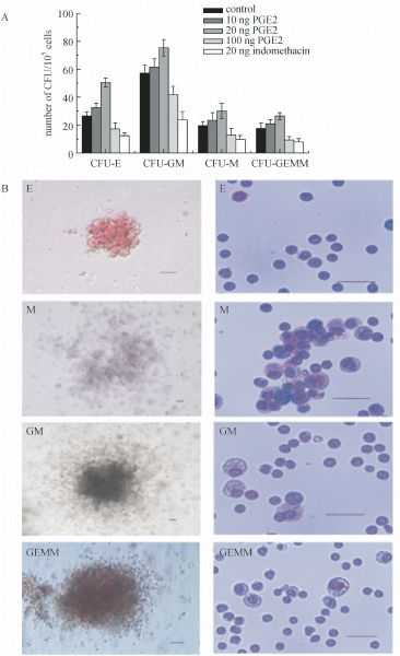
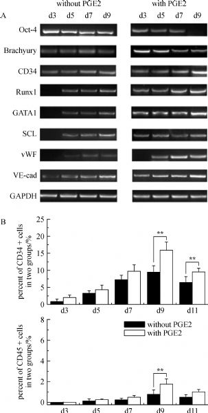
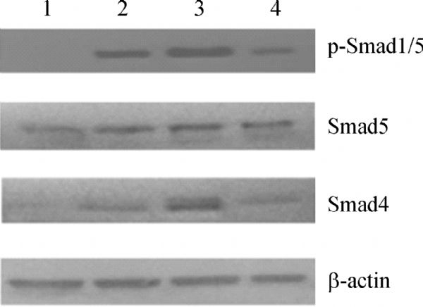
Abstract Recent studies have suggested that prostaglandin (PG) E2 (PGE2) and the prostaglandin pathway are essential for hematopoietic stem cell growth and development. However, similar studies on hematopoietic commitment from human embryonic stem cells (hESCs) are still limited. Here we report that the addition of PGE2 promotes hematopoietic differentiation of hESCs. The induced cells from hESCs/OP9 co-culture and in the presence of PGE2 were characterized by reverse transcription-PCR (RT-PCR), flow cytometry, colony-forming assays and Wright-Giemsa staining. Our results demonstrated that PGE2 exposure could alter the gene expression pattern and morphology of co-cultured hESCs and resulted in a robust hematopoietic differentiation with higher frequencies of CD34+ and CD45+ cells. Furthermore, the Smad signaling pathway may be involved in PGE2 and OP9 induced hematopoietic differentiation of hESCs. This research may improve our knowledge of stem cell regulation and hopefully lead to better stem cell-based therapeutic options.
|
| Keywords
human embryonic stem cells
prostaglandin E2
hematopoiesis
in vitro differentiation
|
|
Corresponding Author(s):
PEI Xue-Tao,Email:shirlylyh@126.com, peixt@nic.bmi.ac.cn
|
|
Issue Date: 01 October 2010
|
|
| 1 |
Alfranca A, López-Oliva J M, Genós L, López-Maderuelo D, Mirones I, Salvado D, Quesada A J, Arroyo A G, Redondo J M (2008). PGE2 induces angiogenesis via MT1-MMP-mediated activation of the TGFbeta/Alk5 signaling pathway. Blood , 112(4): 1120–1128
doi: 10.1182/blood-2007-09-112268
|
| 2 |
Bhatia M, Bonnet D, Wu D, Murdoch B, Wrana J, Gallacher L, Dick J E (1999). Bone morphogenetic proteins regulate the developmental program of human hematopoietic stem cells. J Exp Med , 189(7): 1139–1148
doi: 10.1084/jem.189.7.1139
|
| 3 |
Bhattacharya B, Miura T, Brandenberger R, Mejido J, Luo Y, Yang A X, Joshi B H, Ginis I, Thies R S, Amit M, Lyons I, Condie B G, Itskovitz-Eldor J, Rao M S, Puri R K (2004). Gene expression in human embryonic stem cell lines: unique molecular signature. Blood , 103(8): 2956–2964
doi: 10.1182/blood-2003-09-3314
|
| 4 |
Cha Y I, Kim S H, Solnica-Krezel L, Dubois R N (2005). Cyclooxygenase-1 signaling is required for vascular tube formation during development. Dev Biol , 282(1): 274–283
doi: 10.1016/j.ydbio.2005.03.014
|
| 5 |
Chadwick K, Wang L, Li L, Menendez P, Murdoch B, Rouleau A, Bhatia M (2003). Cytokines and BMP-4 promote hematopoietic differentiation of human embryonic stem cells. Blood , 102(3): 906–915
doi: 10.1182/blood-2003-03-0832
|
| 6 |
Choi J H, Ryu Y S, Kim K H, Lee Y R, Cha K W, Han I S, Kwon B S (2009). In vitro development of a hemangioblast from a human embryonic stem cell, SNUhES#3. Life Sci , 85(1-2): 39–45
|
| 7 |
Frisch B J, Porter R L, Gigliotti B J, Olm-Shipman A J, Weber J M, O’Keefe R J, Jordan C T, Calvi L M (2009). In vivo prostaglandin E2 treatment alters the bone marrow microenvironment and preferentially expands short-term hematopoietic stem cells. Blood , 114(19): 4054–4063
doi: 10.1182/blood-2009-03-205823
|
| 8 |
Gentile P S, Pelus L M (1987). In vivo modulation of myelopoiesis by prostaglandin E2. II. Inhibition of granulocyte-monocyte progenitor cell (CFU-GM) cell-cycle rate. Exp Hematol , 15(2): 119–126
|
| 9 |
Goessling W, North T E, Loewer S, Lord A M, Lee S, Stoick-Cooper C L, Weidinger G, Puder M, Daley G Q, Moon R T, Zon L I (2009). Genetic interaction of PGE2 and Wnt signaling regulates developmental specification of stem cells and regeneration. Cell , 136(6): 1136–1147
|
| 10 |
Hirashima M, Kataoka H, Nishikawa S, Matsuyoshi N, Nishikawa S I (1999). Maturation of embryonic stem cells into endothelial cells in an in vitro model of vasculogenesis. Blood , 93(4): 1253–1263
|
| 11 |
Hoggatt J, Singh P, Sampath J, Pelus L M (2009). Prostaglandin E2 enhances hematopoietic stem cell homing, survival, and proliferation. Blood , 113(22): 5444–5455
doi: 10.1182/blood-2009-01-201335
|
| 12 |
Ji J, Vijayaragavan K, Bosse M, Menendez P, Weisel K, Bhatia M (2008). OP9 stroma augments survival of hematopoietic precursors and progenitors during hematopoietic differentiation from human embryonic stem cells. Stem Cells , 26(10): 2485–2495
doi: 10.1634/stemcells.2008-0642
|
| 13 |
Larsson J, Karlsson S (2005). The role of Smad signaling in hematopoiesis. Oncogene , 24(37): 5676–5692
doi: 10.1038/sj.onc.1208920
|
| 14 |
Leahy K M, Koki A T, Masferrer J L (2000). Role of cyclooxygenases in angiogenesis. Curr Med Chem , 7: 1163–1170
|
| 15 |
Letamendia A, Labbé E, Attisano L (2001). Transcriptional regulation by Smads: crosstalk between the TGF-beta and Wnt pathways. J Bone Joint Surg Am , 83-A(Pt 1 Suppl 1): S31–S39
|
| 16 |
Liou J Y, Ellent D P, Lee S, Goldsby J, Ko B S, Matijevic N, Huang J C, Wu K K (2007). Cyclooxygenase-2-derived prostaglandin e2 protects mouse embryonic stem cells from apoptosis. Stem Cells , 25(5): 1096–1103
doi: 10.1634/stemcells.2006-0505
|
| 17 |
Liu Y X, Ji L, Yue W, Yan Z F, Wang J, Xi J F, Zhang R, Nan X, Bai C X, Chen L, Wang Y F, Pei X T (2009). Cells extract from fetal liver promotes the hematopoietic differentiation of human embryonic stem cells. Cloning Stem Cells , 11(1): 51–60
doi: 10.1089/clo.2008.0049
|
| 18 |
Liu Z, Tang Y, Qiu T, Cao X, Clemens T L (2006). A dishevelled-1/Smad1 interaction couples WNT and bone morphogenetic protein signaling pathways in uncommitted bone marrow stromal cells. J Biol Chem , 281(25): 17156–17163
doi: 10.1074/jbc.M513812200
|
| 19 |
Lu L, Pelus L M, Broxmeyer H E (1984). Modulation of the expression of HLA-DR (Ia) antigens and the proliferation of human erythroid (BFU-E) and multipotential (CFU-GEMM) progenitor cells by prostaglandin E. Exp Hematol , 12(9): 741–748
|
| 20 |
Lu S J, Feng Q, Park J S, Vida L, Lee B S, Strausbauch M, Wettstein P J, Honig G R, Lanza R (2008). Biological properties and enucleation of red blood cells from human embryonic stem cells. Blood , 112: 4475–4484
doi: 10.1182/blood-2008-05-157198
|
| 21 |
Lu S J, Li F, Vida L, Honig G R (2004). CD34+CD38- hematopoietic precursors derived from human embryonic stem cells exhibit an embryonic gene expression pattern. Blood , 103(11): 4134–4141
doi: 10.1182/blood-2003-10-3575
|
| 22 |
Ma F, Ebihara Y, Umeda K, Sakai H, Hanada S, Zhang H, Zaike Y, Tsuchida E, Nakahata T, Nakauchi H, Tsuji K (2008). Generation of functional erythrocytes from human embryonic stem cell-derived definitive hematopoiesis. Proc Natl Acad Sci U S A , 105(35): 13087–13092
doi: 10.1073/pnas.0802220105
|
| 23 |
Miller S B (2006). Prostaglandins in health and disease: an overview. Semin Arthritis Rheum , 36(1): 37–49
doi: 10.1016/j.semarthrit.2006.03.005
|
| 24 |
Naito A T, Shiojima I, Akazawa H, Hidaka K, Morisaki T, Kikuchi A, Komuro I (2006). Developmental stage-specific biphasic roles of Wnt/β-catenin signaling in cardiomyogenesis and hematopoiesis. Proc Natl Acad Sci U S A , 103(52): 19812–19817
doi: 10.1073/pnas.0605768103
|
| 25 |
North T E, Goessling W, Walkley C R, Lengerke C, Kopani K R, Lord A M, Weber G J, BowmanT V, Jang I H, Grosser T, Fitzgerald G A, Daley G Q, Orkin S H, Zon L I (2007). Prostaglandin E2 regulates vertebrate haematopoietic stem cell homeostasis. Nature , 447(7147): 1007–1011
doi: 10.1038/nature05883
|
| 26 |
Smith W L, DeWitt D L, Garavito R M (2000). Cyclooxygenases: structural, cellular, and molecular biology. Annu Rev Biochem , 69: 145–182
doi: 10.1146/annurev.biochem.69.1.145
|
| 27 |
Srivastava A S, Nedelcu E, Esmaeli-AzadB, Mishra R, Carrier E (2007). Thrombopoietin enhances generation of CD34+ cells from human embryonic stem cells. Stem Cells , 25(6): 1456–1461
doi: 10.1634/stemcells.2006-0701
|
| 28 |
Varga A C, Wrana J L (2005). The disparate role of BMP in stem cell biology. Oncogene , 24(37): 5713–5721
doi: 10.1038/sj.onc.1208919
|
| 29 |
Vodyanik M A, Bork J A, Thomson J A, Slukvin I I (2005). Human embryonic stem cell-derived CD34+ cells: efficient production in the coculture with OP9 stromal cells and analysis of lymphohematopoietic potential. Blood , 105(2): 617–626
doi: 10.1182/blood-2004-04-1649
|
| 31 |
WangL, Li L, Menendez P, Cerdan C, Bhatia M (2005). Human embryonic stem cells maintained in the absence of mouse embryonic fibroblasts or conditioned media are capable of hematopoietic development. Blood , 105(12): 4598–4603
doi: 10.1182/blood-2004-10-4065
|
| 32 |
Zhang P, Li J, Tan Z, Wang C, Liu T, Chen L, Yong J, Jiang W, Sun X, Du L, Ding M, Deng H (2008). Short-term BMP-4 treatment initiates mesoderm induction in human embryonic stem cells. Blood , 111(4): 1933–1941
doi: 10.1182/blood-2007-02-074120
|
|
Viewed |
|
|
|
Full text
|
|
|
|
|
Abstract
|
|
|
|
|
Cited |
|
|
|
|
| |
Shared |
|
|
|
|
| |
Discussed |
|
|
|
|