|
|
|
Reactive oxygen species generation is essential for cisplatin-induced accelerated senescence in hepatocellular carcinoma |
Kai Qu1,Ting Lin1,2,Zhixin Wang1,Sinan Liu1,Hulin Chang1,Xinsen Xu1,Fandi Meng1,Lei Zhou1,Jichao Wei1,Minghui Tai1,Yafeng Dong3,*( ),Chang Liu1,2,*() ),Chang Liu1,2,*() |
1. Department of Hepatobiliary Surgery
2. Surgical Intensive Care Unit, the First Affiliated Hospital, School of Medicine, Xi’an Jiaotong University, Xi’an 710061, China
3. Department of Obstetrics and Gynecology, Kansas University, Kansas City, KS 66160, USA |
|
|
|
|
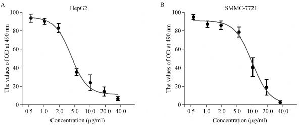
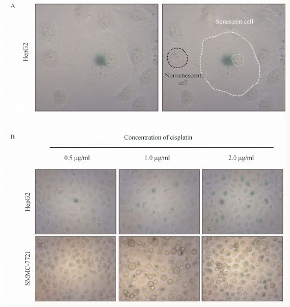
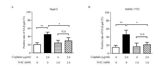
Abstract Accelerated senescence is important because this process is involved in tumor suppression and has been induced by many chemotherapeutic agents. The platinum-based chemotherapeutic agent cisplatin displays a wide range of antitumor activities. However, the molecular mechanism of cisplatin-induced accelerated senescence in hepatocellular carcinoma (HCC) remains unclear. In the present study, the growth inhibitory effect of cisplatin on HepG2 and SMMC-7721 cells was detected by 3-(4,5-dimethylthiazol-2-yl)-2,5-diphenyltetrazolium bromide assay. Cellular senescence was then assessed by β-galactosidase assay. Senescence-related factors, including p53, p21, and p16, were evaluated by quantitative reverse transcription-polymerase chain reaction. Reactive oxygen species (ROS) was analyzed by flow cytometry. Our results revealed that cisplatin reduced the proliferation of HepG2 and SMMC-7721 cells in a dose- and time-dependent manner. Senescent phenotype observed in cisplatin-treated hepatoma cells was dependent on p53 and p21 activation but not on p16 activation. Furthermore, cisplatin-induced accelerated senescence depended on intracellular ROS generation. The ROS scavenger N-acetyl-L-cysteine also significantly suppressed the cisplatin-induced senescence of HepG2 and SMMC-7721 cells. In conclusion, our results revealed a functional link between intracellular ROS generation and cisplatin-induced accelerated senescence, and this link may be used as a potential target of HCC.
|
| Keywords
reactive oxygen species
senescence
cisplatin
hepatocellular carcinoma
|
|
Corresponding Author(s):
Yafeng Dong
|
|
Online First Date: 04 May 2014
Issue Date: 21 May 2014
|
|
| 1 |
HayflickL. The limited in vitro lifetime of human diploid cell strains. Exp Cell Res1965; 37(3): 614-636
doi: 10.1016/0014-4827(65)90211-9 pmid: 14315085
|
| 2 |
DimriGP, LeeX, BasileG, AcostaM, ScottG, RoskelleyC, MedranoEE, LinskensM, RubeljI, Pereira-SmithO. A biomarker that identifies senescent human cells in culture and in aging skin in vivo. Proc Natl Acad Sci USA1995; 92(20): 9363-9367
doi: 10.1073/pnas.92.20.9363 pmid: 7568133
|
| 3 |
ChangBD, BroudeEV, DokmanovicM, ZhuH, RuthA, XuanY, KandelES, LauschE, ChristovK, RoninsonIB. A senescence-like phenotype distinguishes tumor cells that undergo terminal proliferation arrest after exposure to anticancer agents. Cancer Res1999; 59(15): 3761-3767
pmid: 10446993
|
| 4 |
te PoeleRH, OkorokovAL, JardineL, CummingsJ, JoelSP. DNA damage is able to induce senescence in tumor cells in vitro and in vivo. Cancer Res2002; 62(6): 1876-1883
pmid: 11912168
|
| 5 |
FangK, ChiuCC, LiCH, ChangYT, HwangHT. Cisplatin-induced senescence and growth inhibition in human non-small cell lung cancer cells with ectopic transfer of p16INK4a. Oncol Res2007; 16(10): 479-488
doi: 10.3727/096504007783338331 pmid: 18196872
|
| 6 |
WangX, WongSC, PanJ, TsaoSW, FungKH, KwongDL, ShamJS, NichollsJM. Evidence of cisplatin-induced senescent-like growth arrest in nasopharyngeal carcinoma cells. Cancer Res1998; 58(22): 5019-5022
pmid: 9823301
|
| 7 |
PanieriE, GogvadzeV, NorbergE, VenkateshR, OrreniusS, ZhivotovskyB. Reactive oxygen species generated in different compartments induce cell death, survival, or senescence. Free Radic Biol Med2013; 57: 176-187
doi: 10.1016/j.freeradbiomed.2012.12.024 pmid: 23295411
|
| 8 |
LiSK, SmithDK, LeungWY, CheungAM, LamEW, DimriGP, YaoKM. FoxM1c counteracts oxidative stress-induced senescence and stimulates Bmi-1 expression. J Biol Chem2008; 283(24): 16545-16553
doi: 10.1074/jbc.M709604200 pmid: 18408007
|
| 9 |
QuK, XuX, LiuC, WuQ, WeiJ, MengF, ZhouL, WangZ, LeiL, LiuP. Negative regulation of transcription factor FoxM1 by p53 enhances oxaliplatin-induced senescence in hepatocellular carcinoma. Cancer Lett2013; 331(1): 105-114
doi: 10.1016/j.canlet.2012.12.008 pmid: 23262037
|
| 10 |
RobersonRS, KussickSJ, VallieresE, ChenSY, WuDY. Escape from therapy-induced accelerated cellular senescence in p53-null lung cancer cells and in human lung cancers. Cancer Res2005; 65(7): 2795-2803
doi: 10.1158/0008-5472.CAN-04-1270 pmid: 15805280
|
| 11 |
ColavittiR, FinkelT. Reactive oxygen species as mediators of cellular senescence. IUBMB Life2005; 57(4-5): 277-281
doi: 10.1080/15216540500091890 pmid: 16036611
|
| 12 |
IshikawaF. Cellular senescence, an unpopular yet trustworthy tumor suppressor mechanism. Cancer Sci2003; 94(11): 944-947
doi: 10.1111/j.1349-7006.2003.tb01382.x pmid: 14611669
|
| 13 |
ColladoM, GilJ, EfeyanA, GuerraC, SchuhmacherAJ, BarradasM, BenguríaA, ZaballosA, FloresJM, BarbacidM, BeachD, SerranoM. Tumour biology: senescence in premalignant tumours. Nature2005; 436(7051): 642
doi: 10.1038/436642a pmid: 16079833
|
| 14 |
ColladoM, SerranoM. Senescence in tumours: evidence from mice and humans. Nat Rev Cancer2010; 10(1): 51-57
doi: 10.1038/nrc2772 pmid: 20029423
|
| 15 |
XueW, ZenderL, MiethingC, DickinsRA, HernandoE, KrizhanovskyV, Cordon-CardoC, LoweSW. Senescence and tumour clearance is triggered by p53 restoration in murine liver carcinomas. Nature2007; 445(7128): 656-660
doi: 10.1038/nature05529 pmid: 17251933
|
| 16 |
JonesKR, ElmoreLW, Jackson-CookC, DemastersG, PovirkLF, HoltSE, GewirtzDA. p53-Dependent accelerated senescence induced by ionizing radiation in breast tumour cells. Int J Radiat Biol2005; 81(6): 445-458
doi: 10.1080/09553000500168549 pmid: 16308915
|
| 17 |
SantarosaM, Del ColL, ToninE, CaragnanoA, VielA, MaestroR. Premature senescence is a major response to DNA cross-linking agents in BRCA1-defective cells: implication for tailored treatments of BRCA1 mutation carriers. Mol Cancer Ther2009; 8(4): 844-854
doi: 10.1158/1535-7163.MCT-08-0951 pmid: 19372557
|
| 18 |
OzturkN, ErdalE, MumcuogluM, AkcaliKC, YalcinO, SenturkS, Arslan-ErgulA, GurB, YulugI, Cetin-AtalayR, YakicierC, YagciT, TezM, OzturkM. Reprogramming of replicative senescence in hepatocellular carcinoma-derived cells. Proc Natl Acad Sci USA2006; 103(7): 2178-2183
doi: 10.1073/pnas.0510877103 pmid: 16461895
|
| 19 |
PetrosWP, BroadwaterG, BerryD, JonesRB, VredenburghJJ, GilbertCJ, GibbsJP, ColvinOM, PetersWP. Association of high-dose cyclophosphamide, cisplatin, and carmustine pharmacokinetics with survival, toxicity, and dosing weight in patients with primary breast cancer. Clin Cancer Res2002; 8(3): 698-705
pmid: 11895898
|
| 20 |
SchmittCA, FridmanJS, YangM, LeeS, BaranovE, HoffmanRM, LoweSW. A senescence program controlled by p53 and p16INK4a contributes to the outcome of cancer therapy. Cell2002; 109(3): 335-346
doi: 10.1016/S0092-8674(02)00734-1 pmid: 12015983
|
| 21 |
ChangBD, XuanY, BroudeEV, ZhuH, SchottB, FangJ, RoninsonIB. Role of p53 and p21waf1/cip1 in senescence-like terminal proliferation arrest induced in human tumor cells by chemotherapeutic drugs. Oncogene1999; 18(34): 4808-4818
doi: 10.1038/sj.onc.1203078 pmid: 10490814
|
| 22 |
ChenZ, TrotmanLC, ShafferD, LinHK, DotanZA, NikiM, KoutcherJA, ScherHI, LudwigT, GeraldW, Cordon-CardoC, PandolfiPP. Crucial role of p53-dependent cellular senescence in suppression of Pten-deficient tumorigenesis. Nature2005; 436(7051): 725-730
doi: 10.1038/nature03918 pmid: 16079851
|
| 23 |
RayessH, WangMB, SrivatsanES. Cellular senescence and tumor suppressor gene p16. Int J Cancer2012; 130(8): 1715-1725
doi: 10.1002/ijc.27316 pmid: 22025288
|
| 24 |
SteinGH, DrullingerLF, SoulardA, DulićV. Differential roles for cyclin-dependent kinase inhibitors p21 and p16 in the mechanisms of senescence and differentiation in human fibroblasts. Mol Cell Biol1999; 19(3): 2109-2117
pmid: 10022898
|
| 25 |
VigneronA, VousdenKH. p53, ROS and senescence in the control of aging. Aging (Albany NY)2010; 2(8): 471-474
pmid: 20729567
|
| 26 |
LimSC, ChoiJE, KangHS, HanSI. Ursodeoxycholic acid switches oxaliplatin-induced necrosis to apoptosis by inhibiting reactive oxygen species production and activating p53-caspase 8 pathway in HepG2 hepatocellular carcinoma. Int J Cancer2010; 126(7): 1582-1595
pmid: 19728331
|
|
Viewed |
|
|
|
Full text
|
|
|
|
|
Abstract
|
|
|
|
|
Cited |
|
|
|
|
| |
Shared |
|
|
|
|
| |
Discussed |
|
|
|
|