|
|
|
Resveratrol reduces intracellular reactive oxygen species levels by inducing autophagy through the AMPK-mTOR pathway |
Jun Song1,2,3, Yeping Huang1, Wenjian Zheng4, Jing Yan1, Min Cheng5, Ruxing Zhao2, Li Chen2, Cheng Hu1( ), Weiping Jia1() ), Weiping Jia1() |
1. Shanghai Diabetes Institute, Shanghai Key Laboratory of Diabetes Mellitus, Shanghai Key Clinical Center for Metabolic Diseases, Shanghai Jiao Tong University Affiliated Sixth People’s Hospital, Shanghai 200233, China
2. Department of Endocrinology, Qilu Hospital of Shandong University, Jinan 250012, China
3. Department of Endocrinology, Shanghai East Hospital, Tongji University School of Medicine, Shanghai 200120, China
4. Department of Geriatrics, Qingdao Haici Medical Treatment Group, Qingdao 266000, China
5. Huangdao Disease Prevention and Control Center, Qingdao 266555, China |
|
|
|
|
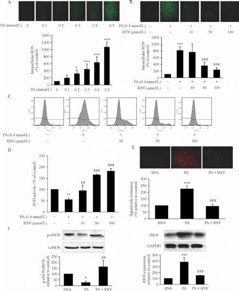
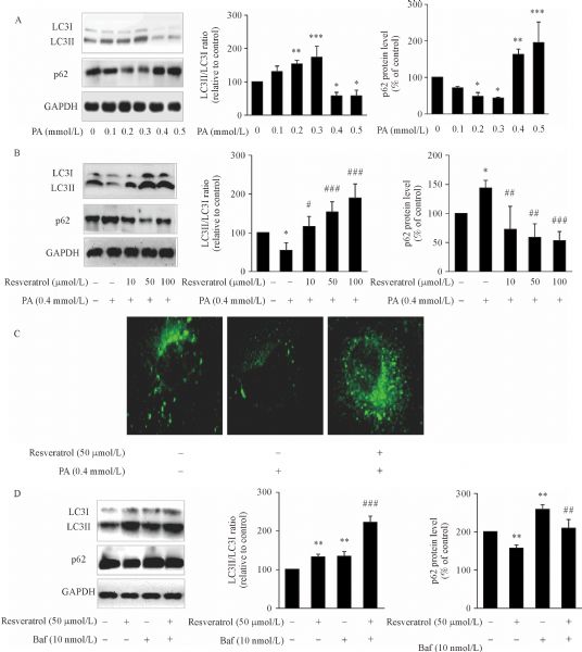
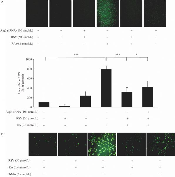
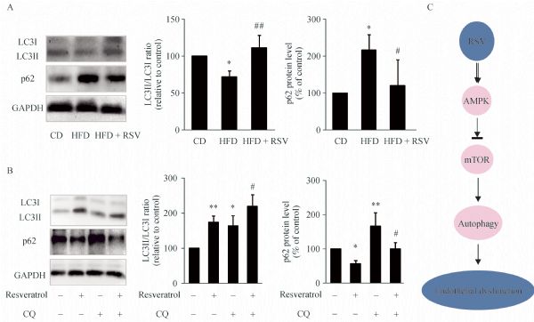
Abstract Oxidative stress induced by free fatty acid aggravates endothelial injury, which leads to diabetic cardiovascular complications. Reduction of intracellular oxidative stress may attenuate these pathogenic processes. The dietary polyphenol resveratrol reportedly exerts potential protective effects against endothelial injury. This study determined whether resveratrol can reduce the palmitic acid (PA)-induced generation of reactive oxygen species (ROS) and further explored the underlying molecular mechanisms. We found that resveratrol significantly reduced the PA-induced endothelial ROS levels in human aortic endothelial cells. Resveratrol also induced endothelial cell autophagy, which mediated the effect of resveratrol on ROS reduction. Resveratrol stimulated autophagy via the AMP-activated protein kinase (AMPK)-mTOR pathway. Taken together, these data suggest that resveratrol prevents PA-induced intracellular ROS by autophagy regulation via the AMPK-mTOR pathway. Thus, the induction of autophagy by resveratrol may provide a novel therapeutic candidate for cardioprotection in metabolic syndrome.
|
| Keywords
resveratrol
reactive oxygen species
AMPK
mTOR
autophagy
|
|
Corresponding Author(s):
Cheng Hu,Weiping Jia
|
|
Just Accepted Date: 12 September 2018
Online First Date: 13 November 2018
Issue Date: 03 December 2018
|
|
| 1 |
Incalza MA, D'Oria R, Natalicchio A, Perrini S, Laviola L, Giorgino F. Oxidative stress and reactive oxygen species in endothelial dysfunction associated with cardiovascular and metabolic diseases. Vascul Pharmacol 2018; 100: 1–19
https://doi.org/10.1016/j.vph.2017.05.005
pmid: 28579545
|
| 2 |
Morales CR, Pedrozo Z, Lavandero S, Hill JA. Oxidative stress and autophagy in cardiovascular homeostasis. Antioxid Redox Signal 2014; 20(3): 507–518
https://doi.org/10.1089/ars.2013.5359
pmid: 23641894
|
| 3 |
Saad MI, Abdelkhalek TM, Saleh MM, Kamel MA, Youssef M, Tawfik SH, Dominguez H. Insights into the molecular mechanisms of diabetes-induced endothelial dysfunction: focus on oxidative stress and endothelial progenitor cells. Endocrine 2015; 50(3): 537–567
https://doi.org/10.1007/s12020-015-0709-4
pmid: 26271514
|
| 4 |
Carrizzo A, Forte M, Damato A, Trimarco V, Salzano F, Bartolo M, Maciag A, Puca AA, Vecchione C. Antioxidant effects of resveratrol in cardiovascular, cerebral and metabolic diseases. Food Chem Toxicol 2013; 61: 215–226
https://doi.org/10.1016/j.fct.2013.07.021
pmid: 23872128
|
| 5 |
Bonnefont-Rousselot D. Resveratrol and cardiovascular diseases. Nutrients 2016; 8(5): E250
https://doi.org/10.3390/nu8050250
pmid: 27144581
|
| 6 |
Baxter RA. Anti-aging properties of resveratrol: review and report of a potent new antioxidant skin care formulation. J Cosmet Dermatol 2008; 7(1): 2–7
https://doi.org/10.1111/j.1473-2165.2008.00354.x
pmid: 18254804
|
| 7 |
Xu M, Xue W, Ma Z, Bai J, Wu S. Resveratrol reduces the incidence of portal vein system thrombosis after splenectomy in a rat fibrosis model. Oxid Med Cell Longev 2016; 2016:7453849
https://doi.org/10.1155/2016/7453849
pmid: 27433290.
|
| 8 |
Han SY, Choi YJ, Kang MK, Park JH, Kang YH. Resveratrol suppresses cytokine production linked to FcεRI-MAPK activation in IgE-antigen complex-exposed basophilic mast cells and mice. Am J Chin Med 2015; 43(8): 1605–1623
https://doi.org/10.1142/S0192415X15500913
pmid: 26621445
|
| 9 |
Diaz-Gerevini GT, Repossi G, Dain A, Tarres MC, Das UN, Eynard AR. Beneficial action of resveratrol: how and why? Nutrition 2016; 32(2): 174–178
https://doi.org/10.1016/j.nut.2015.08.017
pmid: 26706021
|
| 10 |
Novelle MG, Wahl D, Diéguez C, Bernier M, de Cabo R. Resveratrol supplementation: where are we now and where should we go? Ageing Res Rev 2015; 21: 1–15
https://doi.org/10.1016/j.arr.2015.01.002
pmid: 25625901
|
| 11 |
Antonioli M, Di Rienzo M, Piacentini M, Fimia GM. Emerging mechanisms in initiating and terminating autophagy. Trends Biochem Sci 2017; 42(1): 28–41
https://doi.org/10.1016/j.tibs.2016.09.008
pmid: 27765496
|
| 12 |
Kiffin R, Bandyopadhyay U, Cuervo AM. Oxidative stress and autophagy. Antioxid Redox Signal 2006; 8(1-2): 152–162
https://doi.org/10.1089/ars.2006.8.152
pmid: 16487049
|
| 13 |
Gu J, Hu W, Song ZP, Chen YG, Zhang DD, Wang CQ. Resveratrol-induced autophagy promotes survival and attenuates doxorubicin-induced cardiotoxicity. Int Immunopharmacol 2016; 32: 1–7
https://doi.org/10.1016/j.intimp.2016.01.002
pmid: 26774212
|
| 14 |
Nagata D, Mogi M, Walsh K. AMP-activated protein kinase (AMPK) signaling in endothelial cells is essential for angiogenesis in response to hypoxic stress. J Biol Chem 2003; 278(33): 31000–31006
https://doi.org/10.1074/jbc.M300643200
pmid: 12788940
|
| 15 |
He C, Li H, Viollet B, Zou MH, Xie Z. AMPK suppresses vascular inflammation in vivo by inhibiting signal transducer and activator of transcription-1. Diabetes 2015; 64(12): 4285–4297
https://doi.org/10.2337/db15-0107
pmid: 25858560
|
| 16 |
Youn JY, Wang T, Cai H. An ezrin/calpain/PI3K/AMPK/eNOSs1179 signaling cascade mediating VEGF-dependent endothelial nitric oxide production. Circ Res 2009; 104(1): 50–59
https://doi.org/10.1161/CIRCRESAHA.108.178467
pmid: 19038867
|
| 17 |
Zou MH, Hou XY, Shi CM, Nagata D, Walsh K, Cohen RA. Modulation by peroxynitrite of Akt- and AMP-activated kinase-dependent Ser1179 phosphorylation of endothelial nitric oxide synthase. J Biol Chem 2002; 277(36): 32552–32557
https://doi.org/10.1074/jbc.M204512200
pmid: 12107173
|
| 18 |
Gwinn DM, Shackelford DB, Egan DF, Mihaylova MM, Mery A, Vasquez DS, Turk BE, Shaw RJ. AMPK phosphorylation of raptor mediates a metabolic checkpoint. Mol Cell 2008; 30(2): 214–226
https://doi.org/10.1016/j.molcel.2008.03.003
pmid: 18439900
|
| 19 |
Zhang L, Wei J, Ren L, Zhang J, Wang J, Jing L, Yang M, Yu Y, Sun Z, Zhou X. Endosulfan induces autophagy and endothelial dysfunction via the AMPK/mTOR signaling pathway triggered by oxidative stress. Environ Pollut 2017; 220(Pt B): 843–852
https://doi.org/10.1016/j.envpol.2016.10.067
pmid: 27814983
|
| 20 |
Jo HK, Kim GW, Jeong KJ, Kim DY, Chung SH. Eugenol ameliorates hepatic steatosis and fibrosis by down-regulating SREBP1 gene expression via AMPK-mTOR-p70S6K signaling pathway. Biol Pharm Bull 2014; 37(8): 1341–1351
https://doi.org/10.1248/bpb.b14-00281
pmid: 25087956
|
| 21 |
Li XN, Song J, Zhang L, LeMaire SA, Hou X, Zhang C, Coselli JS, Chen L, Wang XL, Zhang Y, Shen YH. Activation of the AMPK-FOXO3 pathway reduces fatty acid-induced increase in intracellular reactive oxygen species by upregulating thioredoxin. Diabetes 2009; 58(10): 2246–2257
https://doi.org/10.2337/db08-1512
pmid: 19592618
|
| 22 |
Koshkin V, Wang X, Scherer PE, Chan CB, Wheeler MB. Mitochondrial functional state in clonal pancreatic β-cells exposed to free fatty acids. J Biol Chem 2003; 278(22): 19709–19715
https://doi.org/10.1074/jbc.M209709200
pmid: 12642585
|
| 23 |
Lee Y, Lee HY, Gustafsson AB. Regulation of autophagy by metabolic and stress signaling pathways in the heart. J Cardiovasc Pharmacol 2012; 60(2): 118–124
https://doi.org/10.1097/FJC.0b013e318256cdd0
pmid: 22472907
|
| 24 |
Elnakish MT, Hassanain HH, Janssen PM, Angelos MG, Khan M. Emerging role of oxidative stress in metabolic syndrome and cardiovascular diseases: important role of Rac/NADPH oxidase. J Pathol 2013; 231(3): 290–300
https://doi.org/10.1002/path.4255
pmid: 24037780
|
| 25 |
Hutcheson R, Rocic P. The metabolic syndrome, oxidative stress, environment, and cardiovascular disease: the great exploration. Exp Diabetes Res 2012; 2012: 271028
https://doi.org/10.1155/2012/271028
pmid: 22829804.
|
| 26 |
Bradamante S, Barenghi L, Villa A. Cardiovascular protective effects of resveratrol. Cardiovasc Drug Rev 2004; 22(3): 169–188
https://doi.org/10.1111/j.1527-3466.2004.tb00139.x
pmid: 15492766
|
| 27 |
Hao HD, He LR. Mechanisms of cardiovascular protection by resveratrol. J Med Food 2004; 7(3): 290–298
https://doi.org/10.1089/jmf.2004.7.290
pmid: 15383221
|
| 28 |
Xia N, Förstermann U, Li H. Resveratrol and endothelial nitric oxide. Molecules 2014; 19(10): 16102–16121
https://doi.org/10.3390/molecules191016102
pmid: 25302702
|
| 29 |
Klionsky DJ, Abdelmohsen K, Abe A, Abedin MJ, Abeliovich H, Acevedo Arozena A, et al.. Guidelines for the use and interpretation of assays for monitoring autophagy (3rd edition). Autophagy 2016; 12(1): 1–222
https://doi.org/10.1080/15548627.2015.1100356
pmid: 26799652
|
| 30 |
Gu J, Hu W, Song ZP, Chen YG, Zhang DD, Wang CQ. Resveratrol-induced autophagy promotes survival and attenuates doxorubicin-induced cardiotoxicity. Int Immunopharmacol 2016; 32: 1–7
https://doi.org/10.1016/j.intimp.2016.01.002
pmid: 26774212
|
| 31 |
Wu SB, Wu YT, Wu TP, Wei YH. Role of AMPK-mediated adaptive responses in human cells with mitochondrial dysfunction to oxidative stress. Biochim Biophys Acta 2014; 1840(4): 1331–1344
https://doi.org/10.1016/j.bbagen.2013.10.034
pmid: 24513455
|
| 32 |
Fan X, Wang J, Hou J, Lin C, Bensoussan A, Chang D, Liu J, Wang B. Berberine alleviates ox-LDL induced inflammatory factors by up-regulation of autophagy via AMPK/mTOR signaling pathway. J Transl Med 2015; 13:92
https://doi.org/10.1186/s12967-015-0450-z
pmid: 25884210
|
| 33 |
Zheng XT, Wu ZH, Wei Y, Dai JJ, Yu GF, Yuan F, Ye LC. Induction of autophagy by salidroside through the AMPK-mTOR pathway protects vascular endothelial cells from oxidative stress-induced apoptosis. Mol Cell Biochem 2017; 425(1-2): 125–138
https://doi.org/10.1007/s11010-016-2868-x
pmid: 27848074
|
|
Viewed |
|
|
|
Full text
|
|
|
|
|
Abstract
|
|
|
|
|
Cited |
|
|
|
|
| |
Shared |
|
|
|
|
| |
Discussed |
|
|
|
|