|
|
|
Microorganism-derived biological macromolecules for tissue engineering |
Naser Amini1,2, Peiman Brouki Milan1,2,3( ), Vahid Hosseinpour Sarmadi1,2, Bahareh Derakhshanmehr2, Ahmad Hivechi1,4, Fateme Khodaei5, Masoud Hamidi6, Sara Ashraf7, Ghazaleh Larijani7, Alireza Rezapour8,9() ), Vahid Hosseinpour Sarmadi1,2, Bahareh Derakhshanmehr2, Ahmad Hivechi1,4, Fateme Khodaei5, Masoud Hamidi6, Sara Ashraf7, Ghazaleh Larijani7, Alireza Rezapour8,9() |
1. Cellular and Molecular Research Center, Iran University of Medical Sciences, Tehran 1591639675, Iran
2. Institutes of Regenerative Medicine, Faculty of Advanced Technologies in Medicine, Iran University of Medical Sciences, Tehran 1449614535, Iran
3. Department of Tissue Engineering and Regenerative Medicine, Faculty of Advanced Technologies in Medicine, Iran University of Medical Sciences, Tehran 1449614535, Iran
4. Department of Pharmaceutics, University of Minnesota, MN 55455, USA
5. Burn Research Center, Department of Plastic and Reconstructive Surgery, Iran University of Medical Sciences, Tehran 1591639675, Iran
6. Department of Medical Biotechnology, Faculty of Paramedicine, Guilan University of Medical Sciences, Rasht 4477166595, Iran
7. Department of Biology, Science and Research Branch, Islamic Azad University, Tehran 1477893855, Iran
8. Cellular and Molecular Research Centre, Qom University of Medical Sciences, Qom 3715835155, Iran
9. Department of Tissue Engineering and Regenerative Medicine, School of Medicine, Qom University of Medical Sciences, Qom 3715835155, Iran |
|
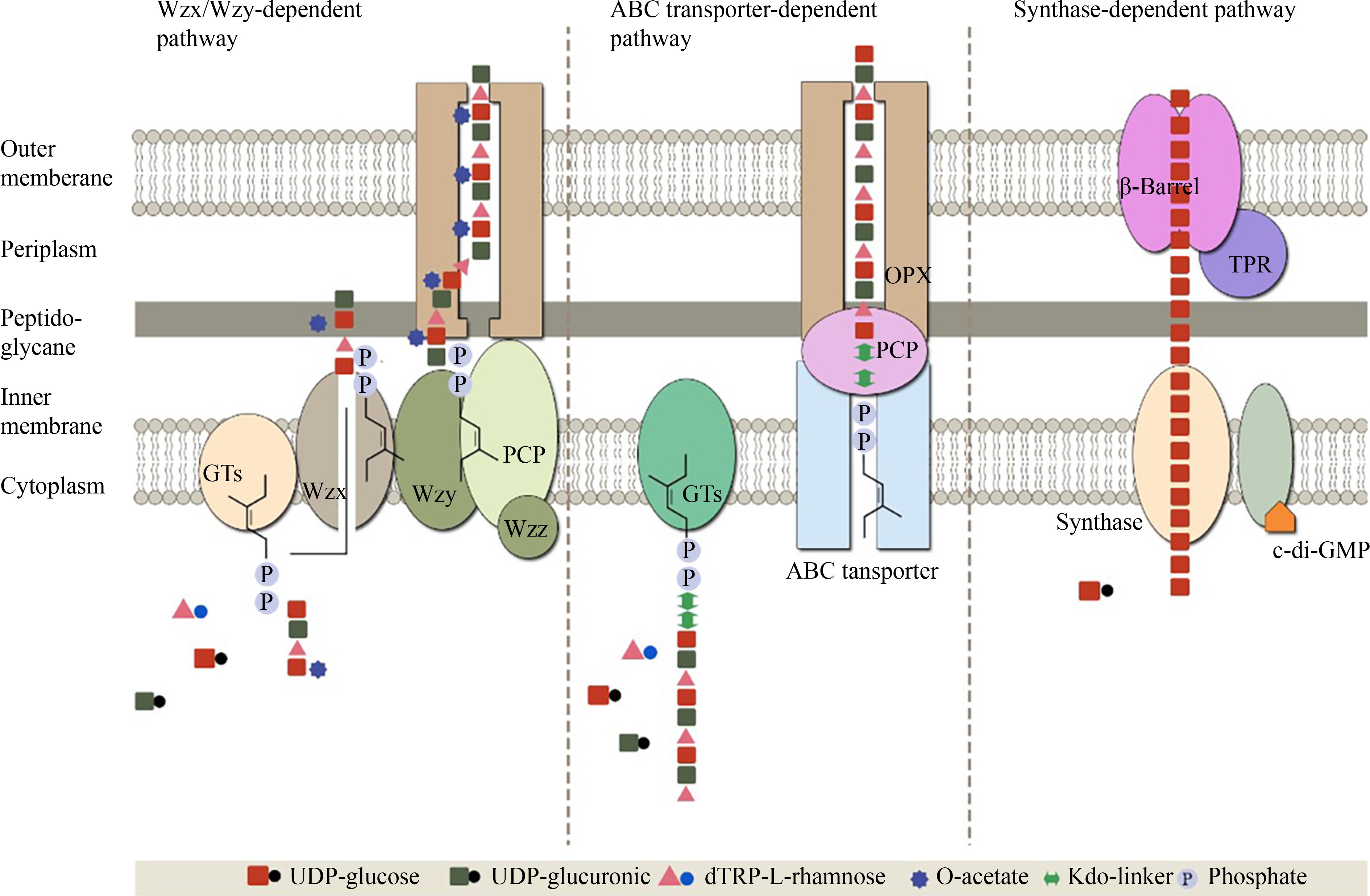
|
|
|
Abstract According to literature, certain microorganism productions mediate biological effects. However, their beneficial characteristics remain unclear. Nowadays, scientists concentrate on obtaining natural materials from live creatures as new sources to produce innovative smart biomaterials for increasing tissue reconstruction in tissue engineering and regenerative medicine. The present review aims to introduce microorganism-derived biological macromolecules, such as pullulan, alginate, dextran, curdlan, and hyaluronic acid, and their available sources for tissue engineering. Growing evidence indicates that these materials can be used as biological material in scaffolds to enhance regeneration in damaged tissues and contribute to cosmetic and dermatological applications. These natural-based materials are attractive in pharmaceutical, regenerative medicine, and biomedical applications. This study provides a detailed overview of natural-based biomaterials, their chemical and physical properties, and new directions for future research and therapeutic applications.
|
| Keywords
biological macromolecules
regenerative medicine
tissue engineering
exopolysaccharide
carbohydrate
|
|
Corresponding Author(s):
Peiman Brouki Milan,Alireza Rezapour
|
|
Just Accepted Date: 26 April 2022
Issue Date: 18 July 2022
|
|
| 1 |
C Staudt, H Horn, DC Hempel, TR Neu. Volumetric measurements of bacterial cells and extracellular polymeric substance glycoconjugates in biofilms. Biotechnol Bioeng 2004; 88( 5): 585– 592
https://doi.org/10.1002/bit.20241
|
| 2 |
UU Nwodo, E Green, AI Okoh. Bacterial exopolysaccharides: functionality and prospects. Int J Mol Sci 2012; 13( 11): 14002– 14015
https://doi.org/10.3390/ijms131114002
|
| 3 |
J Schmid, V Sieber, B Rehm. Bacterial exopolysaccharides: biosynthesis pathways and engineering strategies. Front Microbiol 2015; 6 : 496
https://doi.org/10.3389/fmicb.2015.00496
|
| 4 |
G Geesey. Microbial exopolymers: ecological and economic considerations. Am Soc Microbiol News 1982; 48 : 9– 14
|
| 5 |
A Pal, AK Paul. Microbial extracellular polymeric substances: central elements in heavy metal bioremediation. Indian J Microbiol 2008; 48( 1): 49– 64
https://doi.org/10.1007/s12088-008-0006-5
|
| 6 |
IW Sutherland. Biotechnology of Microbial Exopolysaccharides. Cambridge, UK: Cambridge University Press, 1990
|
| 7 |
MC Roper, LC Greve, JM Labavitch, BC Kirkpatrick. Detection and visualization of an exopolysaccharide produced by Xylella fastidiosa in vitro and in planta. Appl Environ Microbiol 2007; 73( 22): 7252– 7258
https://doi.org/10.1128/AEM.00895-07
|
| 8 |
C Absalon, K Van Dellen, PI Watnick. A communal bacterial adhesin anchors biofilm and bystander cells to surfaces. PLoS Pathog 2011; 7( 8): e1002210
https://doi.org/10.1371/journal.ppat.1002210
|
| 9 |
CD Nadell, BL Bassler. A fitness trade-off between local competition and dispersal in Vibrio cholerae biofilms. Proc Natl Acad Sci USA 2011; 108( 34): 14181– 14185
https://doi.org/10.1073/pnas.1111147108
|
| 10 |
AK Mandal, IK Sen, P Maity, S Chattopadhyay, R Chakraborty, S Roy, SS Islam. Structural elucidation and biological studies of a novel exopolysaccaride from Klebsiella pneumoniae PB12. Int J Biol Macromol 2015; 79 : 413– 422
https://doi.org/10.1016/j.ijbiomac.2015.04.077
|
| 11 |
F Freitas, VD Alves, MA Reis. Advances in bacterial exopolysaccharides: from production to biotechnological applications. Trends Biotechnol 2011; 29( 8): 388– 398
https://doi.org/10.1016/j.tibtech.2011.03.008
|
| 12 |
A Otero, M Vincenzini. Extracellular polysaccharide synthesis by Nostoc strains as affected by N source and light intensity. J Biotechnol 2003; 102( 2): 143– 152
https://doi.org/10.1016/S0168-1656(03)00022-1
|
| 13 |
IW Sutherland. Novel and established applications of microbial polysaccharides. Trends Biotechnol 1998; 16( 1): 41– 46
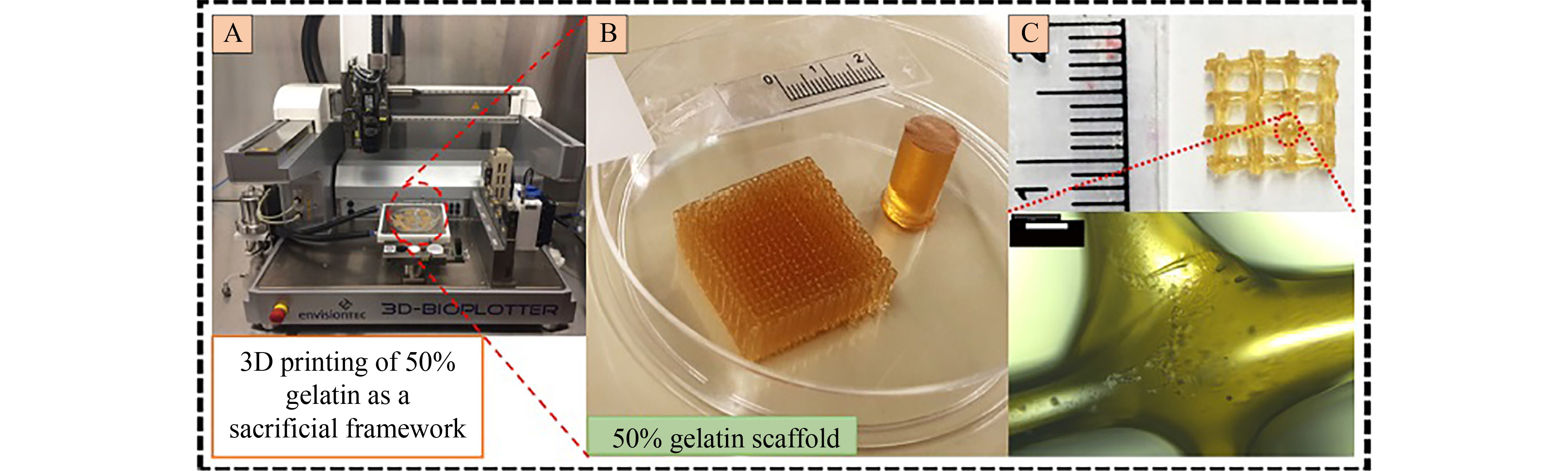
https://doi.org/10.1016/S0167-7799(97)01139-6
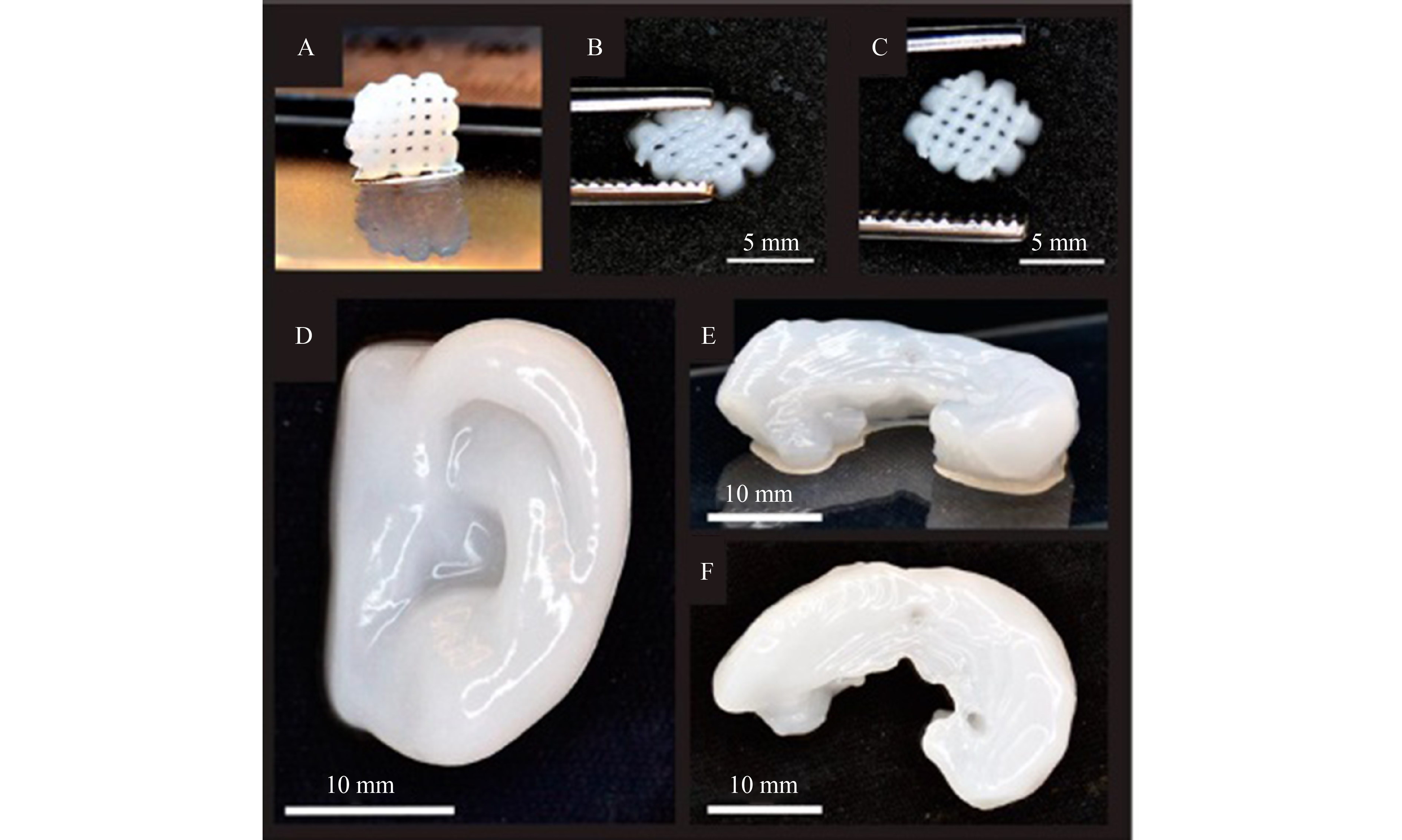
|
| 14 |
BH Rehm. Bacterial polymers: biosynthesis, modifications and applications. Nat Rev Microbiol 2010; 8( 8): 578– 592
https://doi.org/10.1038/nrmicro2354
|
| 15 |
ST Islam, JS Lam. Synthesis of bacterial polysaccharides via the Wzx/Wzy-dependent pathway. Can J Microbiol 2014; 60( 11): 697– 716
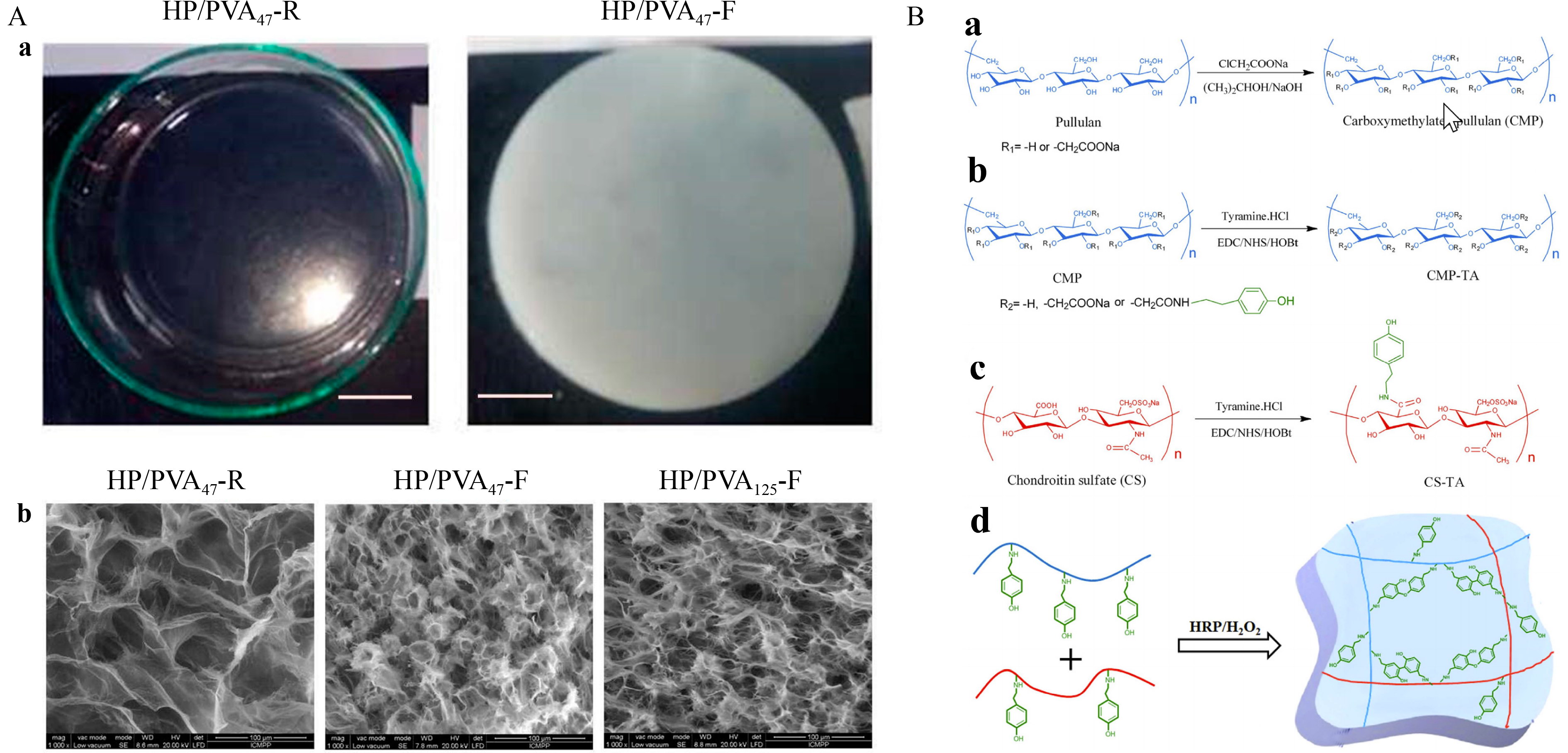
https://doi.org/10.1139/cjm-2014-0595
|
| 16 |
R Morona, L Purins, A Tocilj, A Matte, M Cygler. Sequence-structure relationships in polysaccharide co-polymerase (PCP) proteins. Trends Biochem Sci 2009; 34( 2): 78– 84
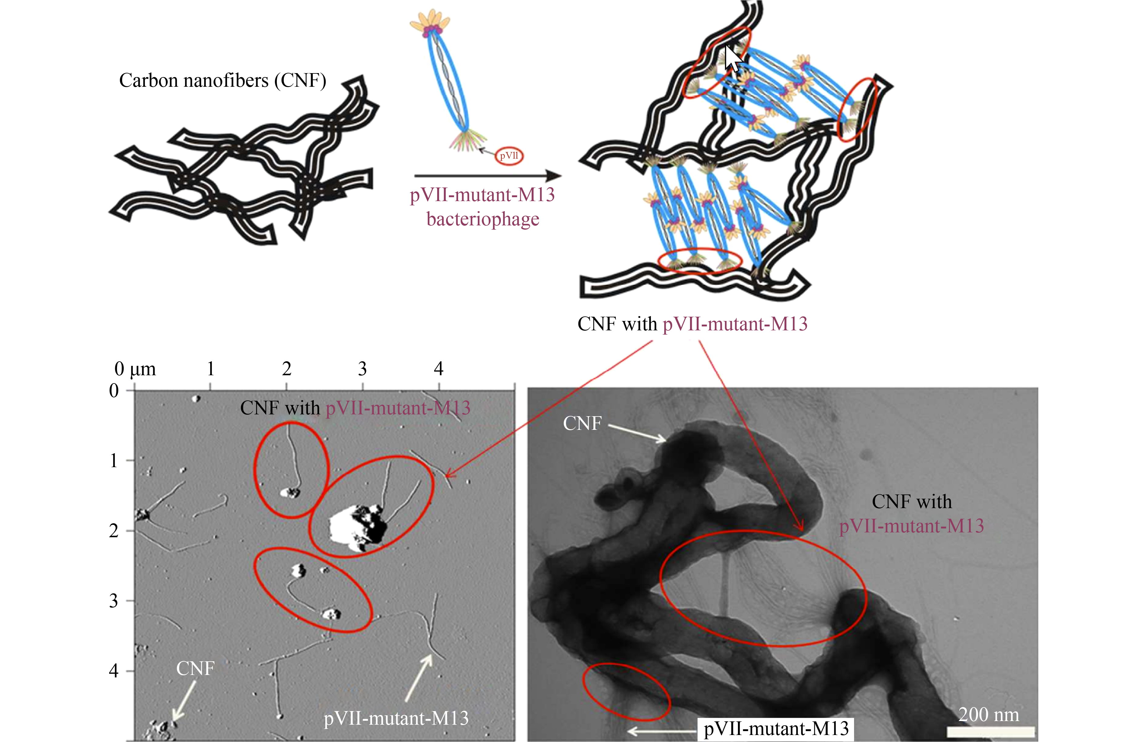
https://doi.org/10.1016/j.tibs.2008.11.001
|
| 17 |
L Cuthbertson, IL Mainprize, JH Naismith, C Whitfield. Pivotal roles of the outer membrane polysaccharide export and polysaccharide copolymerase protein families in export of extracellular polysaccharides in gram-negative bacteria. Microbiol Mol Biol Rev 2009; 73( 1): 155– 177
https://doi.org/10.1128/MMBR.00024-08
|
| 18 |
P Baker, GB Whitfield, PJ Hill, DJ Little, MJ Pestrak, H Robinson, DJ Wozniak, PL Howell. Characterization of the Pseudomonas aeruginosa glycoside hydrolase PslG reveals that its levels are critical for Psl polysaccharide biosynthesis and biofilm formation. J Biol Chem 2015; 290( 47): 28374– 28387
https://doi.org/10.1074/jbc.M115.674929
|
| 19 |
J Schmid, V Sieber. Enzymatic transformations involved in the biosynthesis of microbial exo-polysaccharides based on the assembly of repeat units. ChemBioChem 2015; 16( 8): 1141– 1147
https://doi.org/10.1002/cbic.201500035
|
| 20 |
JC Whitney, PL Howell. Synthase-dependent exopolysaccharide secretion in Gram-negative bacteria. Trends Microbiol 2013; 21( 2): 63– 72
https://doi.org/10.1016/j.tim.2012.10.001
|
| 21 |
LM Willis, C Whitfield. Structure, biosynthesis, and function of bacterial capsular polysaccharides synthesized by ABC transporter-dependent pathways. Carbohydr Res 2013; 378 : 35– 44
https://doi.org/10.1016/j.carres.2013.05.007
|
| 22 |
LM Willis, J Stupak, MR Richards, TL Lowary, J Li, C Whitfield. Conserved glycolipid termini in capsular polysaccharides synthesized by ATP-binding cassette transporter-dependent pathways in Gram-negative pathogens. Proc Natl Acad Sci U S A 2013; 110( 19): 7868– 7873
https://doi.org/10.1073/pnas.1222317110
|
| 23 |
J Schmid. Recent insights in microbial exopolysaccharide biosynthesis and engineering strategies. Curr Opin Biotechnol 2018; 53 : 130– 136
https://doi.org/10.1016/j.copbio.2018.01.005
|
| 24 |
S Comte, G Guibaud, M Baudu. Relations between extraction protocols for activated sludge extracellular polymeric substances (EPS) and EPS complexation properties: Part I. Comparison of the efficiency of eight EPS extraction methods. Enzyme Microb Technol 2006; 38( 1-2): 237– 245
https://doi.org/10.1016/j.enzmictec.2005.06.016
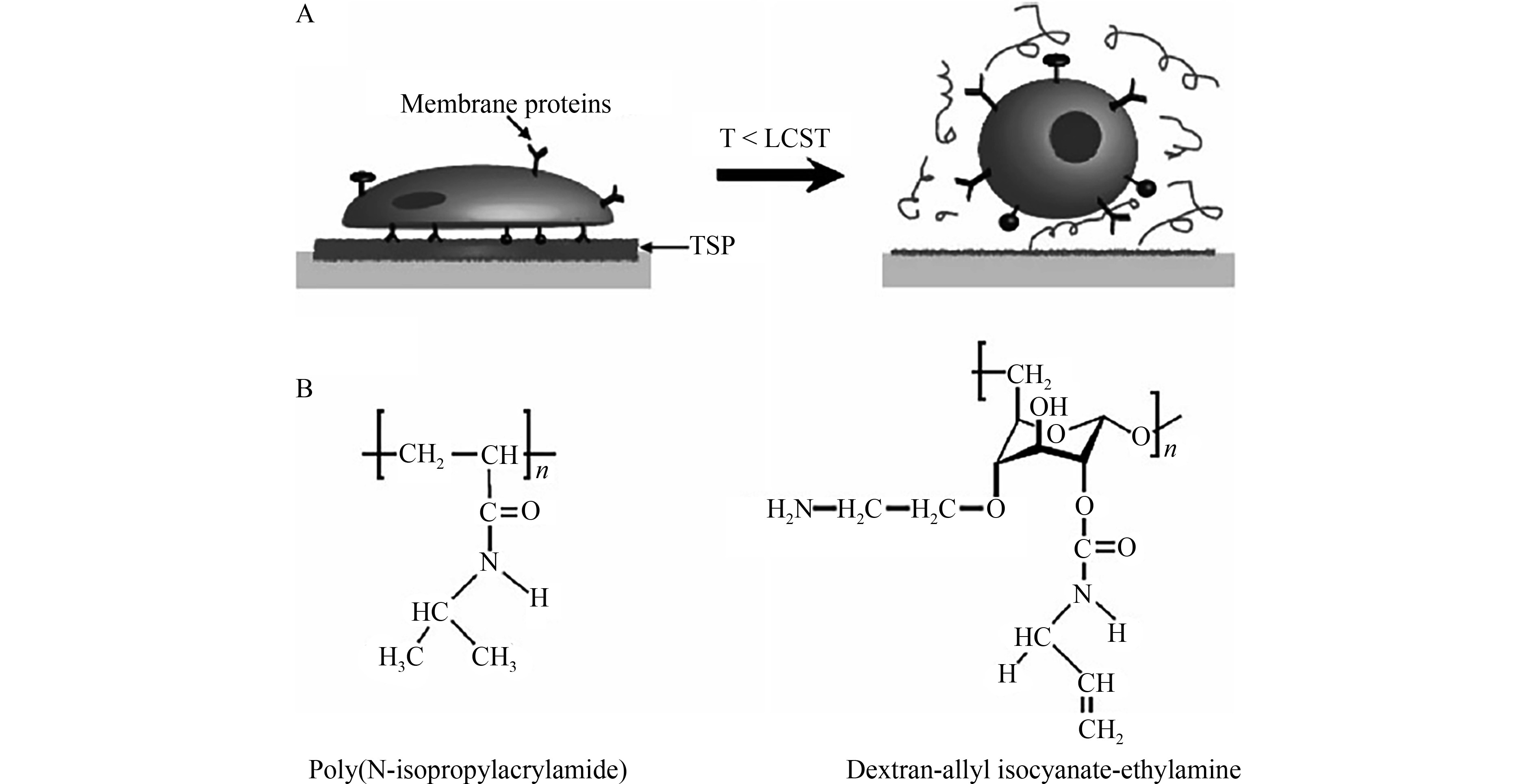
|
| 25 |
H Liu, HH Fang. Extraction of extracellular polymeric substances (EPS) of sludges. J Biotechnol 2002; 95( 3): 249– 256
https://doi.org/10.1016/S0168-1656(02)00025-1
|
| 26 |
GP Sheng, HQ Yu, Z Yu. Extraction of extracellular polymeric substances from the photosynthetic bacterium Rhodopseudomonas acidophila. Appl Microbiol Biotechnol 2005; 67( 1): 125– 130
https://doi.org/10.1007/s00253-004-1704-5
|
| 27 |
K Nouha, RS Kumar, S Balasubramanian, RD Tyagi. Critical review of EPS production, synthesis and composition for sludge flocculation. J Environ Sci (China) 2018; 66 : 225– 245
https://doi.org/10.1016/j.jes.2017.05.020
|
| 28 |
HC Flemming, J Wingender. The biofilm matrix. Nat Rev Microbiol 2010; 8( 9): 623– 633
https://doi.org/10.1038/nrmicro2415
|
| 29 |
R Späth, HC Flemming, S Wuertz. Sorption properties of biofilms. Water Sci Technol 1998; 37( 4-5): 207– 210
https://doi.org/10.2166/wst.1998.0623
|
| 30 |
I Sutherland. Biofilm exopolysaccharides: a strong and sticky framework. Microbiology (Reading) 2001; 147( 1): 3– 9
https://doi.org/10.1099/00221287-147-1-3
|
| 31 |
IW Sutherland. Exopolysaccharides in biofilms, flocs and related structures. Water Sci Technol 2001; 43( 6): 77– 86
https://doi.org/10.2166/wst.2001.0345
|
| 32 |
C Park, JT Novak. Characterization of activated sludge exocellular polymers using several cation-associated extraction methods. Water Res 2007; 41( 8): 1679– 1688
https://doi.org/10.1016/j.watres.2007.01.031
|
| 33 |
PM Joshi, AA Juwarkar. In vivo studies to elucidate the role of extracellular polymeric substances from Azotobacter in immobilization of heavy metals. Environ Sci Technol 2009; 43( 15): 5884– 5889
https://doi.org/10.1021/es900063b
|
| 34 |
J Ha, A Gélabert, AM Spormann, GE Jr Brown. Role of extracellular polymeric substances in metal ion complexation on Shewanella oneidensis: batch uptake, thermodynamic modeling, ATR-FTIR, and EXAFS study. Geochim Cosmochim Acta 2010; 74( 1): 1– 15
https://doi.org/10.1016/j.gca.2009.06.031
|
| 35 |
D Zhang, J Wang, X Pan. Cadmium sorption by EPSs produced by anaerobic sludge under sulfate-reducing conditions. J Hazard Mater 2006; 138( 3): 589– 593
https://doi.org/10.1016/j.jhazmat.2006.05.092
|
| 36 |
JH Priester, SG Olson, SM Webb, MP Neu, LE Hersman, PA Holden. Enhanced exopolymer production and chromium stabilization in Pseudomonas putida unsaturated biofilms. Appl Environ Microbiol 2006; 72( 3): 1988– 1996
https://doi.org/10.1128/AEM.72.3.1988-1996.2006
|
| 37 |
GP Sheng, HQ Yu, XY Li. Extracellular polymeric substances (EPS) of microbial aggregates in biological wastewater treatment systems: a review. Biotechnol Adv 2010; 28( 6): 882– 894
https://doi.org/10.1016/j.biotechadv.2010.08.001
|
| 38 |
ID Hay, Z Ur Rehman, MF Moradali, Y Wang, BH Rehm. Microbial alginate production, modification and its applications. Microb Biotechnol 2013; 6( 6): 637– 650
https://doi.org/10.1111/1751-7915.12076
|
| 39 |
RS Singh, GK Saini, JF Kennedy. Pullulan: microbial sources, production and applications. Carbohydr Polym 2008; 73( 4): 515– 531
https://doi.org/10.1016/j.carbpol.2008.01.003
|
| 40 |
E Díaz-Montes. Dextran: sources, structures, and properties. Polysaccharides 2021; 2( 3): 554– 565
https://doi.org/10.3390/polysaccharides2030033
|
| 41 |
JH Sze, JC Brownlie, CA Love. Biotechnological production of hyaluronic acid: a mini review. 3 Biotech 2016; 6 : 67
https://doi.org/10.1007/s13205-016-0379-9
|
| 42 |
M Shoda, Y Sugano. Recent advances in bacterial cellulose production. Biotechnol Bioprocess Eng 2005; 10( 1): 1– 8
https://doi.org/10.1007/BF02931175
|
| 43 |
F Sarwat, SA Ul Qader, A Aman, N Ahmed. Production & characterization of a unique dextran from an indigenous Leuconostoc mesenteroides CMG713. Int J Biol Sci 2008; 4( 6): 379– 386
https://doi.org/10.7150/ijbs.4.379
|
| 44 |
P Monsan, S Bozonnet, C Albenne, G Joucla, RM Willemot, M Remaud-Siméon. Homopolysaccharides from lactic acid bacteria. Int Dairy J 2001; 11( 9): 675– 685
https://doi.org/10.1016/S0958-6946(01)00113-3
|
| 45 |
M Dols, M Remaud-Simeon, RM Willemot, M Vignon, P Monsan. Characterization of the different dextransucrase activities excreted in glucose, fructose, or sucrose medium by Leuconostoc mesenteroides NRRL B-1299. Appl Environ Microbiol 1998; 64( 4): 1298– 1302
https://doi.org/10.1128/AEM.64.4.1298-1302.1998
|
| 46 |
M Hisamatsu, A Amemura, T Matsuo, H Matsuda, T Harada. Cyclic (1→2)-β-D-glucan and the octasaccharide repeating-unit of succinoglycan produced by Agrobacterium. Microbiology 1982; 128( 8): 1873– 1879
https://doi.org/10.1099/00221287-128-8-1873
|
| 47 |
H Saito, A Misaki, T Harada. A comparison of the structure of curdlan and pachyman. Agric Biol Chem 1968; 32( 10): 1261– 1269
https://doi.org/10.1080/00021369.1968.10859213
|
| 48 |
C Dhiya, IS Benny, V Gunasekar, V Ponnusami. A review on development of fermentative production of curdlan. Int J Chemtech Res 2014; 6 : 2769– 2773
|
| 49 |
TD Leathers. Biotechnological production and applications of pullulan. Appl Microbiol Biotechnol 2003; 62( 5-6): 468– 473
https://doi.org/10.1007/s00253-003-1386-4
|
| 50 |
U.S. Congress, Office of Technology Assessment. Biopolymers: Making Materials Nature’s Way-Background Paper. OTA-BP-E-102. Washington, DC: U.S. Government Printing Office, 1993
|
| 51 |
W Czaja, A Krystynowicz, S Bielecki, RM Jr Brown. Microbial cellulose—the natural power to heal wounds. Biomaterials 2006; 27( 2): 145– 151
https://doi.org/10.1016/j.biomaterials.2005.07.035
|
| 52 |
B Dixon P Abbot P Verger G Pascal M DiNovi. Pullulan. In: Safety Evaluation of Certain Food Additives. Geneva: World Health Organization, 2004: 45– 62
|
| 53 |
K Meyer, JW Palmer. The polysaccharide of the vitreous humor. J Biol Chem 1934; 107( 3): 629– 634
https://doi.org/10.1016/S0021-9258(18)75338-6
|
| 54 |
G Kogan, L Soltés, R Stern, P Gemeiner. Hyaluronic acid: a natural biopolymer with a broad range of biomedical and industrial applications. Biotechnol Lett 2007; 29( 1): 17– 25
https://doi.org/10.1007/s10529-006-9219-z
|
| 55 |
MK Cowman, S Matsuoka. Experimental approaches to hyaluronan structure. Carbohydr Res 2005; 340( 5): 791– 809
https://doi.org/10.1016/j.carres.2005.01.022
|
| 56 |
P Ross, R Mayer, M Benziman. Cellulose biosynthesis and function in bacteria. Microbiol Rev 1991; 55( 1): 35– 58
https://doi.org/10.1128/mr.55.1.35-58.1991
|
| 57 |
D Jones. Crystalline modifications of cellulose. Part V. A crystallographic study of ordered molecular arrangements. J Polym Sci e 1960; 42( 139): 173– 188
https://doi.org/10.1002/pol.1960.1204213920
|
| 58 |
B Bakhshinejad, M Sadeghizadeh. Bacteriophages and development of nanomaterials for neural regeneration. Neural Regen Res 2014; 9( 22): 1955– 1958
https://doi.org/10.4103/1673-5374.145371
|
| 59 |
SY Yoo, M Kobayashi, PP Lee, SW Lee. Early osteogenic differentiation of mouse preosteoblasts induced by collagen-derived DGEA-peptide on nanofibrous phage tissue matrices. Biomacromolecules 2011; 12( 4): 987– 996
https://doi.org/10.1021/bm1013475
|
| 60 |
AS Hoffman. Hydrogels for biomedical applications. Adv Drug Deliv Rev 2012; 64 : 18– 23
https://doi.org/10.1016/j.addr.2012.09.010
|
| 61 |
S Patel, N Kasoju, U Bora, A Goyal. Structural analysis and biomedical applications of dextran produced by a new isolate Pediococcus pentosaceus screened from biodiversity hot spot Assam. Bioresour Technol 2010; 101( 17): 6852– 6855
https://doi.org/10.1016/j.biortech.2010.03.063
|
| 62 |
M Kanke, E Tanabe, H Katayama, Y Koda, H Yoshitomi. Application of curdlan to controlled drug delivery. III. Drug release from sustained release suppositories in vitro. Biol Pharm Bull 1995; 18( 8): 1154– 1158
https://doi.org/10.1248/bpb.18.1154
|
| 63 |
M Kanke, K Koda, Y Koda, H Katayama. Application of curdlan to controlled drug delivery. I. The preparation and evaluation of theophylline-containing curdlan tablets. Pharm Res 1992; 9( 3): 414– 418
https://doi.org/10.1023/A:1015811523426
|
| 64 |
JA Bohn, JN BeMiller. (1→3)-β-D-Glucans as biological response modifiers: a review of structure-functional activity relationships. Carbohydr Polym 1995; 28( 1): 3– 14
https://doi.org/10.1016/0144-8617(95)00076-3
|
| 65 |
PA Williams. Renewable Resources for Functional Polymers and Biomaterials: Polysaccharides, Proteins and Polyesters. Cambridge, UK: Royal Society of Chemistry, 2011
|
| 66 |
M Rekha, CP Sharma. Pullulan as a promising biomaterial for biomedical applications: a perspective. Trends Biomater Artif Organs 2007; 20 : 116– 121
|
| 67 |
PN Danese, LA Pratt, R Kolter. Exopolysaccharide production is required for development of Escherichia coli K-12 biofilm architecture. J Bacteriol 2000; 182( 12): 3593– 3596
https://doi.org/10.1128/JB.182.12.3593-3596.2000
|
| 68 |
C Prigent-Combaret, O Vidal, C Dorel, P Lejeune. Abiotic surface sensing and biofilm-dependent regulation of gene expression in Escherichia coli. J Bacteriol 1999; 181( 19): 5993– 6002
https://doi.org/10.1128/JB.181.19.5993-6002.1999
|
| 69 |
GD Christensen, WA Simpson, AL Bisno, EH Beachey. Adherence of slime-producing strains of Staphylococcus epidermidis to smooth surfaces. Infect Immun 1982; 37( 1): 318– 326
https://doi.org/10.1128/iai.37.1.318-326.1982
|
| 70 |
TE Christensen, O Saxtrup, TI Hansen, BH Kristensen, BL Beck, T Plesner, IM Krogh, V Andersen, S Strandgaard. Familial myoglobinuria. A study of muscle and kidney pathophysiology in three brothers. Dan Med Bull 1983; 30( 2): 112– 115
|
| 71 |
DS Davenport, RM Massanari, MA Pfaller, MJ Bale, SA Streed, WJ Jr Hierholzer. Usefulness of a test for slime production as a marker for clinically significant infections with coagulase-negative staphylococci. J Infect Dis 1986; 153( 2): 332– 339
https://doi.org/10.1093/infdis/153.2.332
|
| 72 |
JL Drury, DJ Mooney. Hydrogels for tissue engineering: scaffold design variables and applications. Biomaterials 2003; 24( 24): 4337– 4351
https://doi.org/10.1016/S0142-9612(03)00340-5
|
| 73 |
P Gupta, K Vermani, S Garg. Hydrogels: from controlled release to pH-responsive drug delivery. Drug Discov Today 2002; 7( 10): 569– 579
https://doi.org/10.1016/S1359-6446(02)02255-9
|
| 74 |
MP Lutolf, JL Lauer-Fields, HG Schmoekel, AT Metters, FE Weber, GB Fields, JA Hubbell. Synthetic matrix metalloproteinase-sensitive hydrogels for the conduction of tissue regeneration: engineering cell-invasion characteristics. Proc Natl Acad Sci USA 2003; 100( 9): 5413– 5418
https://doi.org/10.1073/pnas.0737381100
|
| 75 |
NA Peppas, JZ Hilt, A Khademhosseini, R Langer. Hydrogels in biology and medicine: from molecular principles to bionanotechnology. Adv Mater 2006; 18( 11): 1345– 1360
https://doi.org/10.1002/adma.200501612
|
| 76 |
A Rezapour-Lactoee, H Yeganeh, R Gharibi, PB Milan. Enhanced healing of a full-thickness wound by a thermoresponsive dressing utilized for simultaneous transfer and protection of adipose-derived mesenchymal stem cells sheet. J Mater Sci Mater Med 2020; 31( 11): 101
https://doi.org/10.1007/s10856-020-06433-2
|
| 77 |
MF Cutiongco, MH Tan, MY Ng, C Le Visage, EK Yim. Composite pullulan−dextran polysaccharide scaffold with interfacial polyelectrolyte complexation fibers: a platform with enhanced cell interaction and spatial distribution. Acta Biomater 2014; 10( 10): 4410– 4418
https://doi.org/10.1016/j.actbio.2014.06.029
|
| 78 |
UU Nwodo, E Green, AI Okoh. Bacterial exopolysaccharides: functionality and prospects. Int J Mol Sci 2012; 13( 11): 14002– 14015
https://doi.org/10.3390/ijms131114002
|
| 79 |
AR Unnithan, AR Sasikala, P Murugesan, M Gurusamy, D Wu, CH Park, CS Kim. Electrospun polyurethane-dextran nanofiber mats loaded with estradiol for post-menopausal wound dressing. Int J Biol Macromol 2015; 77 : 1– 8
https://doi.org/10.1016/j.ijbiomac.2015.02.044
|
| 80 |
SG Lévesque, RM Lim, MS Shoichet. Macroporous interconnected dextran scaffolds of controlled porosity for tissue-engineering applications. Biomaterials 2005; 26( 35): 7436– 7446
https://doi.org/10.1016/j.biomaterials.2005.05.054
|
| 81 |
S Lack, V Dulong, L Picton, D Le Cerf, E Condamine. High-resolution nuclear magnetic resonance spectroscopy studies of polysaccharides crosslinked by sodium trimetaphosphate: a proposal for the reaction mechanism. Carbohydr Res 2007; 342( 7): 943– 953
https://doi.org/10.1016/j.carres.2007.01.011
|
| 82 |
C Le Visage, O Gournay, N Benguirat, S Hamidi, L Chaussumier, N Mougenot, JA Flanders, R Isnard, JB Michel, S Hatem, D Letourneur, F Norol. Mesenchymal stem cell delivery into rat infarcted myocardium using a porous polysaccharide-based scaffold: a quantitative comparison with endocardial injection. Tissue Eng Part A 2012; 18( 1-2): 35– 44
https://doi.org/10.1089/ten.tea.2011.0053
|
| 83 |
J Amedee D Letourneur C Le Visage SM Derkaoui JC Fricain S Catros. Porous polysaccharide scaffold comprising nano-hydroxyapatite and use for bone formation. Google Patents; 2017, Universite Victor Segalen Bordeaux 2 Institut National de la Sante et de la RechercheMedicale INSERM Universite Paris Diderot Paris 7. US20130224277A1
|
| 84 |
S Banerjee, M Szepes, N Dibbert, JC Rios-Camacho, A Kirschning, I Gruh, G Dräger. Dextran-based scaffolds for in-situ hydrogelation: use for next generation of bioartificial cardiac tissues. Carbohydr Polym 2021; 262 : 117924
https://doi.org/10.1016/j.carbpol.2021.117924
|
| 85 |
S Naghieh, MD Sarker, E Abelseth, X Chen. Indirect 3D bioprinting and characterization of alginate scaffolds for potential nerve tissue engineering applications. J Mech Behav Biomed Mater 2019; 93 : 183– 193
https://doi.org/10.1016/j.jmbbm.2019.02.014
|
| 86 |
A Hivechi, PB Milan, K Modabberi, M Amoupour, K Ebrahimzadeh, AR Gholipour, F Sedighi, N Amini, SH Bahrami, A Rezapour, M Hamidi, C Delattre. Synthesis and characterization of exopolysaccharide encapsulated PCL/gelatin skin substitute for full-thickness wound regeneration. Polymers (Basel) 2021; 13( 6): 854
https://doi.org/10.3390/polym13060854
|
| 87 |
WK Czaja, DJ Young, M Kawecki, RM Jr Brown. The future prospects of microbial cellulose in biomedical applications. Biomacromolecules 2007; 8( 1): 1– 12
https://doi.org/10.1021/bm060620d
|
| 88 |
PB Milan N Amini M Amoupour A Amadikuchaksaraei A Rezapour F Sefat S Kargozar Kh Ashtari M Mozafari. Scaffolds for regeneration of dermo-epidermal skin tissue. Handbook of Tissue Engineering Scaffolds. Volume 2. Elsevier Science, 2019: 193– 209
|
| 89 |
RS Singh, N Kaur, V Rana, JF Kennedy. Pullulan: a novel molecule for biomedical applications. Carbohydr Polym 2017; 171 : 102– 121
https://doi.org/10.1016/j.carbpol.2017.04.089
|
| 90 |
I Samoila, S Dinescu, GG Pircalabioru, L Marutescu, G Fundueanu, M Aflori, M Constantin. Pullulan/poly(vinyl alcohol) composite hydrogels for adipose tissue engineering. Materials (Basel) 2019; 12( 19): 3220
https://doi.org/10.3390/ma12193220
|
| 91 |
N Kulkarni L Kumar A Sorg. Fast dissolving orally consumable films. Google Patents; 2011, Johnson and Johnson Consumer Inc. US20030206942A1
|
| 92 |
F Chen, S Yu, B Liu, Y Ni, C Yu, Y Su, X Zhu, X Yu, Y Zhou, D Yan. An injectable enzymatically crosslinked carboxymethylated pullulan/chondroitin sulfate hydrogel for cartilage tissue engineering. Sci Rep 2016; 6( 1): 20014
https://doi.org/10.1038/srep20014
|
| 93 |
HE Jin, SW Lee. Engineering of M13 bacteriophage for development of tissue engineering materials. Methods Mol Biol 2018; 1776 : 487– 502
https://doi.org/10.1007/978-1-4939-7808-3_32
|
| 94 |
J Wang, L Wang, M Yang, Y Zhu, A Tomsia, C Mao. Untangling the effects of peptide sequences and nanotopographies in a biomimetic niche for directed differentiation of iPSCs by assemblies of genetically engineered viral nanofibers. Nano Lett 2014; 14( 12): 6850– 6856
https://doi.org/10.1021/nl504358j
|
| 95 |
YC Shin, JH Lee, L Jin, MJ Kim, C Kim, SW Hong, JW Oh, DW Han. Cell-adhesive matrices composed of RGD peptide-displaying M13 bacteriophage/poly(lactic-co-glycolic acid) nanofibers beneficial to myoblast differentiation. J Nanosci Nanotechnol 2015; 15( 10): 7907– 7912
https://doi.org/10.1166/jnn.2015.11214
|
| 96 |
K Szot-Karpińska, P Golec, A Leśniewski, B Pałys, F Marken, J Niedziółka-Jönsson, G Węgrzyn, M Łoś. Modified filamentous bacteriophage as a scaffold for carbon nanofiber. Bioconjug Chem 2016; 27( 12): 2900– 2910
https://doi.org/10.1021/acs.bioconjchem.6b00555
|
| 97 |
MS Aschtgen, CA Brennan, K Nikolakakis, S Cohen, M McFall-Ngai, EG Ruby. Insights into flagellar function and mechanism from the squid-vibrio symbiosis. NPJ Biofilms Microbiomes 2019; 5( 1): 32
https://doi.org/10.1038/s41522-019-0106-5
|
| 98 |
D Li, Y Zhu, T Yang, M Yang, C Mao. Bacterial flagella as an osteogenic differentiation nano-promoter. Nanoscale Horiz 2019; 4( 6): 1286– 1292
https://doi.org/10.1039/C9NH00124G
|
| 99 |
D Li, Y Zhu, T Yang, M Yang, C Mao. Genetically engineered flagella form collagen-like ordered structures for inducing stem cell differentiation. iScience 2019; 17 : 277– 287
https://doi.org/10.1016/j.isci.2019.06.036
|
| 100 |
Y Chen, X Long, W Lin, B Du, H Yin, W Lan, D Zhao, Z Li, J Li, F Luo, H Tan. Bioactive 3D porous cobalt-doped alginate/waterborne polyurethane scaffolds with a coral reef-like rough surface for nerve tissue engineering application. J Mater Chem B Mater Biol Med 2021; 9( 2): 322– 335
https://doi.org/10.1039/D0TB02347G
|
| 101 |
KM Sajesh, R Jayakumar, SV Nair, KP Chennazhi. Biocompatible conducting chitosan/polypyrrole-alginate composite scaffold for bone tissue engineering. Int J Biol Macromol 2013; 62 : 465– 471
https://doi.org/10.1016/j.ijbiomac.2013.09.028
|
| 102 |
B Yang, F Yao, L Ye, T Hao, Y Zhang, L Zhang, D Dong, W Fang, Y Wang, X Zhang, C Wang, J Li. A conductive PEDOT/alginate porous scaffold as a platform to modulate the biological behaviors of brown adipose-derived stem cells. Biomater Sci 2020; 8( 11): 3173– 3185
https://doi.org/10.1039/C9BM02012H
|
| 103 |
H Yuan, X Zheng, W Liu, H Zhang, J Shao, J Yao, C Mao, J Hui, D Fan. A novel bovine serum albumin and sodium alginate hydrogel scaffold doped with hydroxyapatite nanowires for cartilage defects repair. Colloids Surf B Biointerfaces 2020; 192 : 111041
https://doi.org/10.1016/j.colsurfb.2020.111041
|
| 104 |
M Rashtchian, A Hivechi, SH Bahrami, PB Milan, S Simorgh. Fabricating alginate/poly(caprolactone) nanofibers with enhanced bio-mechanical properties via cellulose nanocrystal incorporation. Carbohydr Polym 2020; 233 : 115873
https://doi.org/10.1016/j.carbpol.2020.115873
|
| 105 |
M Tamimi, S Rajabi, M Pezeshki-Modaress. Cardiac ECM/chitosan/alginate ternary scaffolds for cardiac tissue engineering application. Int J Biol Macromol 2020; 164 : 389– 402
https://doi.org/10.1016/j.ijbiomac.2020.07.134
|
| 106 |
R Ghaffari, H Salimi-Kenari, F Fahimipour, SM Rabiee, H Adeli, E Dashtimoghadam. Fabrication and characterization of dextran/nanocrystalline β-tricalcium phosphate nanocomposite hydrogel scaffolds. Int J Biol Macromol 2020; 148 : 434– 448
https://doi.org/10.1016/j.ijbiomac.2020.01.112
|
| 107 |
R Innocenti Malini, J Lesage, C Toncelli, G Fortunato, RM Rossi, F Spano. Crosslinking dextran electrospun nanofibers via borate chemistry: proof of concept for wound patches. Eur Polym J 2019; 110 : 276– 282
https://doi.org/10.1016/j.eurpolymj.2018.11.017
|
| 108 |
AM Moydeen, MS Ali Padusha, EF Aboelfetoh, SS Al-Deyab, MH El-Newehy. Fabrication of electrospun poly(vinyl alcohol)/dextran nanofibers via emulsion process as drug delivery system: kinetics and in vitro release study. Int J Biol Macromol 2018; 116 : 1250– 1259
https://doi.org/10.1016/j.ijbiomac.2018.05.130
|
| 109 |
Y Dong, S Zhao, W Lu, N Chen, D Zhu, Y Li. Preparation and characterization of enzymatically cross-linked gelatin/cellulose nanocrystal composite hydrogels. RSC Advances 2021; 11( 18): 10794– 10803
https://doi.org/10.1039/D1RA00965F
|
| 110 |
S Zhao, O Emery, A Wohlhauser, MM Koebel, C Adlhart, WJ Malfait. Merging flexibility with superinsulation: machinable, nanofibrous pullulan-silica aerogel composites. Mater Des 2018; 160 : 294– 302
https://doi.org/10.1016/j.matdes.2018.09.010
|
| 111 |
N Nosoudi, AJ Oommen, S Stultz, M Jordan, S Aldabel, C Hohne, J Mosser, B Archacki, A Turner, P Turner. Electrospinning live cells using gelatin and pullulan. Bioengineering (Basel) 2020; 7( 1): 21
https://doi.org/10.3390/bioengineering7010021
|
| 112 |
JA Barrera, AA Trotsyuk, ZN Maan, CA Bonham, MR Larson, PA Mittermiller, D Henn, K Chen, CJ Mays, S Mittal, AM Mermin-Bunnell, D Sivaraj, S Jing, M Rodrigues, SH Kwon, C Noishiki, J Padmanabhan, Y Jiang, S Niu, M Inayathullah, J Rajadas, M Januszyk, GC Gurtner. Adipose-derived stromal cells seeded in pullulan-collagen hydrogels improve healing in murine burns. Tissue Eng Part A 2021; 27( 11-12): 844– 856
https://doi.org/10.1089/ten.tea.2020.0320
|
| 113 |
Giustina G Della, A Gandin, L Brigo, T Panciera, S Giulitti, P Sgarbossa, D D’Alessandro, L Trombi, S Danti, G Brusatin. Polysaccharide hydrogels for multiscale 3D printing of pullulan scaffolds. Mater Des 2019; 165 : 107566
https://doi.org/10.1016/j.matdes.2018.107566
|
| 114 |
AD Dalgic, D Atila, A Karatas, A Tezcaner, D Keskin. Diatom shell incorporated PHBV/PCL-pullulan co-electrospun scaffold for bone tissue engineering. Mater Sci Eng C 2019; 100 : 735– 746
https://doi.org/10.1016/j.msec.2019.03.046
|
| 115 |
S Tang, K Chi, H Xu, Q Yong, J Yang, JM Catchmark. A covalently cross-linked hyaluronic acid/bacterial cellulose composite hydrogel for potential biological applications. Carbohydr Polym 2021; 252 : 117123
https://doi.org/10.1016/j.carbpol.2020.117123
|
| 116 |
Q Yang, Z Xie, J Hu, Y Liu. Hyaluronic acid nanofiber mats loaded with antimicrobial peptide towards wound dressing applications. Mater Sci Eng C 2021; 128 : 112319
https://doi.org/10.1016/j.msec.2021.112319
|
| 117 |
Y Cai, M Johnson, S A, Q Xu, H Tai, W Wang. A hybrid injectable and self-healable hydrogel system as 3D cell culture scaffold. Macromol Biosci 2021; 21( 9): e2100079
https://doi.org/10.1002/mabi.202100079
|
| 118 |
AK Lee, YH Lin, CH Tsai, WT Chang, TL Lin, MY Shie. Digital light processing bioprinted human chondrocyte-laden poly (γ-glutamic acid)/hyaluronic acid bio-ink towards cartilage tissue engineering. Biomedicines 2021; 9( 7): 714
https://doi.org/10.3390/biomedicines9070714
|
| 119 |
R Tarrahi, A Khataee, A Karimi, M Golizadeh, F Ebadi Fard Azar. Development of a cellulose-based scaffold for sustained delivery of curcumin. Int J Biol Macromol 2021; 183 : 132– 144
https://doi.org/10.1016/j.ijbiomac.2021.04.123
|
| 120 |
R Guo, J Li, C Chen, M Xiao, M Liao, Y Hu, Y Liu, D Li, J Zou, D Sun, V Torre, Q Zhang, R Chai, M Tang. Biomimetic 3D bacterial cellulose-graphene foam hybrid scaffold regulates neural stem cell proliferation and differentiation. Colloids Surf B Biointerfaces 2021; 200 : 111590
https://doi.org/10.1016/j.colsurfb.2021.111590
|
| 121 |
G Priya, B Madhan, U Narendrakumar, RV Suresh Kumar, I Manjubala. In vitro and in vivo evaluation of carboxymethyl cellulose scaffolds for bone tissue engineering applications. ACS Omega 2021; 6( 2): 1246– 1253
https://doi.org/10.1021/acsomega.0c04551
|
| 122 |
F Wahid, XJ Zhao, XQ Zhao, XF Ma, N Xue, XZ Liu, FP Wang, SR Jia, C Zhong. Fabrication of bacterial cellulose-based dressings for promoting infected wound healing. ACS Appl Mater Interfaces 2021; 13( 28): 32716– 32728
https://doi.org/10.1021/acsami.1c06986
|
| 123 |
Y Han, C Li, Q Cai, X Bao, L Tang, H Ao, J Liu, M Jin, Y Zhou, Y Wan, Z Liu. Studies on bacterial cellulose/poly(vinyl alcohol) hydrogel composites as tissue-engineered corneal stroma. Biomed Mater 2020; 15( 3): 035022
https://doi.org/10.1088/1748-605X/ab56ca
|
| 124 |
K Szot-Karpińska, A Leśniewski, M Jönsson-Niedziółka, F Marken, J Niedziółka-Jönsson. Electrodes modified with bacteriophages and carbon nanofibres for cysteine detection. Sens Actuators B Chem 2019; 287 : 78– 85
https://doi.org/10.1016/j.snb.2019.01.148
|
| 125 |
W Cheng, Z Zhang, R Xu, P Cai, P Kristensen, M Chen, Y Huang. Incorporation of bacteriophages in polycaprolactone/collagen fibers for antibacterial hemostatic dual-function. J Biomed Mater Res B Appl Biomater 2018; 106( 7): 2588– 2595
https://doi.org/10.1002/jbm.b.34075
|
| 126 |
Y Ma, JC Pacan, Q Wang, Y Xu, X Huang, A Korenevsky, PM Sabour. Microencapsulation of bacteriophage felix O1 into chitosan-alginate microspheres for oral delivery. Appl Environ Microbiol 2008; 74( 15): 4799– 4805
https://doi.org/10.1128/AEM.00246-08
|
| 127 |
JY Lee, WJ Chung, G Kim. A mechanically improved virus-based hybrid scaffold for bone tissue regeneration. RSC Advances 2016; 6( 60): 55022– 55032
https://doi.org/10.1039/C6RA07054J
|
| 128 |
N Gallo, H Nasser, L Salvatore, ML Natali, L Campa, M Mahmoud, L Capobianco, A Sannino, M Madaghiele. Hyaluronic acid for advanced therapies: Promises and challenges. Eur Polym J 2019; 117 : 134– 147
https://doi.org/10.1016/j.eurpolymj.2019.05.007
|
| 129 |
G Sun, S Kusuma, S Gerecht. Development of a biodegradable, temperature-sensitive dextran-based polymer as a cell-detaching substrate. Macromol Biosci 2012; 12( 1): 21– 28
https://doi.org/10.1002/mabi.201100258
|
| 130 |
R Curvello, VS Raghuwanshi, G Garnier. Engineering nanocellulose hydrogels for biomedical applications. Adv Colloid Interface Sci 2019; 267 : 47– 61
https://doi.org/10.1016/j.cis.2019.03.002
|
| 131 |
M Szekalska, A Puciłowska, A Szymańska, P Ciosek, K Winnicka. Alginate: current use and future perspectives in pharmaceutical and biomedical applications. Int J Polym Sci 2016; 2016 : 7697031
https://doi.org/10.1155/2016/7697031
|
|
Viewed |
|
|
|
Full text
|
|
|
|
|
Abstract
|
|
|
|
|
Cited |
|
|
|
|
| |
Shared |
|
|
|
|
| |
Discussed |
|
|
|
|