|
|
|
Tooth enamel and enameloid in actinopterygian fish |
I. SASAGAWA1( ), M. ISHIYAMA2, H. YOKOSUKA2, M. MIKAMI3, T. UCHIDA4 ), M. ISHIYAMA2, H. YOKOSUKA2, M. MIKAMI3, T. UCHIDA4 |
| 1. Advanced Research Centre, School of Life Dentistry at Niigata, The Nippon Dental University, Hamaura-cho 1-8, Niigata 951-8580, Japan; 2. Department of Histology, The Nippon Dental University, Hamaura-cho 1-8, Niigata 951-8580, Japan; 3. Department of Microbiology, The Nippon Dental University, Hamaura-cho 1-8, Niigata 951-8580, Japan; 4. Department of Oral Biology, Graduate School of Biomedical Sciences, Hiroshima University, Hiroshima, Japan |
|
|
|
|
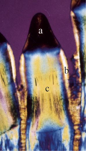
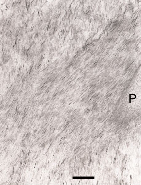
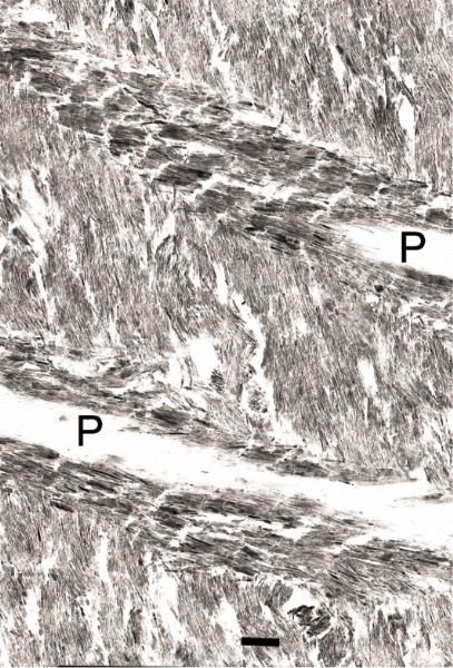
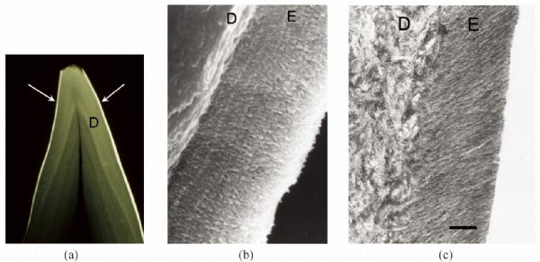
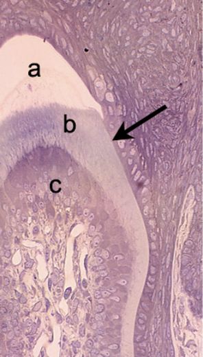
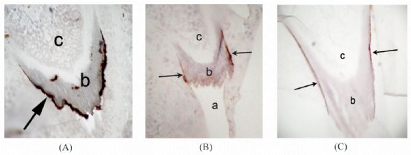
Abstract The morphological features of tooth enamel and enameloid in actinopterygian fish are reviewed to provide basic data concerning the biomineralization of teeth in lower vertebrates. Enameloid, which covers the tooth surface, is a unique well-mineralized tissue and usually has the same functions as mammalian tooth enamel. However, the development of enameloid is different from that of the enamel produced by dental epithelial cells. Enameloid is made by a combination of odontoblasts and dental epithelial cells. An organic matrix that contains collagen is provided by odontoblasts, and then dental epithelial cells dissolve the degenerate matrix and supply inorganic ions during advanced crystal growth in enameloid. It is likely that enameloid is a good model for studying the growth of well-mineralized hard tissues in vertebrates. Some actinopterygian fish possess a collar enamel layer that is situated at the surface of the tooth shaft, indicating that the origin of tooth enamel is found in fish. Collar enamel is thought to be a precursor of mammalian enamel, although it is thin and not well mineralized in comparison with enameloid. In Lepisosteus and Polypterus, both of which are living actinopterygians, both enameloid and enamel are found in the same tooth. Therefore, they are suitable materials for examining the developmental processes of enameloid and enamel and the relationship among them.
|
| Keywords
actinopterygian
enamel
enameloid
fish
tooth
|
|
Corresponding Author(s):
SASAGAWA I.,Email:ichsasgw@ngt.ndu.ac.jp
|
|
Issue Date: 05 June 2009
|
|
| 1 |
?rvig T. Phylogeny of tooth tissue: evolution of some calcified tissues in early vertebrates. In: Miles A E W. Structural and Chemical Organization of Teeth . New York: Academic Press, 1967, 45-110
|
| 2 |
Poole D F G. Phylogeny of tooth tissues: enameloid and enamel in recent vertebrates, with a note on the history of cementum. In: Miles A E W. Structural and Chemical Organization of Teeth . New York: Academic Press, 1967, 111-149
|
| 3 |
Shellis R P, Miles A E W. Autoradiographic study of the formation of enameloid and dentine matrices in teleost fishes using tritiated amino acid. Proceedings of the Royal Society of London B , 1974, 185: 51-72
doi: 10.1098/rspb.1974.0005
|
| 4 |
Sasagawa I, Ishiyama M. The structure and development of the collar enameloid in two teleost fishes, Halichoeres poecilopterus and Pagrus major. Anatomy and Embryology , 1988, 178: 499-511
doi: 10.1007/BF00305037
|
| 5 |
Reif W E. Structural convergences between enameloid of actinopterygian teeth and shark teeth. Scanning Electron Microscopy , 1979, 1979 II: 547-554
|
| 6 |
Sasagawa I. Mechanisms of mineralization in the enameloid of elasmobranches and teleosts. Connective Tissue Research , 1998, 39: 511-518
doi: 10.3109/03008209809023928
|
| 7 |
Sasagawa I. Mineralization patterns in elasmobranch fish. Microscopy Research and Technique , 2002, 59: 396-407
doi: 10.1002/jemt.10219
|
| 8 |
Sasagawa I, Ishiyama M, Akai J. Cellular influence in the formation of enameloid during odontogenesis in bony fishes. Materials Science and Engineering: C , 2006, 26: 630-634
doi: 10.1016/j.msec.2005.04.010
|
| 9 |
Prostak K, Seifert P, Skobe Z. Tooth matrix formation and mineralization in extant fishes. In: Suga S, Nakahara H. Mechanisms and Phylogeny of Mineralization in Biological Systems . Tokyo: Springer-Verlag, 1991, 465-469
|
| 10 |
Shimoda S, Tanabe T, Fukae M, . Degradation of collagen in developing enameloid of sea bream. Journal of Hard Tissue Biology , 1999, 8: 6-8
|
| 11 |
Prostak K, Skobe Z. Effects of colchicines on fish enameloid matrix formation. In: Fearnhead R W, Suga S. Tooth Enamel IV . Amsterdam: Elsevier, 1984, 525-529
|
| 12 |
Prostak K, Skobe Z. The effects of colchicine on the ultrastructure of the dental epithelium and odontoblasts of teleost tooth buds. Journal of Craniofacial Genetics and Developmental Biology , 1985, 5: 75-88
|
| 13 |
Sasagawa I. Fine structure of tooth germs during the formation of enameloid matrix in Tilapia nilotica, a teleost fish. Archives of Oral Biology , 1995, 40: 801-814
doi: 10.1016/0003-9969(95)00050-Y
|
| 14 |
Huysseune A, Takle H, Soenens M, . Unique and conserved characters in salmon tooth development. European Cells and Materials , 2007, 14: 9
|
| 15 |
Kogaya Y. Sulfated glycoconjugates in amelogenesis. Progress in Histochemistry and Cytochemistry , 1994, 29: 1-110
|
| 16 |
Sasagawa I, Ferguson M W J. Fine structure of the organic matrix remaining in the mature cap enameloid in Halichoeres poecilopterus, teleost. Archives of Oral Biology , 1990, 35: 765-770
doi: 10.1016/0003-9969(90)90101-F
|
| 17 |
Ishiyama M, Inage T, Shimokawa H. Abortive secretion of an enamel matrix in the inner enamel epithelial cells during an enameloid formation in the gar-pike, Lepisosteus oculatus (Holostei, Actinopterygii). Archives of Histology and Cytology , 2001, 64: 99-107
doi: 10.1679/aohc.64.99
|
| 18 |
Diekwisch T G H, Berman B J, Anderton X, . Membranes, minerals, and proteins of developing vertebrate enamel. Microscopy Research and Technique , 2002, 59: 373-395
doi: 10.1002/jemt.10218
|
| 19 |
Satchell P G, Anderton X, Ryu O H, . Conservation and variation in enamel protein distribution during vertebrate tooth development. Journal of Experimental Zoology , 2002, 294: 91-106
doi: 10.1002/jez.10148
|
| 20 |
Ishiyama M, Inage T, Shimokawa H, et al. Immunocytochemical detection of enamel proteins in dental matrix of certain fishes. Bulletin de l’Institut Oceanographique, Monaco , 1994, 14: 175-182
|
| 21 |
Sasagawa I. The appearance of matrix vesicles and mineralization during tooth development in three teleost fishes with well-developed enameloid and orthodentine. Archives of Oral Biology , 1988, 33: 75-86
doi: 10.1016/0003-9969(88)90049-0
|
| 22 |
Sasagawa I, Ishiyama M. Fine structural observations of the initial mineralization during enameloid formation in gar-pikes, Lepisosteus oculatus, and polypterus, Polypterus senegalus, bony fish. In: Kobayashi I, Ozawa H. Biomineralization (BIOM2001); formation, diversity, evolution and application. Proceedings of the 8th International Symposium on Biomineralizations . Kanagawa: Tokai University Press, 2003, 381-385
|
| 23 |
Sasagawa I. Fine structure of the cap enameloid and of the dental epithelial cells during enameloid mineralisation and early maturation stages in the tilapia, a teleost. Journal of Anatomy , 1997, 190: 589-600
doi: 10.1046/j.1469-7580.1997.19040589.x
|
| 24 |
Kawasaki K, Fearnhead R W. Comparative histology of tooth enamel and enameloid. In: Suga S. Mechanisms of Tooth Enamel Formation . Tokyo: Quintesessence, 1983, 229-238
|
| 25 |
Suga S, Taki Y, Ogawa M. Iron in the enameloid of perciform fish. Journal of Dental Research , 1992, 71: 1316-1325
|
| 26 |
Miake Y, Aoba T, Moreno E C, . Ultrastructural studies on crystal growth of enameloid minerals in elasmobranch and teleost fish. Calcified Tissue International , 1991, 48: 204-217
doi: 10.1007/BF02570556
|
| 27 |
Shellis R P, Miles A E W. Observations with the electron microscope on enameloid formation in common eel (Anguilla anguilla; Teleostei). Proceedings of the Royal Society of London B , 1976, 194: 253-269
doi: 10.1098/rspb.1976.0077
|
| 28 |
Sasagawa I, Ishiyama M. Fine structural and cytochemical observations on the dental epithelial cells during cap enameloid formation stages in Polypterus senegalus, a bony fish (Actinopterygii). Connective Tissue Research , 2005, 46: 33-52
doi: 10.1080/03008200590935538
|
| 29 |
Sasagawa I, Ishiyama M. Fine structural and cytochemical mapping of enamel organ during the enameloid formation stages in gars, Lepisosteus oculatus, Actinopterygii. Archives of Oral Biology , 2005, 50: 373-391
doi: 10.1016/j.archoralbio.2004.07.013
|
| 30 |
Sasagawa I, Ishiyama M, Yokosuka H, . Immunohistochemical observations on collar enamel in gars (Lepisosteus oculatus). In: Session, Bone and Teeth: Biomineralisation in Fish from Microscopy to Design of Materials, VIIth International Congress on the Biology of Fish. St. John’s, Canada . 2006
|
| 31 |
Sasagawa I, Ishiyama M. Fine structure and Ca-ATPase activity of the stratum intermedium cells during odontogenesis in gars, Lepisosteus, Actinopterygii. Connective Tissue Research , 2002, 43: 505-508
doi: 10.1080/713713532
|
| 32 |
Reif W E. Evolution of dermal skeleton and dentition in vertebrates. The odontode regulation theory. Evolutionary Biology , 1982, 15: 287-368
|
| 33 |
Smith M M. Microstructure and evolution of enamel amongst osteichthyan fishes and early tetrapods. In: Smith P, Tchernov E. Structure, Function and Evolution of Teeth. London: Freund Publishing House , 1992, 73-101
|
| 34 |
Prostak K, Seifert P, Skobe Z. Ultrastructure of developing teeth in the gar pike, (Lepisosteus). In: Fearnhead R W. Tooth Enamel V. Yokohama. Florence , 1989, 188-192
|
| 35 |
Ishiyama M, Inage T, Shimokawa H. An immunocytochemical study of amelogenin proteins in the developing tooth enamel of the gar-pike, Lepisosteus oculatus (Holostei, Actinopterygii). Archives of Histology and Cytology , 1999, 62: 191-197
doi: 10.1679/aohc.62.191
|
| 36 |
Sasagawa I, Yokosuka H, Ishiyama M, . Fine structural and immunocytochemical observations on collar enamel and ganoine in Polypterus, an actinopterygian fish. European Cells and Materials , 2007, 14: 127
|
| 37 |
Sasagawa I, Ishiyama M, Yokosuka H, . Fine structure and development of the collar enamel in gars, Lepisosteus oculatus, Actinopterygii. Frontiers of Materials Science in China , 2008, 2(2): 134-142
doi: 10.1007/s11706-008-0023-7
|
| 38 |
Shellis R P, Poole D F G. The structure of the dental hard tissues of the Coelacanthid fish Latimeria chalumnae Smith. Archives of Oral Biology , 1978, 23: 1105-1113
doi: 10.1016/0003-9969(78)90116-4
|
| 39 |
Smith M M. Enamel in the oral teeth of Latimeria chalumnae (Pisces: Actinistian): A scanning electron microscope study. Journal of Zoology, London , 1978, 185: 355-369
|
| 40 |
Sasagawa I, Ishiyama M, Kodera H. Fine structure of the pharyngeal teeth in the coelacanthid fish (Latimeria chalumnae). In: Fearnhead R W, Suga S. Tooth Enamel IV . Amsterdam: Elsevier, 1984, 462-466
|
| 41 |
Ishiyama M, Teraki Y. The fine structure and formation of hypermineralized petrodentine in the tooth plate of extant lungfish (Lepidosiren paradoxa and Protopterus sp.). Archives of Histology and Cytology , 1990, 53: 307-321
doi: 10.1679/aohc.53.307
|
| 42 |
Kemp A. Ultrastructure of the developing dentition in the Australian lungfish, Neoceratodus forsteri. In: Smith P, Tchernov E. Structure, Function and Evolution of Teeth . London: Freund Publishing House, 1992, 11-33
|
| 43 |
Kemp A. Ultrastructure of developing tooth plates in the Australian lungfish, Neoceratodus forsteri (Osteichthyes: Dipnoi). Tissue and Cell , 2003, 35: 401-426
doi: 10.1016/S0040-8166(03)00066-1
|
| 44 |
Satchell P G, Shuler C F, Diekwisch T G H. True enamel covering in teeth of the Australian lungfish Neoceratodus forsteri. Cell and Tissue Research , 2000, 299: 27-37
doi: 10.1007/s004410050003
|
| 45 |
Smith M M. Distribution and variation in enamel structure in the oral teeth of sarcopterygians: its significance for the evolution of a protoprismatic enamel. Historical Biology , 1989, 3: 97-126
doi: 10.1080/08912968909386516
|
| 46 |
Shimokawa H, Wassmer P, Sobel M E, . Characterization of cell-free translation products of mRNA from bovine ameloblasts by monoclonal and polyclonal antibodies. In: Fearnhead R W, Suga S. Tooth Enamel IV . Amsterdam: Elsevier, 1984, 161-166
|
| 47 |
Uchida T, Tanabe T, Fukae M, . Immunochemical and immunohistochemical studies, using antisera against porcine 25 kDa amelogenin, 89 kDa enamelin and the 13-17 kDa non-amelogenins, on immature enamel of the pig and rat. Histochemistry , 1991, 96: 129-138
doi: 10.1007/BF00315983
|
| 48 |
Toyosawa S, O'hUigin C, Figueroa F, . Identification and characterization of amelogenin genes in monotremes, reptiles, and amphibians. Proceedings of the National Academy of Sciences of the United States of America , 1998, 95: 13056-13061
|
| 49 |
Wang X, Ito Y, Luan X, . Amelogenin sequence and enamel biomineralization in Rana pipiens. Journal of Experimental Zoology , 2005, 304B: 177-186
doi: 10.1002/jez.b.21035
|
| 50 |
Shintani S, Kobata M, Kamakura N, . Identification and characterization of matrix metalloproteinase-20 (MMP20; enamelysin) genes in reptile and amphibian. Gene , 2007, 392: 89-97
doi: 10.1016/j.gene.2006.11.014
|
| 51 |
Kawasaki K, Suzuki T, Weiss K M. Phenogenetic drift in evolution: The changing genetic basis of vertebrate teeth. Proceedings of the National Academy of Sciences of the United States of America , 2005, 102: 18063-18068
doi: 10.1073/pnas.0509263102
|
| 52 |
Kawasaki K, Weiss K M. Evolutionary genetics of vertebrate tissue mineralization: the origin and evolution of the secretory calcium-binding phosphoprotein family. Journal of Experimental Zoology , 2005, 304B: 1-22
|
| 53 |
Kawasaki K, Weiss K M. SCPP gene evolution and the dental mineralization continuum. Journal of Dental Research , 2008, 87: 520-531
doi: 10.1177/154405910808700608
|
| 54 |
Smith M M, Hall B K. Development and evolutionary origins of vertebrate skeletogenic and odontogenic tissues. Biology Reviews , 1990, 65: 277-373
doi: 10.1111/j.1469-185X.1990.tb01427.x
|
| 55 |
Slavkin H C, Diekwish T. Evolution in tooth developmental biology: of morphology and molecules. Anatomical Record , 1996, 245: 131-150
doi: 10.1002/(SICI)1097-0185(199606)245:2<131::AID-AR3>3.0.CO;2-#
|
| 56 |
Meinke D K. A histological and histochemical study of developing teeth in Polypterus (Pisces, Actinopterygii). Archives of Oral Biology , 1982, 27: 197-206
doi: 10.1016/0003-9969(82)90053-X
|
| 57 |
Shellis P. Evolution, dental tissues. In: Osborn J W. Dental Anatomy and Embryology . Oxford: Blackwell Scientific Publications, 1981, 155-165
|
| 58 |
Benton M J. Vertebrate Palaeontology. 2nd Ed. London: Chapman & Hall, 1997, 452
|
|
Viewed |
|
|
|
Full text
|
|
|
|
|
Abstract
|
|
|
|
|
Cited |
|
|
|
|
| |
Shared |
|
|
|
|
| |
Discussed |
|
|
|
|