|
|
|
Effect of incorporating Elaeagnus angustifolia extract in PCL-PEG-PCL nanofibers for bone tissue engineering |
Vahideh R. Hokmabad1, Soodabeh Davaran2, Marziyeh Aghazadeh3,4, Effat Alizadeh3,5, Roya Salehi2( ), Ali Ramazani1() ), Ali Ramazani1() |
1. Department of Chemistry, University of Zanjan, Zanjan, Iran
2. Drug Applied Research Center and Department of Medical Nanotechnology, Faculty of Advanced Medical Science, Tabriz University of Medical Sciences, Tabriz, Iran
3. Stem Cell Research Center, Tabriz University of Medical Sciences, Tabriz, Iran
4. Oral Medicine Department of Dental Faculty, Tabriz University of Medical Sciences, Tabriz, Iran
5. Department of Medical Biotechnology, Faculty of Advanced Medical Science, Tabriz University of Medical Sciences, Tabriz, Iran |
|
|
|
|
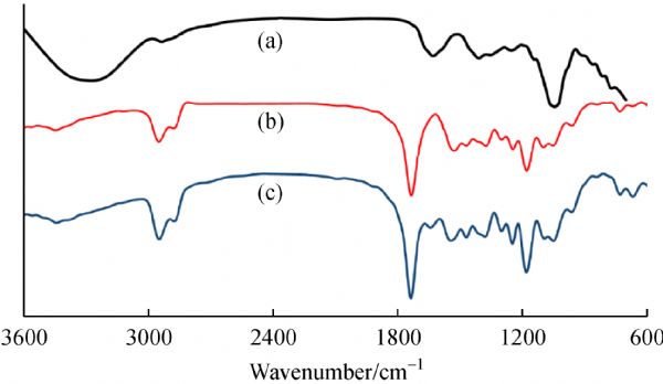
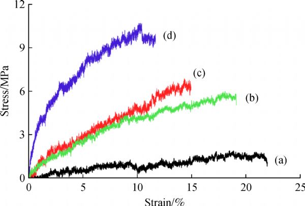
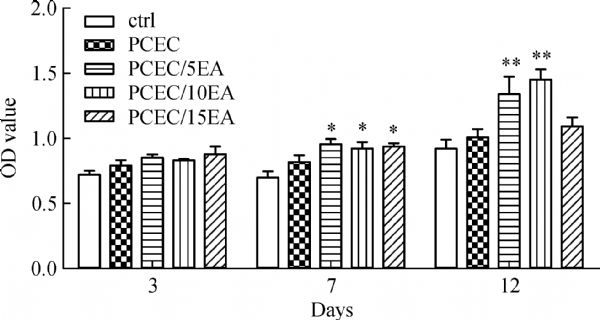
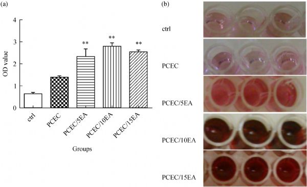
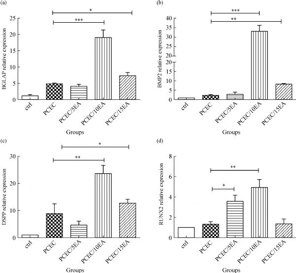
Abstract Plants have been used for medicinal purposes for thousands of years but they are still finding new uses in modern times. For example, Elaeagnus angustifolia (EA) is a medicinal herb with antinociceptive, anti-inflammatory, antibacterial and antioxidant properties and it is widely used in the treatment of rheumatoid arthritis and osteoarthritis. EA extract was loaded onto poly(ϵ-caprolactone)-poly(ethylene glycol)-poly(ϵ-caprolactone) (PCL-PEG-PCL/EA) nanofibers and their potential applications for bone tissue engineering were studied. The morphology and chemical properties of the fibers were evaluated using Fourier transform infrared spectroscopy, field emission scanning electron microscopy, contact angle measurements and mechanical tests. All the samples had bead-free morphologies with average diameters ranging from 100 to 200 nm. The response of human cells to the PCL-PEG-PCL/EA nanofibers was evaluated using human dental pulp stem cells (hDPSCs). The hDPSCs had better adhesion and proliferation capacity on the EA loaded nanofibers than on the pristine PCL-PEG-PCL nanofibers. An alizarin red S assay and the alkaline phosphatase activity confirmed that the nanofibrous scaffolds induced osteoblastic performance in the hDPSCs. The quantitative real time polymerase chain reaction results confirmed that the EA loaded nanofibrous scaffolds had significantly upregulated gene expression correlating to osteogenic differentiation. These results suggest that PCL-PEG-PCL/EA nanofibers might have potential applications for bone tissue engineering.
|
| Keywords
Elaeagnus angustifolia
scaffold
electrospinning
human dental pulp stem cell
tissue engineering
|
|
Corresponding Author(s):
Roya Salehi,Ali Ramazani
|
|
Just Accepted Date: 14 May 2018
Online First Date: 23 January 2019
Issue Date: 25 February 2019
|
|
| 1 |
MKikuchi, S Itoh, SIchinose, KShinomiya, JTanaka. Self-organization mechanism in a bone-like hydroxyapatite/collagen nanocomposite synthesized in vitro and its biological reaction in vivo. Biomaterials, 2001, 22(13): 1705–1711
https://doi.org/10.1016/S0142-9612(00)00305-7
pmid: 11396873
|
| 2 |
M SChapekar. Tissue engineering: Challenges and opportunities. Journal of Biomedical Materials Research: Part A, 2000, 53(6): 617–620
https://doi.org/10.1002/1097-4636(2000)53:6<617::AID-JBM1>3.0.CO;2-C
pmid: 11074418
|
| 3 |
NShadjou, M Hasanzadeh. Graphene and its nanostructure derivatives for use in bone tissue engineering: Recent advances. Journal of Biomedical Materials Research: Part A, 2016, 104(5): 1250–1275
https://doi.org/10.1002/jbm.a.35645
pmid: 26748447
|
| 4 |
MDiba, M Kharaziha, MFathi, MGholipourmalekabadi, ASamadikuchaksaraei. Preparation and characterization of polycaprolactone/forsterite nanocomposite porous scaffolds designed for bone tissue regeneration. Composites Science and Technology, 2012, 72(6): 716–723
https://doi.org/10.1016/j.compscitech.2012.01.023
|
| 5 |
YYu, B Sun, CYi, XMo. Stem cell homing-based tissue engineering using bioactive materials. Frontiers of Materials Science, 2017, 11(2): 93–105
https://doi.org/10.1007/s11706-017-0373-0
|
| 6 |
KVerma, R Bains, V KBains, MRawtiya, KLoomba, S CSrivastava. Therapeutic potential of dental pulp stem cells in regenerative medicine: An overview. Dental Research Journal, 2014, 11(3): 302–308
pmid: 25097638
|
| 7 |
P DPotdar, Y D Jethmalani. Human dental pulp stem cells: Applications in future regenerative medicine. World Journal of Stem Cells, 2015, 7(5): 839–851
https://doi.org/10.4252/wjsc.v7.i5.839
pmid: 26131314
|
| 8 |
Y YJo, H J Lee, S Y Kook, H W Choung, J Y Park, J H Chung, Y H Choung, E S Kim, H C Yang, P H Choung. Isolation and characterization of postnatal stem cells from human dental tissues. Tissue Engineering, 2007, 13(4): 767–773
https://doi.org/10.1089/ten.2006.0192
pmid: 17432951
|
| 9 |
SGronthos, J Brahim, WLi, L WFisher, NCherman, ABoyde, PDenBesten, P GRobey, SShi. Stem cell properties of human dental pulp stem cells. Journal of Dental Research, 2002, 81(8): 531–535
https://doi.org/10.1177/154405910208100806
pmid: 12147742
|
| 10 |
RHass, C Kasper, SBöhm, RJacobs. Different populations and sources of human mesenchymal stem cells (MSC): A comparison of adult and neonatal tissue-derived MSC. Cell Communication and Signaling, 2011, 9(1): 12–25
https://doi.org/10.1186/1478-811X-9-12
pmid: 21569606
|
| 11 |
RKhanna-Jain, B Mannerström, AVuorinen, G KSándor, RSuuronen, SMiettinen. Osteogenic differentiation of human dental pulp stem cells on β-tricalcium phosphate/poly (L-lactic acid/caprolactone) three-dimensional scaffolds. Journal of Tissue Engineering, 2012, 3(1): 2041731412467998
https://doi.org/10.1177/2041731412467998
pmid: 23316276
|
| 12 |
FPaduano, M Marrelli, L JWhite, K MShakesheff, MTatullo. Odontogenic differentiation of human dental pulp stem cells on hydrogel scaffolds derived from decellularized bone extracellular matrix and collagen type I. PLoS One, 2016, 11(2): e0148225
https://doi.org/10.1371/journal.pone.0148225
pmid: 26882351
|
| 13 |
MMiura, S Gronthos, MZhao, BLu, L W Fisher, P G Robey, S Shi. SHED: Stem cells from human exfoliated deciduous teeth. Proceedings of the National Academy of Sciences of the United States of America, 2003, 100(10): 5807–5812
https://doi.org/10.1073/pnas.0937635100
pmid: 12716973
|
| 14 |
JKarbanová, TSoukup, JSuchánek, JMokrý. Osteogenic differentiation of human dental pulp-derived stem cells under various ex-vivo culture conditions. Acta Medica, 2010, 53(2): 79–84
https://doi.org/10.14712/18059694.2016.64
pmid: 20672743
|
| 15 |
FAsghari, R Salehi, MAgazadeh, EAlizadeh, KAdibkia, MSamiei, AAkbarzadeh, N AAval, SDavaran. The odontogenic differentiation of human dental pulp stem cells on hydroxyapatite-coated biodegradable nanofibrous scaffolds. International Journal of Polymeric Materials and Polymeric Biomaterials, 2016, 65(14): 720–728
https://doi.org/10.1080/00914037.2016.1163564
|
| 16 |
SFu, P Ni, BWang, BChu, J Peng, LZheng, XZhao, F Luo, YWei, ZQian. In vivo biocompatibility and osteogenesis of electrospun poly(ε-caprolactone)-poly(ethylene glycol)-poly(ε-caprolactone)/nano-hydroxyapatite composite scaffold. Biomaterials, 2012, 33(33): 8363–8371
https://doi.org/10.1016/j.biomaterials.2012.08.023
pmid: 22921926
|
| 17 |
W JLi, C T Laurencin, E J Caterson, R S Tuan, F K Ko. Electrospun nanofibrous structure: A novel scaffold for tissue engineering. Journal of Biomedical Materials Research: Part A, 2002, 60(4): 613–621
https://doi.org/10.1002/jbm.10167
pmid: 11948520
|
| 18 |
CLi, C Vepari, H JJin, H JKim, D LKaplan. Electrospun silk-BMP-2 scaffolds for bone tissue engineering. Biomaterials, 2006, 27(16): 3115–3124
https://doi.org/10.1016/j.biomaterials.2006.01.022
pmid: 16458961
|
| 19 |
RZhang, P X Ma. Poly(α-hydroxyl acids)/hydroxyapatite porous composites for bone-tissue engineering. I. Preparation and morphology. Journal of Biomedical Materials Research, 1999, 44(4): 446–455
https://doi.org/10.1002/(SICI)1097-4636(19990315)44:4<446::AID-JBM11>3.0.CO;2-F
pmid: 10397949
|
| 20 |
Y BKim, G H Kim. PCL/alginate composite scaffolds for hard tissue engineering: Fabrication, characterization, and cellular activities. ACS Combinatorial Science, 2015, 17(2): 87–99
https://doi.org/10.1021/co500033h
pmid: 25541639
|
| 21 |
IMaitlo, S Ali, M YAkram, F KShehzad, JNie. Binary phase solid-state photopolymerization of acrylates: Design, characterization and biomineralization of 3D scaffolds for tissue engineering. Frontiers of Materials Science, 2017, 11(4): 307–317
https://doi.org/10.1007/s11706-017-0394-8
|
| 22 |
D HReneker, I Chun. Nanometre diameter fibres of polymer, produced by electrospinning. Nanotechnology, 1996, 7(3): 216–223
https://doi.org/10.1088/0957-4484/7/3/009
|
| 23 |
XZong, H Bien, C YChung, LYin, D Fang, B SHsiao, BChu, E Entcheva. Electrospun fine-textured scaffolds for heart tissue constructs. Biomaterials, 2005, 26(26): 5330–5338
https://doi.org/10.1016/j.biomaterials.2005.01.052
pmid: 15814131
|
| 24 |
T JSill, H A von Recum. Electrospinning: Applications in drug delivery and tissue engineering. Biomaterials, 2008, 29(13): 1989–2006
https://doi.org/10.1016/j.biomaterials.2008.01.011
pmid: 18281090
|
| 25 |
NOgata, S Yamaguchi, NShimada, GLu, T Iwata, KNakane, TOgihara. Poly(lactide) nanofibers produced by a melt-electrospinning system with a laser melting device. Journal of Applied Polymer Science, 2007, 104(3): 1640–1645
https://doi.org/10.1002/app.25782
|
| 26 |
M RForoughi, S Karbasi, MKhoroushi, A AKhademi. Polyhydroxybutyrate/chitosan/bioglass nanocomposite as a novel electrospun scaffold: Fabrication and characterization. Journal of Porous Materials, 2017, 24(6): 1447–1460
https://doi.org/10.1007/s10934-017-0385-2
|
| 27 |
S RBhattarai, NBhattarai, H KYi, P HHwang, D ICha, H YKim. Novel biodegradable electrospun membrane: Scaffold for tissue engineering. Biomaterials, 2004, 25(13): 2595–2602
https://doi.org/10.1016/j.biomaterials.2003.09.043
pmid: 14751745
|
| 28 |
DPuppi, A M Piras, N Detta, DDinucci, FChiellini. Poly(lactic-co-glycolic acid) electrospun fibrous meshes for the controlled release of retinoic acid. Acta Biomaterialia, 2010, 6(4): 1258–1268
https://doi.org/10.1016/j.actbio.2009.08.015
pmid: 19683605
|
| 29 |
GChouzouri, M Xanthos. In vitro bioactivity and degradation of polycaprolactone composites containing silicate fillers. Acta Biomaterialia, 2007, 3(5): 745–756
https://doi.org/10.1016/j.actbio.2007.01.005
pmid: 17392042
|
| 30 |
YHosseini, R Emadi, MKharaziha, ADoostmohammadi. Reinforcement of electrospun poly(ε-caprolactone) scaffold using diopside nanopowder to promote biological and physical properties. Journal of Applied Polymer Science, 2017, 134(6): 44433–44441
https://doi.org/10.1002/app.44433
|
| 31 |
YDu, X Chen, Y HKoh, BLei. Facilely fabricating PCL nanofibrous scaffolds with hierarchical pore structure for tissue engineering. Materials Letters, 2014, 122(22): 62–65
https://doi.org/10.1016/j.matlet.2014.02.031
|
| 32 |
J MMiszuk, T Xu, QYao, FFang, J D Childs, Z Hong, JTao, HFong, H Sun. Functionalization of PCL-3D electrospun nanofibrous scaffolds for improved BMP2-induced bone formation. Applied Materials Today, 2018, 10(1): 194–202
https://doi.org/10.1016/j.apmt.2017.12.004
pmid: 29577064
|
| 33 |
AValizadeh, M Bakhtiary, AAkbarzadeh, RSalehi, S MFrakhani, OEbrahimi, MRahmati-Yamchi, SDavaran. Preparation and characterization of novel electrospun poly(ε-caprolactone)-based nanofibrous scaffolds. Artificial Cells, Nanomedicine, and Biotechnology, 2016, 44(2): 504–509
https://doi.org/10.3109/21691401.2014.965310
pmid: 25307268
|
| 34 |
PNi, Q Ding, MFan, JLiao, Z Qian, JLuo, XLi, F Luo, ZYang, YWei. Injectable thermosensitive PEG-PCL-PEG hydrogel/acellular bone matrix composite for bone regeneration in cranial defects. Biomaterials, 2014, 35(1): 236–248
https://doi.org/10.1016/j.biomaterials.2013.10.016
pmid: 24138826
|
| 35 |
M BSá, M TRalph, D C ONascimento, C SRamos, I M SBarbosa, F BSá, JLima-Filho. Phytochemistry and preliminary assessment of the antibacterial activity of chloroform extract of Amburana cearensis (Allemão) AC Sm. against Klebsiella pneumoniae carbapenemase-producing strains. Evidence-Based Complementary and Alternative Medicine, 2014, 2014: 1–7
|
| 36 |
ANageeb, A Al-Tawashi, A HMohammad Emwas, Z Abdel-Halim Al-Talla, NAl-Rifai. Comparison of artemisia annua bioactivities between traditional medicine and chemical extracts. Current Bioactive Compounds, 2013, 9(4): 324–332
https://doi.org/10.2174/157340720904140404151439
pmid: 24761137
|
| 37 |
M SSabir, D S Ahmad, I M Hussain, K M Tahir. Antibacterial activity of Elaeagnus umbellata (Thunb.) a medicinal plant from Pakistan. Saudi Medical Journal, 2007, 28(2): 259–263
pmid: 17268691
|
| 38 |
SSuganya, J Venugopal, SRamakrishna, BLakshmi, VGiri Dev. Herbally derived polymeric nanofibrous scaffolds for bone tissue regeneration. Journal of Applied Polymer Science, 2014, 131(3): 39835–39845
https://doi.org/10.1002/app.39835
|
| 39 |
D KKim, J I Kim, T I Hwang, B R Sim, G Khang. Bioengineered osteoinductive Broussonetia kazinoki/silk fibroin composite scaffolds for bone tissue regeneration. ACS Applied Materials & Interfaces, 2017, 9(2): 1384–1394
https://doi.org/10.1021/acsami.6b14351
pmid: 28001353
|
| 40 |
SSuganya, T Senthil Ram, BLakshmi, VGiridev. Herbal drug incorporated antibacterial nanofibrous mat fabricated by electrospinning: An excellent matrix for wound dressings. Journal of Applied Polymer Science, 2011, 121(5): 2893–2899
https://doi.org/10.1002/app.33915
|
| 41 |
TTalaei-Khozani, Z Vojdani, FDehghani, EHeidari, EKharazinejad, M RPanjehshahin. Toxic effects of Elaeagnus angustifolia fruit extract on chondrogenesis and osteogenesis in mouse limb buds. Tokai Journal of Experimental and Clinical Medicine, 2011, 36(3): 63–70
pmid: 21932186
|
| 42 |
AKhodakarm-Tafti, D Mehrabani, LHomafar, GFarjanikish. Healing effects of Elaeagnus angustifolia extract in experimentally induced ulcerative colitis in rats. Journal of Pharmacologicaly Toxicologicaly, 2015, 10(1): 29–35
https://doi.org/10.3923/jpt.2015.29.35
|
| 43 |
M HFarzaei, R Bahramsoltani, ZAbbasabadi, RRahimi. A comprehensive review on phytochemical and pharmacological aspects of Elaeagnus angustifolia L. Journal of Pharmacy and Pharmacology, 2015, 67(11): 1467–1480
https://doi.org/10.1111/jphp.12442
pmid: 26076872
|
| 44 |
HHosseinzadeh, R Rahimi. Anti-inflammatory effects of Elaeagnus angustifolia L. fruits in mice and rats. Iranian Journal of Medical Sciences, 1999, 24: 143–147
|
| 45 |
MRamezani, H Hosseinzadeh, NDaneshmand. Antinociceptive effect of Elaeagnus angustifolia fruit seeds in mice. Fitoterapia, 2001, 72(3): 255–262
https://doi.org/10.1016/S0367-326X(00)00290-2
pmid: 11295301
|
| 46 |
HHosseinzadeh, M Ramezani, NNamjo. Muscle relaxant activity of Elaeagnus angustifolia L. fruit seeds in mice. Journal of Ethnopharmacology, 2003, 84(2-3): 275–278
https://doi.org/10.1016/S0378-8741(02)00331-8
pmid: 12648826
|
| 47 |
AAhmadiani, J Hosseiny, SSemnanian, MJavan, FSaeedi, MKamalinejad, SSaremi. Antinociceptive and anti-inflammatory effects of Elaeagnus angustifolia fruit extract. Journal of Ethnopharmacology, 2000, 72(1-2): 287–292
https://doi.org/10.1016/S0378-8741(00)00222-1
pmid: 10967484
|
| 48 |
IGürbüz, OUstün, EYesilada, ESezik, OKutsal. Anti-ulcerogenic activity of some plants used as folk remedy in Turkey. Journal of Ethnopharmacology, 2003, 88(1): 93–97
https://doi.org/10.1016/S0378-8741(03)00174-0
pmid: 12902057
|
| 49 |
MMehrabani Natanzi, PPasalar, MKamalinejad, A RDehpour, S MTavangar, RSharifi, NGhanadian, MRahimi-Balaei, SGerayesh-Nejad. Effect of aqueous extract of Elaeagnus angustifolia fruit on experimental cutaneous wound healing in rats. Acta Medica Iranica, 2012, 50(9): 589–596
pmid: 23165807
|
| 50 |
YPanahi, G H Alishiri, N Bayat, S MHosseini, ASahebkar. Efficacy of Elaeagnus Angustifolia extract in the treatment of knee osteoarthritis: A randomized controlled trial. EXCLI Journal, 2016, 15: 203–210
pmid: 27330526
|
| 51 |
RHamidpour, S Hamidpour, MHamidpour, MShahlari, MSohraby, NShahlari, RHamidpour. Russian olive (Elaeagnus angustifolia L.): From a variety of traditional medicinal applications to its novel roles as active antioxidant, anti-inflammatory, anti-mutagenic and analgesic agent. Journal of Traditional and Complementary Medicine, 2016, 7(1): 24–29
https://doi.org/10.1016/j.jtcme.2015.09.004
pmid: 28053884
|
| 52 |
ZAmiri Tehranizadeh, ABaratian, HHosseinzadeh. Russian olive (Elaeagnus angustifolia) as a herbal healer. BioImpacts, 2016, 6(3): 155–167
https://doi.org/10.15171/bi.2016.22
pmid: 27853679
|
| 53 |
MMofid, S H Sadraie, H Imani, GTorkaman, GKaka, M R Naghii, G Alishiri, M HAsadi. The effect of mesenchymal stem cells and aqueous extract of Elaeagnus angustifolia on the mechanical properties of articular cartilage in an experimental model of rat osteoarthritis. Anatomical Sciences Journal, 2015, 12(2): 68–74
|
| 54 |
M HDabbaghmanesh, ANoorafshan, PTalezadeh, NTanideh, FKoohpeyma, AIraji, MBakhshayeshkaram, NMontazeri-Najafabady. Stereological investigation of the effect of Elaeagnus angustifolia fruit hydroalcoholic extract on osteoporosis in ovariectomized rats. Avicenna Journal of Phytomedicine, 2017, 7(3): 261–274
pmid: 28748173
|
| 55 |
LJunqueira, J Carneiro, RKelley. Adipose tissue. In: Malley J, Lebowiz H, Boyle P J, eds. Basic Histology Text & Atlas. 11th ed. New York: McGraw-Hill, 2005: 123–127
|
| 56 |
RGarcía-Villalba, MLarrosa, SPossemiers, F ATomás-Barberán, J CEspín. Bioavailability of phenolics from an oleuropein-rich olive (Olea europaea) leaf extract and its acute effect on plasma antioxidant status: Comparison between pre- and postmenopausal women. European Journal of Nutrition, 2014, 53(4): 1015–1027
https://doi.org/10.1007/s00394-013-0604-9
pmid: 24158653
|
| 57 |
J RChen, O P Lazarenko, X Wu, JKang, M LBlackburn, KShankar, T MBadger, M JRonis. Dietary-induced serum phenolic acids promote bone growth via p38 MAPK/β-catenin canonical Wnt signaling. Journal of Bone and Mineral Research, 2010, 25(11): 2399–2411
https://doi.org/10.1002/jbmr.137
pmid: 20499363
|
| 58 |
MBakhtiari, R Salehi, AAkbarzadeh, SDavaran. Development of novel doxorubicin loaded biodegradable polymeric nanofibers as the anticancer drug delivery systems. BioNanoScience, 2018, 8(1): 60–66
https://doi.org/10.1007/s12668-017-0421-3
|
| 59 |
FAjalloueian, H Tavanai, JHilborn, ODonzel-Gargand, KLeifer, AWickham, AArpanaei. Emulsion electrospinning as an approach to fabricate PLGA/chitosan nanofibers for biomedical applications. BioMed Research International, 2014, 2014: 1–13
|
| 60 |
J RVenugopal, SLow, A T Choon, A B Kumar, S Ramakrishna. Nanobioengineered electrospun composite nanofibers and osteoblasts for bone regeneration. Artificial Organs, 2008, 32(5): 388–397
https://doi.org/10.1111/j.1525-1594.2008.00557.x
pmid: 18471168
|
| 61 |
MAghazadeh, M Samiei, EAlizadeh, PPorkar, MBakhtiyari, RSalehi. Towards osteogenic bioengineering of dental pulp stem induced by sodium fluoride on hydroxyapatite based biodegradable polymeric scaffold. Fibers and Polymers, 2017, 18(8): 1468–1477
https://doi.org/10.1007/s12221-017-7120-0
|
| 62 |
D CRio, M Ares Jr, G JHannon, T WNilsen. Purification of RNA using TRIzol (TRI reagent). Cold Spring Harbor Protocols, 2010, 2010(6): t5439
https://doi.org/10.1101/pdb.prot5439
pmid: 20516177
|
| 63 |
S JCho, S M Jung, M Kang, H SShin, J HYouk. Preparation of hydrophilic PCL nanofiber scaffolds via electrospinning of PCL/PVP-b-PCL block copolymers for enhanced cell biocompatibility. Polymer, 2015, 69(14): 95–102
https://doi.org/10.1016/j.polymer.2015.05.037
|
| 64 |
M FCanbolat, A Celebioglu, TUyar. Drug delivery system based on cyclodextrin-naproxen inclusion complex incorporated in electrospun polycaprolactone nanofibers. Colloids and Surfaces B: Biointerfaces, 2014, 115(3): 15–21
https://doi.org/10.1016/j.colsurfb.2013.11.021
pmid: 24316584
|
| 65 |
QChen, J Chen, HDu, QLi, J Chen, GZhang, HLiu, J Wang. Structural characterization and antioxidant activities of polysaccharides extracted from the pulp of Elaeagnus angustifolia L. International Journal of Molecular Sciences, 2014, 15(7): 11446–11455
https://doi.org/10.3390/ijms150711446
pmid: 24972139
|
| 66 |
VRaeisdasteh Hokmabad, SDavaran, ARamazani, RSalehi. Design and fabrication of porous biodegradable scaffolds: A strategy for tissue engineering. Journal of Biomaterials Science. Polymer Edition, 2017, 28(16): 1797–1825
https://doi.org/10.1080/09205063.2017.1354674
pmid: 28707508
|
| 67 |
VLeung, F Ko. Biomedical applications of nanofibers. Polymers for Advanced Technologies, 2011, 22(3): 350–365
https://doi.org/10.1002/pat.1813
|
| 68 |
VZijah, R Salehi, MAghazadeh, MSamiei, EAlizadeh, SDavaran. Towards optimization of odonto/osteogenic bioengineering: in vitro comparison of simvastatin, sodium fluoride, melanocyte-stimulating hormone. In vitro Cellular & Developmental Biology. Animal, 2017, 53(6): 502–512
https://doi.org/10.1007/s11626-017-0141-6
pmid: 28342024
|
| 69 |
DLi, H Sun, XHu, YLin, B Xu. Facile method to prepare PLGA/hydroxyapatite composite scaffold for bone tissue engineering. Materials Technology, 2013, 28(6): 316–323
https://doi.org/10.1179/1753555712Y.0000000054
|
| 70 |
CPisani, E Rascol, CDorandeu, CCharnay, YGuari, JChopineau, J MDevoisselle, OPrat. Biocompatibility assessment of functionalized magnetic mesoporous silica nanoparticles in human HepaRG cells. Nanotoxicology, 2017, 11(7): 871–890
https://doi.org/10.1080/17435390.2017.1378749
pmid: 28937306
|
| 71 |
SPreethi Soundarya, VSanjay, AHaritha Menon, SDhivya, NSelvamurugan. Effects of flavonoids incorporated biological macromolecules based scaffolds in bone tissue engineering. International Journal of Biological Macromolecules, 2018, 110(6): 74–87
https://doi.org/10.1016/j.ijbiomac.2017.09.014
pmid: 28893682
|
| 72 |
J FZhang, G Li, C YChan, C LMeng, M CLin, Y CChen, M LHe, P CLeung, H FKung. Flavonoids of Herba epimedii regulate osteogenesis of human mesenchymal stem cells through BMP and Wnt/β-catenin signaling pathway. Molecular and Cellular Endocrinology, 2010, 314(1): 70–74
https://doi.org/10.1016/j.mce.2009.08.012
pmid: 19703516
|
| 73 |
EAlizadeh, N Zarghami, M BEslaminejad, AAkbarzadeh, ABarzegar, S AMohammadi. The effect of dimethyl sulfoxide on hepatic differentiation of mesenchymal stem cells. Artificial Cells, Nanomedicine, and Biotechnology, 2016, 44(1): 157–164
https://doi.org/10.3109/21691401.2014.928778
pmid: 24978442
|
| 74 |
B BShotorbani, EAlizadeh, RSalehi, ABarzegar. Adhesion of mesenchymal stem cells to biomimetic polymers: A review. Materials Science and Engineering C, 2017, 71(80): 1192–1200
https://doi.org/10.1016/j.msec.2016.10.013
pmid: 27987676
|
| 75 |
SHoseinzadeh, A Atashi, MSoleimani, EAlizadeh, NZarghami. MiR-221-inhibited adipose tissue-derived mesenchymal stem cells bioengineered in a nano-hydroxy apatite scaffold. In vitro Cellular & Developmental Biology. Animal, 2016, 52(4): 479–487
https://doi.org/10.1007/s11626-015-9992-x
pmid: 26822432
|
| 76 |
MSamiei, M Aghazadeh, EAlizadeh, NAslaminabadi, SDavaran, SShirazi, FAshrafi, RSalehi. Osteogenic/odontogenic bioengineering with co-administration of simvastatin and hydroxyapatite on poly caprolactone based nanofibrous scaffold. Advanced Pharmaceutical Bulletin, 2016, 6(3): 353–365
https://doi.org/10.15171/apb.2016.047
pmid: 27766219
|
|
Viewed |
|
|
|
Full text
|
|
|
|
|
Abstract
|
|
|
|
|
Cited |
|
|
|
|
| |
Shared |
|
|
|
|
| |
Discussed |
|
|
|
|