|
|
|
Single-cell RNA-seq data analysis on the receptor ACE2 expression reveals the potential risk of different human organs vulnerable to 2019-nCoV infection |
Xin Zou1, Ke Chen1, Jiawei Zou1, Peiyi Han2, Jie Hao1( ), Zeguang Han1() ), Zeguang Han1() |
1. Key Laboratory of Systems Biomedicine (Ministry of Education), Shanghai Centre for Systems Biomedicine, Shanghai Jiao Tong University, Shanghai 200240, China
2. Ruijin Hospital Affiliated to Shanghai Jiao Tong University School of Medicine, Shanghai 200025, China |
|
|
|
|
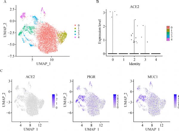
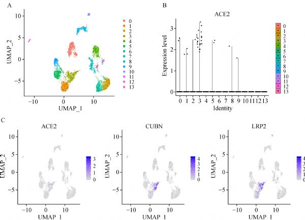
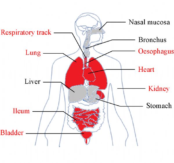
Abstract It has been known that, the novel coronavirus, 2019-nCoV, which is considered similar to SARS-CoV, invades human cells via the receptor angiotensin converting enzyme II (ACE2). Moreover, lung cells that have ACE2 expression may be the main target cells during 2019-nCoV infection. However, some patients also exhibit non-respiratory symptoms, such as kidney failure, implying that 2019-nCoV could also invade other organs. To construct a risk map of different human organs, we analyzed the single-cell RNA sequencing (scRNA-seq) datasets derived from major human physiological systems, including the respiratory, cardiovascular, digestive, and urinary systems. Through scRNA-seq data analyses, we identified the organs at risk, such as lung, heart, esophagus, kidney, bladder, and ileum, and located specific cell types (i.e., type II alveolar cells (AT2), myocardial cells, proximal tubule cells of the kidney, ileum and esophagus epithelial cells, and bladder urothelial cells), which are vulnerable to 2019-nCoV infection. Based on the findings, we constructed a risk map indicating the vulnerability of different organs to 2019-nCoV infection. This study may provide potential clues for further investigation of the pathogenesis and route of 2019-nCoV infection.
|
| Keywords
2019-nCoV
ACE2
single-cell RNA-seq
|
|
Corresponding Author(s):
Jie Hao,Zeguang Han
|
|
Just Accepted Date: 08 February 2020
Online First Date: 13 March 2020
Issue Date: 09 May 2020
|
|
| 1 |
WHO. Statement on the second meeting of the International Health Regulations (2005) Emergency Committee regarding the outbreak of novel coronavirus (2019-nCoV). 2020.
|
| 2 |
X Xu, P Chen, J Wang, J Feng, H Zhou, X Li, W Zhong, P Hao. Evolution of the novel coronavirus from the ongoing Wuhan outbreak and modeling of its spike protein for risk of human transmission. Sci China Life Sci 2020 Jan 21. [Epub ahead of print] doi:10.1007/s11427-020-1637-5
https://doi.org/10.1007/s11427-020-1637-5
pmid: 32009228
|
| 3 |
P Zhou, XL Yang, XG Wang, B Hu, L Zhang, W Zhang, HR Si, Y Zhu, B Li, CL Huang, HD Chen, J Chen, Y Luo, H Guo, RD Jiang, MQ Liu, Y Chen, XR Shen, X Wang, XS Zheng, K Zhao, QJ Chen, F Deng, LL Liu, B Yan, FX Zhan, YY Wang, GF Xiao, ZL Shi. A pneumonia outbreak associated with a new coronavirus of probable bat origin. Nature 2020 Feb 3. [Epub ahead of print] doi: 10.1038/s41586-020-2012-7
https://doi.org/10.1038/s41586-020-2012-7
pmid: 32015507
|
| 4 |
PA Reyfman, JM Walter, N Joshi, KR Anekalla, AC McQuattie-Pimentel, S Chiu, R Fernandez, M Akbarpour, CI Chen, Z Ren, R Verma, H Abdala-Valencia, K Nam, M Chi, S Han, FJ Gonzalez-Gonzalez, S Soberanes, S Watanabe, KJN Williams, AS Flozak, TT Nicholson, VK Morgan, DR Winter, M Hinchcliff, CL Hrusch, RD Guzy, CA Bonham, AI Sperling, R Bag, RB Hamanaka, GM Mutlu, AV Yeldandi, SA Marshall, A Shilatifard, LAN Amaral, H Perlman, JI Sznajder, AC Argento, CT Gillespie, J Dematte, M Jain, BD Singer, KM Ridge, AP Lam, A Bharat, SM Bhorade, CJ Gottardi, GRS Budinger, AV Misharin. Single-cell transcriptomic analysis of human lung provides insights into the pathobiology of pulmonary fibrosis. Am J Respir Crit Care Med 2019; 199(12): 1517–1536
https://doi.org/10.1164/rccm.201712-2410OC
pmid: 30554520
|
| 5 |
S Ruiz García, M Deprez, K Lebrigand, A Cavard, A Paquet, MJ Arguel, V Magnone, M Truchi, I Caballero, S Leroy, CH Marquette, B Marcet, P Barbry, LE Zaragosi. Novel dynamics of human mucociliary differentiation revealed by single-cell RNA sequencing of nasal epithelial cultures. Development 2019; 146(20): 146
https://doi.org/10.1242/dev.177428
pmid: 31558434
|
| 6 |
Y Cui, Y Zheng, X Liu, L Yan, X Fan, J Yong, Y Hu, J Dong, Q Li, X Wu, S Gao, J Li, L Wen, J Qiao, F Tang. Single-cell transcriptome analysis maps the developmental track of the human heart. Cell Rep 2019; 26(7): 1934–1950.e5
https://doi.org/10.1016/j.celrep.2019.01.079
pmid: 30759401
|
| 7 |
E Madissoon, A Wilbrey-Clark, RJ Miragaia, K Saeb-Parsy, KT Mahbubani, N Georgakopoulos, P Harding, K Polanski, N Huang, K Nowicki-Osuch, RC Fitzgerald, KW Loudon, JR Ferdinand, MR Clatworthy, A Tsingene, S van Dongen, M Dabrowska, M Patel, MJT Stubbington, SA Teichmann, O Stegle, KB Meyer. scRNA-seq assessment of the human lung, spleen, and esophagus tissue stability after cold preservation. Genome Biol 2020; 21(1): 1
https://doi.org/10.1186/s13059-019-1906-x
pmid: 31892341
|
| 8 |
P Zhang, M Yang, Y Zhang, S Xiao, X Lai, A Tan, S Du, S Li. Dissecting the single-cell transcriptome network underlying gastric premalignant lesions and early gastric cancer. Cell Rep 2019; 27(6): 1934–1947.e5
https://doi.org/10.1016/j.celrep.2019.04.052
pmid: 31067475
|
| 9 |
SA MacParland, JC Liu, XZ Ma, BT Innes, AM Bartczak, BK Gage, J Manuel, N Khuu, J Echeverri, I Linares, R Gupta, ML Cheng, LY Liu, D Camat, SW Chung, RK Seliga, Z Shao, E Lee, S Ogawa, M Ogawa, MD Wilson, JE Fish, M Selzner, A Ghanekar, D Grant, P Greig, G Sapisochin, N Selzner, N Winegarden, O Adeyi, G Keller, GD Bader, ID McGilvray. Single cell RNA sequencing of human liver reveals distinct intrahepatic macrophage populations. Nat Commun 2018; 9(1): 4383
https://doi.org/10.1038/s41467-018-06318-7
pmid: 30348985
|
| 10 |
JC Martin, C Chang, G Boschetti, R Ungaro, M Giri, JA Grout, K Gettler, LS Chuang, S Nayar, AJ Greenstein, M Dubinsky, L Walker, A Leader, JS Fine, CE Whitehurst, ML Mbow, S Kugathasan, LA Denson, JS Hyams, JR Friedman, PT Desai, HM Ko, I Laface, G Akturk, EE Schadt, H Salmon, S Gnjatic, AH Rahman, M Merad, JH Cho, E Kenigsberg. Single-cell analysis of Crohn’s disease lesions identifies a pathogenic cellular module associated with resistance to anti-TNF therapy. Cell 2019; 178(6): 1493–1508.e20
https://doi.org/10.1016/j.cell.2019.08.008
pmid: 31474370
|
| 11 |
H Wu, AF Malone, EL Donnelly, Y Kirita, K Uchimura, SM Ramakrishnan, JP Gaut, BD Humphreys. Single-cell transcriptomics of a human kidney allograft biopsy specimen defines a diverse inflammatory response. J Am Soc Nephrol 2018; 29(8): 2069–2080
https://doi.org/10.1681/ASN.2018020125
pmid: 29980650
|
| 12 |
Z Yu, J Liao, Y Chen, C Zou, H Zhang, J Cheng, D Liu, T Li, Q Zhang, J Li, X Yang, Y Ye, Z Huang, X Long, R Yang, Z Mo. Single-cell transcriptomic map of the human and mouse bladders. J Am Soc Nephrol 2019; 30(11): 2159–2176
https://doi.org/10.1681/ASN.2019040335
pmid: 31462402
|
| 13 |
E Becht, L McInnes, J Healy, CA Dutertre, IWH Kwok, LG Ng, F Ginhoux, EW Newell. Dimensionality reduction for visualizing single-cell data using UMAP. Nat Biotechnol 2019; 37: 38–44
https://doi.org/10.1038/nbt.4314
pmid: 30531897
|
| 14 |
I Hamming, W Timens, MLC Bulthuis, AT Lely, G Navis, H van Goor. Tissue distribution of ACE2 protein, the functional receptor for SARS coronavirus. A first step in understanding SARS pathogenesis. J Pathol 2004; 203(2): 631–637
https://doi.org/10.1002/path.1570
pmid: 15141377
|
| 15 |
MA Crackower, R Sarao, GY Oudit, C Yagil, I Kozieradzki, SE Scanga, AJ Oliveira-dos-Santos, J da Costa, L Zhang, Y Pei, J Scholey, CM Ferrario, AS Manoukian, MC Chappell, PH Backx, Y Yagil, JM Penninger. Angiotensin-converting enzyme 2 is an essential regulator of heart function. Nature 2002; 417(6891): 822–828
https://doi.org/10.1038/nature00786
pmid: 12075344
|
| 16 |
S Kowalczuk, A Bröer, N Tietze, JM Vanslambrouck, JEJ Rasko, S Bröer. A protein complex in the brush-border membrane explains a Hartnup disorder allele. FASEB J 2008; 22(8): 2880–2887
https://doi.org/10.1096/fj.08-107300
pmid: 18424768
|
| 17 |
M Donoghue, F Hsieh, E Baronas, K Godbout, M Gosselin, N Stagliano, M Donovan, B Woolf, K Robison, R Jeyaseelan, RE Breitbart, S Acton. A novel angiotensin-converting enzyme-related carboxypeptidase (ACE2) converts angiotensin I to angiotensin 1-9. Circ Res 2000; 87(5): E1–E9
https://doi.org/10.1161/01.RES.87.5.e1
pmid: 10969042
|
| 18 |
D Harmer, M Gilbert, R Borman, KL Clark. Quantitative mRNA expression profiling of ACE 2, a novel homologue of angiotensin converting enzyme. FEBS Lett 2002; 532(1-2): 107–110
https://doi.org/10.1016/S0014-5793(02)03640-2
pmid: 12459472
|
| 19 |
LM Burrell, J Risvanis, E Kubota, RG Dean, PS MacDonald, S Lu, C Tikellis, SL Grant, RA Lew, AI Smith, ME Cooper, CI Johnston. Myocardial infarction increases ACE2 expression in rat and humans. Eur Heart J 2005; 26(4): 369–375, discussion 322–324
https://doi.org/10.1093/eurheartj/ehi114
pmid: 15671045
|
|
Viewed |
|
|
|
Full text
|
|
|
|
|
Abstract
|
|
|
|
|
Cited |
|
|
|
|
| |
Shared |
|
|
|
|
| |
Discussed |
|
|
|
|