|
|
|
Degradation of SARS-CoV-2 receptor ACE2 by the E3 ubiquitin ligase Skp2 in lung epithelial cells |
Guizhen Wang1,2, Qun Zhao1,3, Hui Zhang4, Fan Liang1,2, Chen Zhang1,2, Jun Wang5, Zhenyin Chen1,6, Ran Wu1,6, Hong Yu7, Beibei Sun1, Hua Guo1, Ruie Feng4, Kaifeng Xu5, Guangbiao Zhou1( ) ) |
1. State Key Laboratory of Molecular Oncology, National Cancer Center/National Clinical Research Center for Cancer/Cancer Hospital, Chinese Academy of Medical Sciences and Peking Union Medical College, Beijing 100021, China
2. State Key Laboratory of Membrane Biology, Institute of Zoology, Chinese Academy of Sciences & University of Chinese Academy of Sciences, Beijing 100101, China
3. Hubei University of Medicine, Shiyan 442000, China
4. Department of Pathology, Peking Union Medical College Hospital, Chinese Academy of Medical Sciences, Beijing 100730, China
5. Department of Pulmonary and Critical Care Medicine, Peking Union Medical College Hospital, Chinese Academy of Medical Sciences, Beijing 100730, China
6. Guizhou University School of Medicine, Guiyang 550025, China
7. School of Chinese Materia Medica, Beijing University of Chinese Medicine, Beijing 100029, China |
|
|
|
|
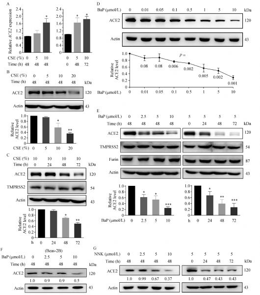
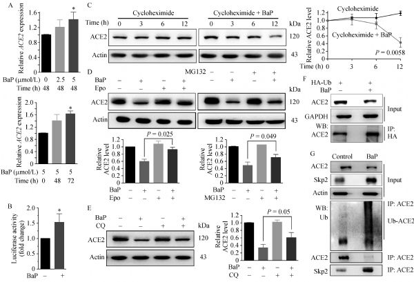
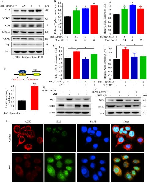
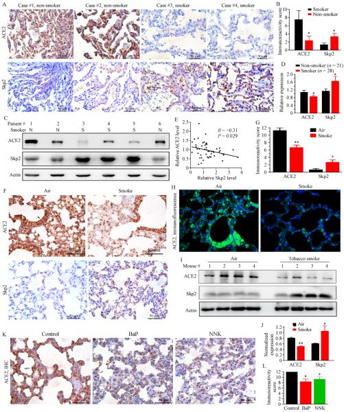
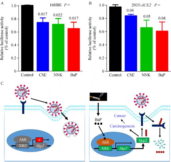
Abstract An unexpected observation among the COVID-19 pandemic is that smokers constituted only 1.4%−18.5% of hospitalized adults, calling for an urgent investigation to determine the role of smoking in SARS-CoV-2 infection. Here, we show that cigarette smoke extract (CSE) and carcinogen benzo(a)pyrene (BaP) increase ACE2 mRNA but trigger ACE2 protein catabolism. BaP induces an aryl hydrocarbon receptor (AhR)-dependent upregulation of the ubiquitin E3 ligase Skp2 for ACE2 ubiquitination. ACE2 in lung tissues of non-smokers is higher than in smokers, consistent with the findings that tobacco carcinogens downregulate ACE2 in mice. Tobacco carcinogens inhibit SARS-CoV-2 spike protein pseudovirions infection of the cells. Given that tobacco smoke accounts for 8 million deaths including 2.1 million cancer deaths annually and Skp2 is an oncoprotein, tobacco use should not be recommended and cessation plan should be prepared for smokers in COVID-19 pandemic.
|
| Keywords
SARS-CoV-2
tobacco smoke
benzo(a)pyrene
ACE2
Skp2
|
|
Corresponding Author(s):
Guangbiao Zhou
|
|
Just Accepted Date: 18 December 2020
Online First Date: 03 February 2021
Issue Date: 23 April 2021
|
|
| 1 |
COVID-19 Dashboard by the Center for Systems Science and Engineering (CSSE) at Johns Hopkins University. (JHU). 2020. (accessed December 8, 2020)
|
| 2 |
World Health Organization. Smoking and COVID-19. 2020. (accessed December 8, 2020)
|
| 3 |
G Lippi, BM Henry. Active smoking is not associated with severity of coronavirus disease 2019 (COVID-19). Eur J Intern Med 2020; 75:107–108
https://doi.org/10.1016/j.ejim.2020.03.014
pmid: 32192856
|
| 4 |
K Farsalinos, A Barbouni, R Niaura. Systematic review of the prevalence of current smoking among hospitalized COVID-19 patients in China: could nicotine be a therapeutic option? Intern Emerg Med 2020; 15(5): 845–852
https://doi.org/10.1007/s11739-020-02355-7
pmid: 32385628
|
| 5 |
CDC COVID-19 Response Team. Preliminary estimates of the prevalence of selected underlying health conditions among patients with coronavirus disease 2019—United States, February 12−March 28, 2020. MMWR Morb Mortal Wkly Rep 2020; 69(13): 382–386
https://doi.org/10.15585/mmwr.mm6913e2
pmid: 32240123
|
| 6 |
WJ Guan, ZY Ni, Y Hu, WH Liang, CQ Ou, JX He, L Liu, H Shan, CL Lei, DSC Hui, B Du, LJ Li, G Zeng, KY Yuen, RC Chen, CL Tang, T Wang, PY Chen, J Xiang, SY Li, JL Wang, ZJ Liang, YX Peng, L Wei, Y Liu, YH Hu, P Peng, JM Wang, JY Liu, Z Chen, G Li, ZJ Zheng, SQ Qiu, J Luo, CJ Ye, SY Zhu, NS; China Medical Treatment Expert Group for Covid-19 Zhong. Clinical characteristics of coronavirus disease 2019 in China. N Engl J Med 2020; 382(18): 1708–1720
https://doi.org/10.1056/NEJMoa2002032
pmid: 32109013
|
| 7 |
M Miyara, F Tubach, V Pourcher, C Morelot-Panzini, J Pernet, J Haroche, S Lebbah, E Morawiec, G Gorochov, E Caumes, P Hausfater, A Combes, T Similowski, Z Amoura. Low incidence of daily active tobacco smoking in patients with symptomatic COVID-19 infection. Qeios 2020; 10.32388/WPP19W
https://doi.org/10.32388/WPP19W.4
|
| 8 |
CI Vardavas, K Nikitara. COVID-19 and smoking: a systematic review of the evidence. Tob Induc Dis 2020; 18: 20
https://doi.org/10.18332/tid/119324
pmid: 32206052
|
| 9 |
S Gebel, S Diehl, J Pype, B Friedrichs, H Weiler, J Schüller, H Xu, K Taguchi, M Yamamoto, T Müller. The transcriptome of Nrf2−/− mice provides evidence for impaired cell cycle progression in the development of cigarette smoke-induced emphysematous changes. Toxicol Sci 2010; 115(1): 238–252
https://doi.org/10.1093/toxsci/kfq039
pmid: 20133372
|
| 10 |
GZ Wang, X Cheng, B Zhou, ZS Wen, YC Huang, HB Chen, GF Li, ZL Huang, YC Zhou, L Feng, MM Wei, LW Qu, Y Cao, GB Zhou. The chemokine CXCL13 in lung cancers associated with environmental polycyclic aromatic hydrocarbons pollution. eLife 2015; 4: e09419
https://doi.org/10.7554/eLife.09419
pmid: 26565418
|
| 11 |
S Carnevali, S Petruzzelli, B Longoni, R Vanacore, R Barale, M Cipollini, F Scatena, P Paggiaro, A Celi, C Giuntini. Cigarette smoke extract induces oxidative stress and apoptosis in human lung fibroblasts. Am J Physiol Lung Cell Mol Physiol 2003; 284(6): L955–L963
https://doi.org/10.1152/ajplung.00466.2001
pmid: 12547733
|
| 12 |
GZ Wang, X Cheng, XC Li, YQ Liu, XQ Wang, X Shi, ZY Wang, YQ Guo, ZS Wen, YC Huang, GB Zhou. Tobacco smoke induces production of chemokine CCL20 to promote lung cancer. Cancer Lett 2015; 363(1): 60–70
https://doi.org/10.1016/j.canlet.2015.04.005
pmid: 25864589
|
| 13 |
Z Liu, L Ma, ZS Wen, Z Hu, FQ Wu, W Li, J Liu, GB Zhou. Cancerous inhibitor of PP2A is targeted by natural compound celastrol for degradation in non-small-cell lung cancer. Carcinogenesis 2014; 35(4): 905–914
https://doi.org/10.1093/carcin/bgt395
pmid: 24293411
|
| 14 |
GZ Wang, L Zhang, XC Zhao, SH Gao, LW Qu, H Yu, WF Fang, YC Zhou, F Liang, C Zhang, YC Huang, Z Liu, YX Fu, GB Zhou. The Aryl hydrocarbon receptor mediates tobacco-induced PD-L1 expression and is associated with response to immunotherapy. Nat Commun 2019; 10(1): 1125
https://doi.org/10.1038/s41467-019-08887-7
pmid: 30850589
|
| 15 |
M Hoffmann, H Kleine-Weber, S Schroeder, N Krüger, T Herrler, S Erichsen, TS Schiergens, G Herrler, NH Wu, A Nitsche, MA Müller, C Drosten, S Pöhlmann. SARS-CoV-2 cell entry depends on ACE2 and TMPRSS2 and is blocked by a clinically proven protease inhibitor. Cell 2020; 181(2): 271–280
https://doi.org/DOI: 10.1016/j.cell.2020.02.052
pmid: 32142651
|
| 16 |
X Zou, K Chen, J Zou, P Han, J Hao, Z. Han Single-cell RNA-seq data analysis on the receptor ACE2 expression reveals the potential risk of different human organs vulnerable to 2019-nCoV infection. Front Med 2020; 14(2): 185–192
https://doi.org/10.1007/s11684-020-0754-0
pmid: 32170560
|
| 17 |
G Cai, Y Bossé, F Xiao, F Kheradmand, CI Amos. Tobacco smoking increases the lung gene expression of ACE2, the receptor of SARS-CoV-2. Am J Respir Crit Care Med 2020; 201(12): 1557–1559
https://doi.org/10.1164/rccm.202003-0693LE
pmid: 32329629
|
| 18 |
JC Smith, EL Sausville, V Girish, ML Yuan, A Vasudevan, KM John, JM Sheltzer. Cigarette smoke exposure and inflammatory signaling increase the expression of the SARS-CoV-2 receptor ACE2 in the respiratory tract. Dev Cell 2020; 53(5): 514–529.e3
https://doi.org/10.1016/j.devcel.2020.05.012
pmid: 32425701
|
| 19 |
G Zhou, S Chen, Z. Chen Advances in COVID-19: the virus, the pathogenesis, and evidence-based control and therapeutic strategies. Front Med 2020; 14(2): 117–125
https://doi.org/10.1007/s11684-020-0773-x
pmid: 32318975
|
| 20 |
M Gheblawi, K Wang, A Viveiros, Q Nguyen, JC Zhong, AJ Turner, MK Raizada, MB Grant, GY Oudit. Angiotensin-converting enzyme 2: SARS-CoV-2 receptor and regulator of the renin-angiotensin system: celebrating the 20th anniversary of the discovery of ACE2. Circ Res 2020; 126(10): 1456–1474
https://doi.org/10.1161/CIRCRESAHA.120.317015
pmid: 32264791
|
| 21 |
T Kamitani, K Kito, HP Nguyen, ETH Yeh. Characterization of NEDD8, a developmentally down-regulated ubiquitin-like protein. J Biol Chem 1997; 272(45): 28557–28562
https://doi.org/10.1074/jbc.272.45.28557
pmid: 9353319
|
| 22 |
H Zhang, R Kobayashi, K Galaktionov, D Beach. p19Skp1 and p45Skp2 are essential elements of the cyclin A-CDK2 S phase kinase. Cell 1995; 82(6): 915–925
https://doi.org/10.1016/0092-8674(95)90271-6
pmid: 7553852
|
| 23 |
JJ Tsay, KM Tchou-Wong, AK Greenberg, H Pass, WN Rom. Aryl hydrocarbon receptor and lung cancer. Anticancer Res 2013; 33(4): 1247–1256
pmid: 23564762
|
| 24 |
X Ou, Y Liu, X Lei, P Li, D Mi, L Ren, L Guo, R Guo, T Chen, J Hu, Z Xiang, Z Mu, X Chen, J Chen, K Hu, Q Jin, J Wang, Z Qian. Characterization of spike glycoprotein of SARS-CoV-2 on virus entry and its immune cross-reactivity with SARS-CoV. Nat Commun 2020; 11(1): 1620
https://doi.org/10.1038/s41467-020-15562-9
pmid: 32221306
|
| 25 |
World Health Organization. Tobacco fact sheet. 2020. (accessed December 8, 2020)
|
| 26 |
BD Carter, CC Abnet, D Feskanich, ND Freedman, P Hartge, CE Lewis, JK Ockene, RL Prentice, FE Speizer, MJ Thun, EJ Jacobs. Smoking and mortality—beyond established causes. N Engl J Med 2015; 372(7): 631–640
https://doi.org/10.1056/NEJMsa1407211
pmid: 25671255
|
| 27 |
Centers for Disease Control and Prevention (US), National Center for Chronic Disease Prevention and Health Promotion (US), Office on Smoking and Health (US). How tobacco smoke causes disease: the biology and behavioral basis for smoking-attributable disease: a report of the surgeon general. Atlanta (GA): Centers for Disease Control and Prevention (US). 2010. (accessed December 8, 2020)
|
| 28 |
G Zhou. Tobacco, air pollution, environmental carcinogenesis, and thoughts on conquering strategies of lung cancer. Cancer Biol Med 2019; 16(4): 700–713
pmid: 31908889
|
| 29 |
J Liu, Y Peng, J Zhang, J Long, J Liu, W Wei. Targeting SCF E3 ligases for cancer therapies. Adv Exp Med Biol 2020; 1217: 123–146
https://doi.org/10.1007/978-981-15-1025-0_9
pmid: 31898226
|
| 30 |
JY Xu, C Zhang, X Wang, L Zhai, Y Ma, Y Mao, K Qian, C Sun, Z Liu, S Jiang, M Wang, L Feng, L Zhao, P Liu, B Wang, X Zhao, H Xie, X Yang, L Zhao, Y Chang, J Jia, X Wang, Y Zhang, Y Wang, Y Yang, Z Wu, L Yang, B Liu, T Zhao, S Ren, A Sun, Y Zhao, W Ying, F Wang, G Wang, Y Zhang, S Cheng, J Qin, X Qian, Y Wang, J Li, F He, T Xiao, M Tan. Integrative proteomic characterization of human lung adenocarcinoma. Cell 2020; 182(1): 245–261.e17
https://doi.org/10.1016/j.cell.2020.05.043
pmid: 32649877
|
| 31 |
MR Deshotels, H Xia, S Sriramula, E Lazartigues, CM Filipeanu. Angiotensin II mediates angiotensin converting enzyme type 2 internalization and degradation through an angiotensin II type I receptor-dependent mechanism. Hypertension 2014; 64(6): 1368–1375
https://doi.org/10.1161/HYPERTENSIONAHA.114.03743
pmid: 25225202
|
|
Viewed |
|
|
|
Full text
|
|
|
|
|
Abstract
|
|
|
|
|
Cited |
|
|
|
|
| |
Shared |
|
|
|
|
| |
Discussed |
|
|
|
|