|
|
|
Dynamic changes of protein corona compositions on the surface of zinc oxide nanoparticle in cell culture media |
Vo-Van Giau1, Yoon-Hee Park2, Kyu-Hwan Shim1, Sang-Wook Son2( ), Seong-Soo A. An1() ), Seong-Soo A. An1() |
1. Department of Bionanotechnology and Gachon Medical Research Institute, Gachon University and Gachon Medical Research Institute, Seongnam, Korea
2. Laboratory of Cell Signaling and Nanomedicine, Department of Dermatology and Division of Brain Korea 21 Project for Biomedical Science, Korea University College of Medicine, Seoul, Korea |
|
|
|
|
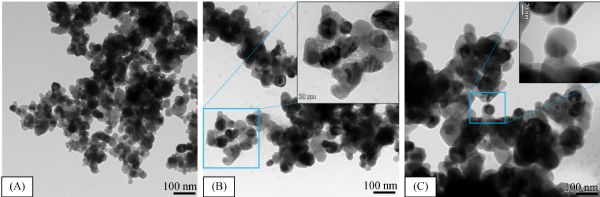
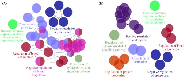
Abstract The potential applications of nanomaterials used in nanomedicine as ingredients in drug delivery systems and in other products continue to expand. When nanomaterials are introduced into physiological environments and driven by energetics, they readily associate proteins forming a protein corona (PC) on their surface. This PC could result in an alteration of the nanomaterial’s surface characteristics, affecting their interaction with cells due to conformational changes in adsorbed protein molecules. However, our current understanding of nanobiological interactions is still very limited. Utilizing a liquid chromatography–mass spectroscopy/mass spectroscopy technology and a Cytoscape plugin (ClueGO) approach, we examined the composition of the PC for a set of zinc oxide nanoparticles (ZnONP) from cell culture media typically and further analyzed the biological interaction of identified proteins, respectively. In total, 36 and 33 common proteins were investigated as being bound to ZnONP at 5 min and 60 min, respectively. These proteins were further analyzed with ClueGO, a Cytoscape plugin, which provided gene ontology and the biological interaction processes of identified proteins. Proteins bound to the surface of nanoparticles that may modify the structure, therefore the function of the adsorbed protein could be consequently affect the complicated biological processes.
|
| Keywords
ZnONPs
nanoparticles
protein corona
ClueGO
|
|
Corresponding Author(s):
Sang-Wook Son,Seong-Soo A. An
|
|
Just Accepted Date: 27 July 2018
Online First Date: 25 January 2019
Issue Date: 25 February 2019
|
|
| 1 |
TBlunk, D F Hochstrasser, J C Sanchez, B W Muller, R H Muller. Colloidal carriers for intravenous drug targeting: Plasma protein adsorption patterns on surface-modified latex particles evaluated by two-dimensional polyacrylamide gel electrophoresis. Electrophoresis, 1993, 14(12): 1382–1387
https://doi.org/10.1002/elps.11501401214
|
| 2 |
M SEhrenberg, A EFriedman, J NFinkelstein, GOberdorster, J LMcGrath. The influence of protein adsorption on nanoparticle association with cultured endothelial cells. Biomaterials, 2009, 30(4): 603–610
https://doi.org/10.1016/j.biomaterials.2008.09.050
|
| 3 |
MLundqvist, C Augustsson, MLilja, KLundkvist, BDahlbäck, SLinse, TCedervall. The nanoparticle protein corona formed in human blood or human blood fractions. PLoS One, 2017, 12(4): e0175871
https://doi.org/10.1371/journal.pone.0175871
|
| 4 |
ABeduneau, Z Ma, C BGrotepas, AKabanov, B ERabinow, NGong, R L Mosley, H Dou, M DBoska, H EGendelman. Facilitated monocyte-macrophage uptake and tissue distribution of superparmagnetic iron-oxide nanoparticles. PLoS One, 2009, 4(2): e4343
https://doi.org/10.1371/journal.pone.0004343
|
| 5 |
M JClift, S Bhattacharjee, D MBrown, VStone. The effects of serum on the toxicity of manufactured nanoparticles. Toxicology Letters, 2010, 198(3): 358–365
https://doi.org/10.1016/j.toxlet.2010.08.002
|
| 6 |
PKhanna, C Ong, B HBay, G HBaeg. Nanotoxicity: An interplay of oxidative stress, inflammation and cell death. Nanomaterials (Basel, Switzerland), 2015, 5(3): 1163–1180
https://doi.org/10.3390/nano5031163
|
| 7 |
LLartigue, C Wilhelm, JServais, CFactor, ADencausse, J CBacri, NLuciani, FGazeau. Nanomagnetic sensing of blood plasma protein interactions with iron oxide nanoparticles: Impact on macrophage uptake. ACS Nano, 2012, 6(3): 2665–2678
https://doi.org/10.1021/nn300060u
|
| 8 |
OLunov, T Syrovets, CLoos, JBeil, M Delacher, KTron, G UNienhaus, AMusyanovych, VMailänder, KLandfester, et al. Differential uptake of functionalized polystyrene nanoparticles by human macrophages and a monocytic cell line. ACS Nano, 2011, 5(3): 1657–1669
https://doi.org/10.1021/nn2000756
|
| 9 |
C DWalkey, J B Olsen, F Song, RLiu, RLiu, H Guo, D WOlsen, YCohen, AEmili, W CChan. Protein corona fingerprinting predicts the cellular interaction of gold and silver nanoparticles. ACS Nano, 2014, 8(3): 2439–2455
https://doi.org/10.1021/nn406018q
|
| 10 |
ZGu, Z Yang, YChong, CGe, J K Weber, D R Bell, R Zhou. Surface curvature relation to protein adsorption for carbon-based nanomaterials. Scientific Reports, 2015, 5(1): 10886
https://doi.org/10.1038/srep10886
|
| 11 |
AMarucco, E Gazzano, DGhigo, EEnrico, IFenoglio. Fibrinogen enhances the inflammatory response of alveolar macrophages to TiO2, SiO2 and carbon nanomaterials. Nanotoxicology, 2016, 10(1): 1–9
|
| 12 |
CGe, J Du, LZhao, LWang, Y Liu, DLi, YYang, R Zhou, YZhao, ZChai, C Chen. Binding of blood proteins to carbon nanotubes reduces cytotoxicity. Proceedings of the National Academy of Sciences of the United States of America, 2011, 108(41): 16968–16973
https://doi.org/10.1073/pnas.1105270108
|
| 13 |
M JHajipour, J Raheb, OAkhavan, SArjmand, OMashinchian, MRahman, MAbdolahad, VSerpooshan, LLaurentj, MMahmoudi. Personalized disease-specific protein corona influences the therapeutic impact of graphene oxide. Nanoscale, 2015, 7(19): 8978–8994
https://doi.org/10.1039/C5NR00520E
|
| 14 |
ASalvati, A S Pitek, M P Monopoli, K Prapainop, F BBombelli, D RHristov, P MKelly, CÅberg, EMahon, K ADawson. Transferrin-functionalized nanoparticles lose their targeting capabilities when a biomolecule corona adsorbs on the surface. Nature Nanotechnology, 2013, 8(2): 137–143
https://doi.org/10.1038/nnano.2012.237
|
| 15 |
Z JDeng, M Liang, MMonteiro, IToth, R F Minchin. Nanoparticle-induced unfolding of fibrinogen promotes Mac-1 receptor activation and inflammation. Nature Nanotechnology, 2011, 6(1): 39–44
https://doi.org/10.1038/nnano.2010.250
|
| 16 |
JSaikia, M Yazdimamaghani, M. S PHadipour, HGhandehari. Differential protein adsorption and cellular uptake of silica nanoparticles based on size and porosity. ACS Applied Materials & Interfaces, 2016, 8(50): 34820–34832
https://doi.org/10.1021/acsami.6b09950
|
| 17 |
C BAnders, J E Eixenberger, N A Franco, R J Hermann, K D Rainey, J J Chess, A Punnooseb, D GWingett. ZnO nanoparticle preparation route influences surface reactivity, dissolution and cytotoxicity. Environmental Science. Nano, 2018, 5(2): 572–588
https://doi.org/10.1039/C7EN00888K
|
| 18 |
J WRasmussen, EMartinez, PLouka, GDenise, D GWingett. Zinc oxide nanoparticles for selective destruction of tumor cells and potential for drug delivery applications. Expert Opinion on Drug Delivery, 2010, 7(9): 1063–1077
https://doi.org/10.1517/17425247.2010.502560
|
| 19 |
AScherzad, T Meyer, NKleinsasser, SHackenberg. Molecular mechanisms of zinc oxide nanoparticle-induced genotoxicity short running title: Genotoxicity of ZnO NPs. Materials (Basel), 2017, 10(12): e1427
https://doi.org/10.3390/ma10121427
|
| 20 |
NSingh, B Manshian, G JJenkins, S MGriffiths, P MWilliams, T GMaffeis, C JWright, S HDoak. Nanogenotoxicology: The DNA damaging potential of engineered nanomaterials. Biomaterials, 2009, 30(23-24): 3891–3914
https://doi.org/10.1016/j.biomaterials.2009.04.009
|
| 21 |
V DRajput, T M Minkina, A Behal, S NSushkova, SMandzhieva, RSingh, AGorovtsov, V STsitsuashvili, W OPurvis, K AGhazaryan, et al. Effects of zinc-oxide nanoparticles on soil, plants, animals and soil organisms: A review. Environmental Nanotechnology, Monitoring & Management, 2018, 9: 76–84
https://doi.org/10.1016/j.enmm.2017.12.006
|
| 22 |
RWahab, M A Siddiqui, Q Saquib, SDwivedi, JAhmad, JMusarrat, A AAl-Khedhairy, H SShin. ZnO nanoparticles induced oxidative stress and apoptosis in HepG2 and MCF-7 cancer cells and their antibacterial activity. Colloids and Surfaces. B, Biointerfaces, 2014, 117(1): 267–276
https://doi.org/10.1016/j.colsurfb.2014.02.038
|
| 23 |
MChevallet, B Gallet, AFuchs, P HJouneau, KUm, E Mintz, IMichaud-Soret. Metal homeostasis disruption and mitochondrial dysfunction in hepatocytes exposed to sub-toxic doses of zinc oxide nanoparticles. Nanoscale, 2016, 8(43): 18495–18506
https://doi.org/10.1039/C6NR05306H
|
| 24 |
WZhang, Y Zhao, FLi, LLi, Y Feng, LMin, DMa, S Yu, JLiu, HZhang, et al. Zinc oxide nanoparticle caused plasma metabolomic perturbations correlate with hepatic steatosis. Frontiers in Pharmacology, 2018, 9: 57
https://doi.org/10.3389/fphar.2018.00057
|
| 25 |
V RLeite-Silva, D CLiu, W YSanchez, HStudier, Y HMohammed, AHolmes, WBecker, J EGrice, H ABenson, M SRoberts. Effect of flexing and massage on in vivo human skin penetration and toxicity of zinc oxide nanoparticles. Nanomedicine (London), 2016, 11(10): 1193–1205
https://doi.org/10.2217/nnm-2016-0010
|
| 26 |
C MSayes, K L Reed, D B Warheit. Assessing toxicity of fine and nanoparticles: Comparing in vitro measurements to in vivo pulmonary toxicity profiles. Toxicological Sciences: An Official Journal of the Society of Toxicology, 2007, 97(1): 163–180
https://doi.org/10.1093/toxsci/kfm018
|
| 27 |
TXia, Y Zhao, TSager, SGeorge, SPokhrel, NLi, D Schoenfeld, HMeng, SLin, X Wang, et al. Decreased dissolution of ZnO by iron doping yields nanoparticles with reduced toxicity in the rodent lung and zebrafish embryos. ACS Nano, 2011, 5(2): 1223–1235
https://doi.org/10.1021/nn1028482
|
| 28 |
D BWarheit, C M Sayes, K L Reed. Nanoscale and fine zinc oxide particles: Can in vitro assays accurately forecast lung hazards following inhalation exposures? Environmental Science & Technology, 2009, 43(20): 7939–7945
https://doi.org/10.1021/es901453p
|
| 29 |
V VGiau, S S An. Emergence of exosomal miRNAs as a diagnostic biomarker for Alzheimer’s disease. Journal of the Neurological Sciences, 2016, 15(360): 141–152
https://doi.org/10.1016/j.jns.2015.12.005
|
| 30 |
M NMana, P A Fougères, Y H Leung, F Liu, A BDjurišić, J PGiesy, K M YLeung. Physicochemical characteristics and toxicity of surface-modified zinc oxide nanoparticles to freshwater and marine microalgae. Scientific Reports, 2017, 7(1): 15909
https://doi.org/10.1038/s41598-017-15988-0
|
| 31 |
SMilani, F B Bombelli, A S Pitek, K A Dawson, J Rädler. Reversible versus irreversible binding of transferrin to polystyrene nanoparticles: Soft and hard corona. ACS Nano, 2012, 6(3): 2532–2541
https://doi.org/10.1021/nn204951s
|
| 32 |
FPederzoli, G Tosi, M AVandelli, DBelletti, FForni, BRuozi. Protein corona and nanoparticles: How can we investigate on? Wiley Interdisciplinary Reviews. Nanomedicine and Nanobiotechnology, 2017, 9(6): e1467
https://doi.org/10.1002/wnan.1467
|
| 33 |
ARao, H Long, AHarley-Trochimczyk, TPham, AZettl, CCarraro, RMaboudian. In situ localized growth of ordered metal oxide hollow sphere array on microheater platform for sensitive, ultra-fast gas sensing. ACS Applied Materials & Interfaces, 2017, 9(3): 2634–2641
https://doi.org/10.1021/acsami.6b12677
|
| 34 |
SSchöttler, K Landfester, VMailänder. Controlling the stealth effect of nanocarriers through understanding the protein corona. Angewandte Chemie International Edition in English, 2016, 55(31): 8806–8815
https://doi.org/10.1002/anie.201602233
|
| 35 |
STenzer, D Docter, JKuharev, AMusyanovych, VFetz, R Hecht, FSchlenk, DFischer, KKiouptsi, CReinhardt, et al. Rapid formation of plasma protein corona critically affects nanoparticle pathophysiology. Nature Nanotechnology, 2013, 8(10): 772–781
https://doi.org/10.1038/nnano.2013.181
|
| 36 |
KSaha, M Rahimi, MYazdani, S TKim, D FMoyano, SHou, R Das, RMout, FRezaee, MMahmoudi, et al. Regulation of macrophage recognition through the interplay of nanoparticle surface functionality and protein corona. ACS Nano, 2016, 10(4): 4421–4430
https://doi.org/10.1021/acsnano.6b00053
|
| 37 |
J HShannahan, J MBrown, RChen, P C Ke, X Lai, SMitra, F AWitzmann. Comparison of nanotube-protein corona composition in cell culture media. Small, 2013, 9(12): 2171–2181
https://doi.org/10.1002/smll.201202243
|
| 38 |
J HShannahan, XLai, P C Ke, R Podila, J MBrown, F AWitzmann. Silver nanoparticle protein corona composition in cell culture media. PLoS One, 2013, 8(9): e74001
https://doi.org/10.1371/journal.pone.0074001
|
| 39 |
K MKim, M H Choi, J K Lee, J Jeong, Y RKim, M KKim, S MPaek, J MOh. Physicochemical properties of surface charge-modified ZnO nanoparticles with different particle sizes. International Journal of Nanomedicine, 2014, 9(Suppl 2): 41–56
|
| 40 |
K HShim, J Hulme, E HMaeng, M KKim. An S S A. Analysis of zinc oxide nanoparticles binding proteins in rat blood and brain homogenate. International Journal of Nanomedicine, 2014, 9(Suppl 2): 217–224
|
| 41 |
TCedervall, I Lynch, MFoy, TBerggård, S CDonnelly, GCagney, SLinse, K ADawson. Detailed identification of plasma proteins adsorbed on copolymer nanoparticles. Angewandte Chemie International Edition in English, 2007, 46(30): 5754–5756
https://doi.org/10.1002/anie.200700465
|
| 42 |
SLara, F Alnasser, EPolo, DGarry, M C LGiudice, D RHristov, LRocks, ASalvati, YYan, N K A Dawso. Identification of receptor binding to the biomolecular corona of nanoparticles. ACS Nano, 2017, 11(2): 1884–1893
https://doi.org/10.1021/acsnano.6b07933
|
| 43 |
JKreuter, D Shamenkov, VPetrov, PRamge, KCychutek, CKoch-Brandt, RAlyautdin. Apolipoprotein-mediated transport of nanoparticle-bond drugs across the blood-brain barrier. Journal of Drug Targeting, 2002, 10(4): 317–325
https://doi.org/10.1080/10611860290031877
|
| 44 |
SPalchetti, V Colapicchioni, LDigiacomo, GCaracciolo, DPozzi, A LCapriotti, GLa Barbera, ALaganà. The protein corona of circulating PEGylated liposomes. Biochimica et Biophysica Acta, 2016, 1858(2): 189–196
https://doi.org/10.1016/j.bbamem.2015.11.012
|
| 45 |
SRitz, S Schöttler, NKotman, GBaier, AMusyanovych, JKuharev, KLandfester, HSchild, OJahn, S Tenzer, et al. Protein corona of nanoparticles: Distinct proteins regulate the cellular uptake. Biomacromolecules, 2015, 16(4): 1311–1321
https://doi.org/10.1021/acs.biomac.5b00108
|
| 46 |
MLundqvist, J Stigler, GElia, ILynch, TCedervall, K ADawson. Nanoparticle size and surface properties determine the protein corona with possible implications for biological impacts. Proceedings of the National Academy of Sciences of the United States of America, 2008, 105(38): 14265–14270
https://doi.org/10.1073/pnas.0805135105
|
| 47 |
J LTownson, Y S Lin, J O Agola, E C Carnes, H S Leong, J D Lewis, C L Haynes, C J Brinker. Differing in vivo characteristics of size- and charge-matched mesoporous silica nanoparticles. Journal of the American Chemical Society, 2013, 135(43): 16030–16033
https://doi.org/10.1021/ja4082414
|
| 48 |
CRöcker, M Pötzl, FZhang, W JParak, G UNienhaus. A quantitative fluorescence study of protein monolayer formation on colloidal nanoparticles. Nature Nanotechnology, 2009, 4(9): 577–580
https://doi.org/10.1038/nnano.2009.195
|
| 49 |
Z JDeng, G Mortimer, TSchiller, AMusumeci, DMartin, R FMinchin. Differential plasma protein binding to metal oxide nanoparticles. Nanotechnology, 2009, 20(45): 455101
https://doi.org/10.1088/0957-4484/20/45/455101
|
| 50 |
ALesniak, A Campbell, M PMonopoli, ILynch, ASalvati, K ADawson. Serum heat inactivation affects protein corona composition and nanoparticle uptake. Biomaterials, 2010, 31(36): 9511–5918
https://doi.org/10.1016/j.biomaterials.2010.09.049
|
| 51 |
ALesniak, F Fenaroli, M PMonopoli, CÅberg, K ADawson, ASalvati. Effects of the presence or absence of a protein corona on silica nanoparticle uptake and impact on cells. ACS Nano, 2012, 6(7): 5845–5857
https://doi.org/10.1021/nn300223w
|
| 52 |
ALesniak, A Salvati, M JSantos-Martinez, M WRadomski, K ADawson, CÅberg. Nanoparticle adhesion to the cell membrane and its effect on nanoparticle uptake efficiency. Journal of the American Chemical Society, 2013, 135(4): 1438–1444
https://doi.org/10.1021/ja309812z
|
| 53 |
R CMurdock, L Braydich-Stolle, A MSchrand, J JSchlager, S MHussain. Characterization of nanomaterial dispersion in solution prior to in vitro exposure using dynamic light scattering technique. Toxicological Sciences: An Official Journal of the Society of Toxicology, 2008, 101(2): 239–253
https://doi.org/10.1093/toxsci/kfm240
|
| 54 |
GMaiorano, S Sabella, BSorce, VBrunetti, M AMalvindi, RCingolani, P PPompa. Effects of cell culture media on the dynamic formation of protein-nanoparticle complexes and influence on the cellular response. ACS Nano, 2010, 4(12): 7481–7491
https://doi.org/10.1021/nn101557e
|
| 55 |
JWang, U B Jensen, G V Jensen, S Shipovskov, V SBalakrishnan, DOtzen, J SPedersen, FBesenbacher, D SSutherland. Soft interactions at nanoparticles alter protein function and conformation in a size dependent manner. Nano Letters, 2011, 11(11): 4985–4991
https://doi.org/10.1021/nl202940k
|
| 56 |
YZhou, X Fang, YGong, AXiao, Y Xie, LLiu, YCao. The interactions between ZnO nanoparticles (NPs) and α-linolenic acid (LNA) complexed to BSA did not influence the toxicity of ZnO NPs on HepG2 cells. Journal of the American Chemical Society, 2017, 135(4): 1438–1444
|
| 57 |
M RGo, J Yu, S HBae, H JKim, S JChoi. Effects of interactions between ZnO nanoparticles and saccharides on biological responses. International Journal of Molecular Sciences, 2018, 19(2): 486
https://doi.org/10.3390/ijms19020486
|
|
Viewed |
|
|
|
Full text
|
|
|
|
|
Abstract
|
|
|
|
|
Cited |
|
|
|
|
| |
Shared |
|
|
|
|
| |
Discussed |
|
|
|
|