|
|
|
Liver cell therapies: cellular sources and grafting strategies |
Wencheng Zhang1,2,3, Yangyang Cui1,2,3,4, Yuan Du1,5, Yong Yang1,5, Ting Fang1,2,3, Fengfeng Lu1,2,3, Weixia Kong6, Canjun Xiao7, Jun Shi5,7, Lola M. Reid8( ), Zhiying He1,2,3() ), Zhiying He1,2,3() |
1. Institute for Regenerative Medicine, Ji’an Hospital, Shanghai East Hospital, School of Life Sciences and Technology, Tongji University, Shanghai 200123, China
2. Shanghai Engineering Research Center of Stem Cells Translational Medicine, Shanghai 200335, China
3. Shanghai Institute of Stem Cell Research and Clinical Translation, Shanghai 200120, China
4. Postgraduate Training Base of Shanghai East Hospital, Jinzhou Medical University, Jinzhou 121001, China
5. The First Affiliated Hospital of Nanchang University, Nanchang 330006, China
6. Graduate School of Frontier Biosciences, Osaka University, Suita, Osaka, 565-0871, Japan
7. Department of General Surgery, Ji’an Hospital, Shanghai East Hospital, School of Medicine, Tongji University, Ji’an 343006, China
8. Department of Cell Biology and Physiology and Program in Molecular Biology and Biotechnology; UNC School of Medicine, Chapel Hill, NC 27599, USA |
|
|
|
|
Abstract The liver has a complex cellular composition and a remarkable regenerative capacity. The primary cell types in the liver are two parenchymal cell populations, hepatocytes and cholangiocytes, that perform most of the functions of the liver and that are helped through interactions with non-parenchymal cell types comprising stellate cells, endothelia and various hemopoietic cell populations. The regulation of the cells in the liver is mediated by an insoluble complex of proteins and carbohydrates, the extracellular matrix, working synergistically with soluble paracrine and systemic signals. In recent years, with the rapid development of genetic sequencing technologies, research on the liver’s cellular composition and its regulatory mechanisms during various conditions has been extensively explored. Meanwhile breakthroughs in strategies for cell transplantation are enabling a future in which there can be a rescue of patients with end-stage liver diseases, offering potential solutions to the chronic shortage of livers and alternatives to liver transplantation. This review will focus on the cellular mechanisms of liver homeostasis and how to select ideal sources of cells to be transplanted to achieve liver regeneration and repair. Recent advances are summarized for promoting the treatment of end-stage liver diseases by forms of cell transplantation that now include grafting strategies.
|
| Keywords
liver regeneration
hepatocytes
cholangiocytes
stem cells
organoids
regulatory mechanisms
transplantation/grafting strategies
|
|
Corresponding Author(s):
Lola M. Reid,Zhiying He
|
|
Just Accepted Date: 05 June 2023
Online First Date: 30 June 2023
Issue Date: 28 July 2023
|
|
| 1 |
GK Michalopoulos. Hepatostat: liver regeneration and normal liver tissue maintenance. Hepatology 2017; 65(4): 1384–1392
https://doi.org/10.1002/hep.28988
|
| 2 |
L Campana, H Esser, M Huch, S Forbes. Liver regeneration and inflammation: from fundamental science to clinical applications. Nat Rev Mol Cell Biol 2021; 22(9): 608–624
https://doi.org/10.1038/s41580-021-00373-7
|
| 3 |
VL Gadd, N Aleksieva, SJ Forbes. Epithelial plasticity during liver injury and regeneration. Cell Stem Cell 2020; 27(4): 557–573
https://doi.org/10.1016/j.stem.2020.08.016
|
| 4 |
SH SigalS BrillAS FiorinoLM Reid. The liver as a stem cell and lineage system. Am J Physiol 1992; 263(2 Pt 1): G139–G148
pmid: 1325126
|
| 5 |
SH Sigal, S Gupta, DF Jr Gebhard, P Holst, D Neufeld, LM Reid. Evidence for a terminal differentiation process in the rat liver. Differentiation 1995; 59(1): 35–42
https://doi.org/10.1046/j.1432-0436.1995.5910035.x
|
| 6 |
W ZhangA AllenE WauthierX YiH Hani P SethupathyD GerberV CardinaleG CarpinoJ Dominguez-BendalaG LanzoniD AlvaroE Gaudio L Reid. Stem cell-fueled maturational lineages in hepatic and pancreatic organogenesis. In: Arias IM, Alter HJ, Boyer JL, Cohen DE, Shafritz DA, Thorgeirsson SS, Wolkoff AW. The Liver: Biology and Pathobiology. John Wiley & Sons Ltd. 2020. 523–538
|
| 7 |
V Cardinale, Y Wang, G Carpino, G Mendel, G Alpini, E Gaudio, LM Reid, D Alvaro. The biliary tree—a reservoir of multipotent stem cells. Nat Rev Gastroenterol Hepatol 2012; 9(4): 231–240
https://doi.org/10.1038/nrgastro.2012.23
|
| 8 |
D Alvaro, E Gaudio. Liver capsule: biliary tree stem cell subpopulations. Hepatology 2016; 64(2): 644
https://doi.org/10.1002/hep.28546
|
| 9 |
G Carpino, V Cardinale, P Onori, A Franchitto, PB Berloco, M Rossi, Y Wang, R Semeraro, M Anceschi, R Brunelli, D Alvaro, LM Reid, E Gaudio. Biliary tree stem/progenitor cells in glands of extrahepatic and intraheptic bile ducts: an anatomical in situ study yielding evidence of maturational lineages. J Anat 2012; 220(2): 186–199
https://doi.org/10.1111/j.1469-7580.2011.01462.x
|
| 10 |
G Zajicek, R Oren, M Jr Weinreb. The streaming liver. Liver 1985; 5(6): 293–300
https://doi.org/10.1111/j.1600-0676.1985.tb00252.x
|
| 11 |
N Arber, G Zajicek, I Ariel. The streaming liver. II. Hepatocyte life history. Liver 1988; 8(2): 80–87
https://doi.org/10.1111/j.1600-0676.1988.tb00972.x
|
| 12 |
SH Sigal, P Rajvanshi, GR Gorla, RP Sokhi, R Saxena, DR Jr Gebhard, LM Reid, S Gupta. Partial hepatectomy-induced polyploidy attenuates hepatocyte replication and activates cell aging events. Am J Physiol 1999; 276(5): G1260–G1272
|
| 13 |
SH Sigal, P Rajvanshi, LM Reid, S Gupta. Demonstration of differentiation in hepatocyte progenitor cells using dipeptidyl peptidase IV deficient mutant rats. Cell Mol Biol Res 1995; 41(1): 39–47
|
| 14 |
LM Reid. Stem cell biology, hormone/matrix synergies and liver differentiation. Curr Opin Cell Biol 1990; 2(1): 121–130
https://doi.org/10.1016/S0955-0674(05)80042-0
|
| 15 |
LM Reid, AS Fiorino, SH Sigal, S Brill, PA Holst. Extracellular matrix gradients in the space of Disse: relevance to liver biology. Hepatology 1992; 15(6): 1198–1203
https://doi.org/10.1002/hep.1840150635
|
| 16 |
S Brill, P Holst, S Sigal, I Zvibel, A Fiorino, A Ochs, U Somasundaran, LM Reid. Hepatic progenitor populations in embryonic, neonatal, and adult liver. Proc Soc Exp Biol Med 1993; 204(3): 261–269
https://doi.org/10.3181/00379727-204-43662
|
| 17 |
S Brill, I Zvibel, LM Reid. Maturation-dependent changes in the regulation of liver-specific gene expression in embryonal versus adult primary liver cultures. Differentiation 1995; 59(2): 95–102
https://doi.org/10.1046/j.1432-0436.1995.5920095.x
|
| 18 |
S Brill, I Zvibel, LM Reid. Expansion conditions for early hepatic progenitor cells from embryonal and neonatal rat livers. Dig Dis Sci 1999; 44(2): 364–371
https://doi.org/10.1023/A:1026666820422
|
| 19 |
ASL XuTL LuntzJM MacdonaldH KubotaE Hsu RE LondonLM Reid. Lineage biology and liver. In: Lanza R, Langer R, Vacanti J. Principles of Tissue Engineering. 2nd Ed. Academic Press, 2000. 559–597
|
| 20 |
R Susick, N Moss, H Kubota, E Lecluyse, G Hamilton, T Luntz, J Ludlow, J Fair, D Gerber, K Bergstrand, J White, A Bruce, O Drury, S Gupta, LM Reid. Hepatic progenitors and strategies for liver cell therapies. Ann N Y Acad Sci 2001; 944(1): 398–419
https://doi.org/10.1111/j.1749-6632.2001.tb03851.x
|
| 21 |
H Kubota, LM Reid. Clonogenic hepatoblasts, common precursors for hepatocytic and biliary lineages, are lacking classical major histocompatibility complex class I antigen. Proc Natl Acad Sci USA 2000; 97(22): 12132–12137
https://doi.org/10.1073/pnas.97.22.12132
|
| 22 |
H Kubota, RW Storms, LM Reid. Variant forms of alpha-fetoprotein transcripts expressed in human hematopoietic progenitors. Implications for their developmental potential towards endoderm. J Biol Chem 2002; 277(31): 27629–27635
https://doi.org/10.1074/jbc.M202117200
|
| 23 |
H Kubota, HL Yao, LM Reid. Identification and characterization of vitamin A-storing cells in fetal liver: implications for functional importance of hepatic stellate cells in liver development and hematopoiesis. Stem Cells 2007; 25(9): 2339–2349
https://doi.org/10.1634/stemcells.2006-0316
|
| 24 |
P Rajvanshi, D Liu, M Ott, S Gagandeep, ML Schilsky, S Gupta. Fractionation of rat hepatocyte subpopulations with varying metabolic potential, proliferative capacity, and retroviral gene transfer efficiency. Exp Cell Res 1998; 244(2): 405–419
https://doi.org/10.1006/excr.1998.4223
|
| 25 |
M Okabe, Y Tsukahara, M Tanaka, K Suzuki, S Saito, Y Kamiya, T Tsujimura, K Nakamura, A Miyajima. Potential hepatic stem cells reside in EpCAM+ cells of normal and injured mouse liver. Development 2009; 136(11): 1951–1960
https://doi.org/10.1242/dev.031369
|
| 26 |
K Kaneko, K Kamimoto, A Miyajima, T Itoh. Adaptive remodeling of the biliary architecture underlies liver homeostasis. Hepatology 2015; 61(6): 2056–2066
https://doi.org/10.1002/hep.27685
|
| 27 |
M Tanaka, M Okabe, K Suzuki, Y Kamiya, Y Tsukahara, S Saito, A Miyajima. Mouse hepatoblasts at distinct developmental stages are characterized by expression of EpCAM and DLK1: drastic change of EpCAM expression during liver development. Mech Dev 2009; 126(8–9): 665–676
https://doi.org/10.1016/j.mod.2009.06.939
|
| 28 |
A Katoonizadeh, H Poustchi, R Malekzadeh. Hepatic progenitor cells in liver regeneration: current advances and clinical perspectives. Liver Int 2014; 34(10): 1464–1472
https://doi.org/10.1111/liv.12573
|
| 29 |
T Itoh, A Miyajima. Liver regeneration by stem/progenitor cells. Hepatology 2014; 59(4): 1617–1626
https://doi.org/10.1002/hep.26753
|
| 30 |
A Miyajima, M Tanaka, T Itoh. Stem/progenitor cells in liver development, homeostasis, regeneration, and reprogramming. Cell Stem Cell 2014; 14(5): 561–574
https://doi.org/10.1016/j.stem.2014.04.010
|
| 31 |
M Huch, SF Boj, H Clevers. Lgr5(+) liver stem cells, hepatic organoids and regenerative medicine. Regen Med 2013; 8(4): 385–387
https://doi.org/10.2217/rme.13.39
|
| 32 |
M Tanaka, T Itoh, N Tanimizu, A Miyajima. Liver stem/progenitor cells: their characteristics and regulatory mechanisms. J Biochem 2011; 149(3): 231–239
https://doi.org/10.1093/jb/mvr001
|
| 33 |
N Barker, JH van Es, J Kuipers, P Kujala, M van den Born, M Cozijnsen, A Haegebarth, J Korving, H Begthel, PJ Peters, H Clevers. Identification of stem cells in small intestine and colon by marker gene Lgr5. Nature 2007; 449(7165): 1003–1007
https://doi.org/10.1038/nature06196
|
| 34 |
M Huch, C Dorrell, SF Boj, JH van Es, VS Li, M van de Wetering, T Sato, K Hamer, N Sasaki, MJ Finegold, A Haft, RG Vries, M Grompe, H Clevers. In vitro expansion of single Lgr5+ liver stem cells induced by Wnt-driven regeneration. Nature 2013; 494(7436): 247–250
https://doi.org/10.1038/nature11826
|
| 35 |
E Schmelzer, E Wauthier, LM Reid. The phenotypes of pluripotent human hepatic progenitors. Stem Cells 2006; 24(8): 1852–1858
https://doi.org/10.1634/stemcells.2006-0036
|
| 36 |
E Schmelzer, LM Reid. Human telomerase activity, telomerase and telomeric template expression in hepatic stem cells and in livers from fetal and postnatal donors. Eur J Gastroenterol Hepatol 2009; 21(10): 1191–1198
https://doi.org/10.1097/MEG.0b013e32832973fc
|
| 37 |
E Schmelzer, L Zhang, A Bruce, E Wauthier, J Ludlow, HL Yao, N Moss, A Melhem, R McClelland, W Turner, M Kulik, S Sherwood, T Tallheden, N Cheng, ME Furth, LM Reid. Human hepatic stem cells from fetal and postnatal donors. J Exp Med 2007; 204(8): 1973–1987
https://doi.org/10.1084/jem.20061603
|
| 38 |
L Zhang, N Theise, M Chua, LM Reid. The stem cell niche of human livers: symmetry between development and regeneration. Hepatology 2008; 48: 1598–1607
https://doi.org/10.1002/hep.22516
|
| 39 |
WS Turner, E Schmelzer, R McClelland, E Wauthier, W Chen, LM Reid. Human hepatoblast phenotype maintained by hyaluronan hydrogels. J Biomed Mater Res B Appl Biomater 2007; 82(1): 156–168
https://doi.org/10.1002/jbm.b.30717
|
| 40 |
WS Turner, C Seagle, JA Galanko, O Favorov, GD Prestwich, JM Macdonald, LM Reid. Nuclear magnetic resonance metabolomic footprinting of human hepatic stem cells and hepatoblasts cultured in hyaluronan-matrix hydrogels. Stem Cells 2008; 26(6): 1547–1555
https://doi.org/10.1634/stemcells.2007-0863
|
| 41 |
B Wang, L Zhao, M Fish, CY Logan, R Nusse. Self-renewing diploid Axin2(+) cells fuel homeostatic renewal of the liver. Nature 2015; 524(7564): 180–185
https://doi.org/10.1038/nature14863
|
| 42 |
T Sun, S Annunziato, S Bergling, C Sheng, V Orsini, P Forcella, M Pikiolek, V Kancherla, S Holwerda, D Imanci, F Wu, LC Meylan, LF Puehringer, A Waldt, M Oertli, S Schuierer, LM Terracciano, S Reinker, H Ruffner, T Bouwmeester, AW Sailer, E George, G Roma, A de Weck, S Piscuoglio, F Lohmann, U Naumann, P Liberali, F Cong, JS Tchorz. ZNRF3 and RNF43 cooperate to safeguard metabolic liver zonation and hepatocyte proliferation. Cell Stem Cell 2021; 28(10): 1822–1837.e10
https://doi.org/10.1016/j.stem.2021.05.013
|
| 43 |
V Cardinale, Y Wang, G Carpino, CB Cui, M Gatto, M Rossi, PB Berloco, A Cantafora, E Wauthier, ME Furth, L Inverardi, J Dominguez-Bendala, C Ricordi, D Gerber, E Gaudio, D Alvaro, L Reid. Multipotent stem/progenitor cells in human biliary tree give rise to hepatocytes, cholangiocytes, and pancreatic islets. Hepatology 2011; 54(6): 2159–2172
https://doi.org/10.1002/hep.24590
|
| 44 |
G Carpino, L Nevi, D Overi, V Cardinale, WY Lu, S Di Matteo, S Safarikia, PB Berloco, R Venere, P Onori, A Franchitto, SJ Forbes, D Alvaro, E Gaudio. Peribiliary gland niche participates in biliary tree regeneration in mouse and in human primary sclerosing cholangitis. Hepatology 2020; 71(3): 972–989
https://doi.org/10.1002/hep.30871
|
| 45 |
G Carpino, A Renzi, A Franchitto, V Cardinale, P Onori, L Reid, D Alvaro, E Gaudio. Stem/progenitor cell niches involved in hepatic and biliary regeneration. Stem Cells Int 2016; 2016: 3658013
https://doi.org/10.1155/2016/3658013
|
| 46 |
JK Sicklick, YX Li, A Melhem, E Schmelzer, M Zdanowicz, J Huang, M Caballero, JH Fair, JW Ludlow, RE McClelland, LM Reid, AM Diehl. Hedgehog signaling maintains resident hepatic progenitors throughout life. Am J Physiol Gastrointest Liver Physiol 2006; 290(5): G859–G870
https://doi.org/10.1152/ajpgi.00456.2005
|
| 47 |
G Carpino, V Cardinale, T Folseraas, D Overi, K Grzyb, D Costantini, PB Berloco, S Di Matteo, TH Karlsen, D Alvaro, E Gaudio. Neoplastic transformation of the peribiliary stem cell niche in cholangiocarcinoma arisen in primary sclerosing cholangitis. Hepatology 2019; 69(2): 622–638
https://doi.org/10.1002/hep.30210
|
| 48 |
G Carpino, V Cardinale, A Renzi, JR Hov, PB Berloco, M Rossi, TH Karlsen, D Alvaro, E Gaudio. Activation of biliary tree stem cells within peribiliary glands in primary sclerosing cholangitis. J Hepatol 2015; 63(5): 1220–1228
https://doi.org/10.1016/j.jhep.2015.06.018
|
| 49 |
V Cardinale, G Carpino, A Cantafora, LM Reid, E Gaudio, D Alvaro. Metabolic oxidation controls the hepatic stem cells (HpSCs) fate and the hepatic lineage organization in physiologic and pathologic conditions. Hepatology 2012; 56(5): 2006–2007
https://doi.org/10.1002/hep.25825
|
| 50 |
V Cardinale, G Carpino, L Reid, E Gaudio, D Alvaro. Multiple cells of origin in cholangiocarcinoma underlie biological, epidemiological and clinical heterogeneity. World J Gastrointest Oncol 2012; 4(5): 94–102
https://doi.org/10.4251/wjgo.v4.i5.94
|
| 51 |
G Carpino, V Cardinale, R Gentile, P Onori, R Semeraro, A Franchitto, Y Wang, D Bosco, A Iossa, C Napoletano, A Cantafora, G D’Argenio, M Nuti, N Caporaso, P Berloco, R Venere, T Oikawa, L Reid, D Alvaro, E Gaudio. Evidence for multipotent endodermal stem/progenitor cell populations in human gallbladder. J Hepatol 2014; 60(6): 1194–1202
https://doi.org/10.1016/j.jhep.2014.01.026
|
| 52 |
G Carpino, A Renzi, V Cardinale, A Franchitto, P Onori, D Overi, M Rossi, PB Berloco, D Alvaro, LM Reid, E Gaudio. Progenitor cell niches in the human pancreatic duct system and associated pancreatic duct glands: an anatomical and immunophenotyping study. J Anat 2016; 228(3): 474–486
https://doi.org/10.1111/joa.12418
|
| 53 |
D OveriG CarpinoM MorettiA FranchittoL Nevi P OnoriE De SmaeleL FedericiD SantorelliM Maroder LM ReidV CardinaleD AlvaroE Gaudio. Islet regeneration and pancreatic duct glands in human and experimental diabetes. Front Cell Dev Biol 2022; 10: 814165 doi:
https://doi.org/10.3389/fcell.2022.814165
pmid: 35186929" target="_blank">35186929
|
| 54 |
W ZhangX WangG LanzoniE WauthierS Simpson JA EzzellA AllenC SuittJ KrolikA Jhirad J Dominguez-BendalaV CardinaleD Alvaro D OveriE GaudioP SethupathyG CarpinoC Adin J PiedrahitaK MathewsZ HeLM Reid. A postnatal network of co-hepato/pancreatic stem/progenitors in the biliary trees of pigs and humans. NPJ Regen Med 2023; [Epub ahead of print] doi:
https://doi.org/10.1038/s41536-023-00303-5
|
| 55 |
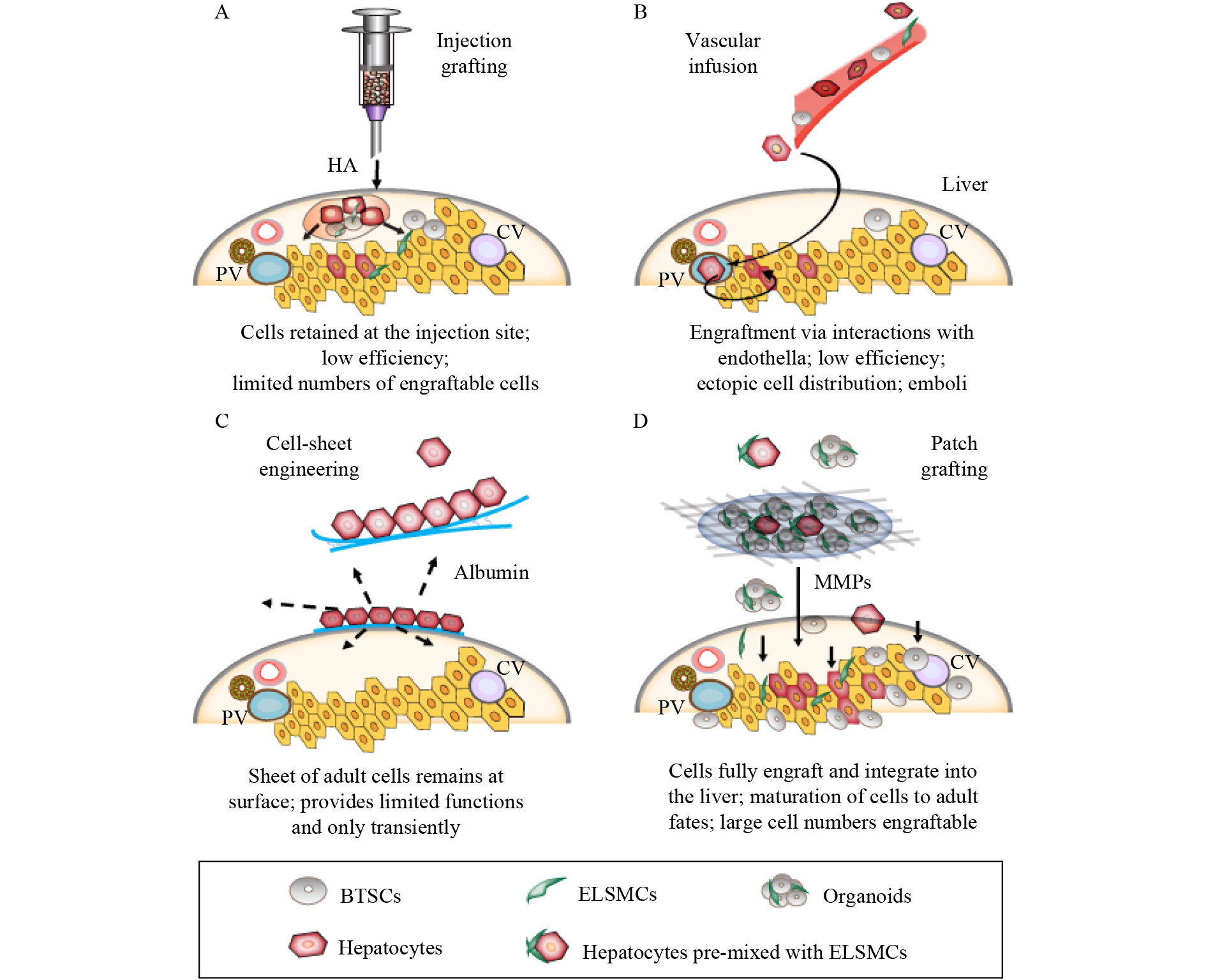
W Zhang, E Wauthier, G Lanzoni, H Hani, X Yi, D Overi, L Shi, S Simpson, A Allen, C Suitt, JA Ezzell, D Alvaro, V Cardinale, E Gaudio, G Carpino, G Prestwich, J Dominguez-Bendala, D Gerber, K Mathews, J Piedrahita, C Adin, P Sethupathy, Z He, LM Reid. Patch grafting of organoids of stem/progenitors into solid organs can correct genetic-based disease states. Biomaterials 2022; 288: 121647
https://doi.org/10.1016/j.biomaterials.2022.121647
|
| 56 |
W Zhang, G Lanzoni, H Hani, D Overi, V Cardinale, S Simpson, W Pitman, A Allen, X Yi, X Wang, D Gerber, G Prestwich, O Lozoya, E Gaudio, D Alvaro, D Tokaz, J Dominguez-Bendala, C Adin, J Piedrahita, K Mathews, P Sethupathy, G Carpino, Z He, E Wauthier, LM Reid. Patch grafting, strategies for transplantation of organoids into solid organs such as liver. Biomaterials 2021; 277: 121067
https://doi.org/10.1016/j.biomaterials.2021.121067
|
| 57 |
V Cardinale, G Carpino, D Overi, S Safarikia, W Zhang, M Kanke, A Franchitto, D Costantini, O Riccioni, L Nevi, M Chiappetta, P Onori, M Franchitto, S Bini, YH Hung, Q Lai, I Zizzari, M Nuti, C Nicoletti, S Checquolo, L Di Magno, MV Giuli, M Rossi, P Sethupathy, LM Reid, D Alvaro, E Gaudio. Human duodenal submucosal glands contain a defined stem/progenitor subpopulation with liver-specific regenerative potential. J Hepatol 2023; 78(1): 165–179
https://doi.org/10.1016/j.jhep.2022.08.037
|
| 58 |
J Font-Burgada, S Shalapour, S Ramaswamy, B Hsueh, D Rossell, A Umemura, K Taniguchi, H Nakagawa, MA Valasek, L Ye, JL Kopp, M Sander, H Carter, K Deisseroth, IM Verma, M Karin. Hybrid periportal hepatocytes regenerate the injured liver without giving rise to cancer. Cell 2015; 162(4): 766–779
https://doi.org/10.1016/j.cell.2015.07.026
|
| 59 |
T Sun, M Pikiolek, V Orsini, S Bergling, S Holwerda, L Morelli, PS Hoppe, L Planas-Paz, Y Yang, H Ruffner, T Bouwmeester, F Lohmann, LM Terracciano, G Roma, F Cong, JS Tchorz. AXIN2+ pericentral hepatocytes have limited contributions to liver homeostasis and regeneration. Cell Stem Cell 2020; 26(1): 97–107.e6
https://doi.org/10.1016/j.stem.2019.10.011
|
| 60 |
F Chen, RJ Jimenez, K Sharma, HY Luu, BY Hsu, A Ravindranathan, BA Stohr, H Willenbring. Broad distribution of hepatocyte proliferation in liver homeostasis and regeneration. Cell Stem Cell 2020; 26(1): 27–33.e4
https://doi.org/10.1016/j.stem.2019.11.001
|
| 61 |
T Matsumoto, L Wakefield, BD Tarlow, M Grompe. In vivo lineage tracing of polyploid hepatocytes reveals extensive proliferation during liver regeneration. Cell Stem Cell 2020; 26(1): 34–47.e3
https://doi.org/10.1016/j.stem.2019.11.014
|
| 62 |
Y Wei, YG Wang, Y Jia, L Li, J Yoon, S Zhang, Z Wang, Y Zhang, M Zhu, T Sharma, YH Lin, MH Hsieh, JH Albrecht, PT Le, CJ Rosen, T Wang, H Zhu. Liver homeostasis is maintained by midlobular zone 2 hepatocytes. Science 2021; 371(6532): eabb1625
https://doi.org/10.1126/science.abb1625
|
| 63 |
L He, W Pu, X Liu, Z Zhang, M Han, Y Li, X Huang, X Han, Y Li, K Liu, M Shi, L Lai, R Sun, QD Wang, Y Ji, JS Tchorz, B Zhou. Proliferation tracing reveals regional hepatocyte generation in liver homeostasis and repair. Science 2021; 371(6532): eabc4346
https://doi.org/10.1126/science.abc4346
|
| 64 |
EN Tafaleng, A Mukherjee, A Bell, K Morita, J Guzman-Lepe, N Haep, RM Florentino, R Diaz-Aragon, C Frau, A Ostrowska, JR Schultz, PGV Martini, A Soto-Gutierrez, IJ Fox. Hepatocyte nuclear factor 4 alpha 2 messenger RNA reprograms liver-enriched transcription factors and functional proteins in end-stage cirrhotic human hepatocytes. Hepatol Commun 2021; 5(11): 1911–1926
https://doi.org/10.1002/hep4.1763
|
| 65 |
KA Soltys, K Setoyama, EN Tafaleng, Gutiérrez A Soto, J Fong, K Fukumitsu, T Nishikawa, M Nagaya, R Sada, K Haberman, R Gramignoli, K Dorko, V Tahan, A Dreyzin, K Baskin, JJ Crowley, MA Quader, M Deutsch, C Ashokkumar, BL Shneider, RH Squires, S Ranganathan, M Reyes-Mugica, SF Dobrowolski, G Mazariegos, R Elango, DB Stolz, SC Strom, G Vockley, J Roy-Chowdhury, M Cascalho, C Guha, R Sindhi, JL Platt, IJ Fox. Host conditioning and rejection monitoring in hepatocyte transplantation in humans. J Hepatol 2017; 66(5): 987–1000
https://doi.org/10.1016/j.jhep.2016.12.017
|
| 66 |
C Mitchell, H Willenbring. A reproducible and well-tolerated method for 2/3 partial hepatectomy in mice. Nat Protoc 2008; 3(7): 1167–1170
https://doi.org/10.1038/nprot.2008.80
|
| 67 |
Y Miyaoka, A Miyajima. To divide or not to divide: revisiting liver regeneration. Cell Div 2013; 8(1): 8
https://doi.org/10.1186/1747-1028-8-8
|
| 68 |
M Mito, M Kusano, Y Kawaura. Hepatocyte transplantation in man. Transplant Proc 1992; 24(6): 3052–3053
|
| 69 |
G Lanzoni, T Oikawa, Y Wang, CB Cui, G Carpino, V Cardinale, D Gerber, M Gabriel, J Dominguez-Bendala, ME Furth, E Gaudio, D Alvaro, L Inverardi, LM Reid. Concise review: clinical programs of stem cell therapies for liver and pancreas. Stem Cells 2013; 31(10): 2047–2060
https://doi.org/10.1002/stem.1457
|
| 70 |
A Dhawan, J Puppi, RD Hughes, RR Mitry. Human hepatocyte transplantation: current experience and future challenges. Nat Rev Gastroenterol Hepatol 2010; 7(5): 288–298
https://doi.org/10.1038/nrgastro.2010.44
|
| 71 |
J Puppi, SC Strom, RD Hughes, S Bansal, JV Castell, I Dagher, EC Ellis, G Nowak, BG Ericzon, IJ Fox, MJ Gómez-Lechón, C Guha, S Gupta, RR Mitry, K Ohashi, M Ott, LM Reid, J Roy-Chowdhury, E Sokal, A Weber, A Dhawan. Improving the techniques for human hepatocyte transplantation: report from a consensus meeting in London. Cell Transplant 2012; 21(1): 1–10
https://doi.org/10.3727/096368911X566208
|
| 72 |
IJ Fox, SC Strom. To be or not to be: generation of hepatocytes from cells outside the liver. Gastroenterology 2008; 134(3): 878–881
https://doi.org/10.1053/j.gastro.2008.01.065
|
| 73 |
FN Smets, X Stephenne, G Debray, R Menten, R Reding, M Najimi, EM Sokal. Hepatocyte transplantation transforms severe phenylketonuria to mild hyperphenylalaninemia. Gastroenterology 2011; 140(5): s967
https://doi.org/10.1016/S0016-5085(11)64003-1
|
| 74 |
RA Fisher, SC Strom. Human hepatocyte transplantation: worldwide results. Transplantation 2006; 82(4): 441–449
https://doi.org/10.1097/01.tp.0000231689.44266.ac
|
| 75 |
A Trounson, C McDonald. Stem cell therapies in clinical trials: progress and challenges. Cell Stem Cell 2015; 17(1): 11–22
https://doi.org/10.1016/j.stem.2015.06.007
|
| 76 |
AI Caplan, D Correa. The MSC: an injury drugstore. Cell Stem Cell 2011; 9(1): 11–15
https://doi.org/10.1016/j.stem.2011.06.008
|
| 77 |
E El Agha, R Kramann, RK Schneider, X Li, W Seeger, BD Humphreys, S Bellusci. Mesenchymal stem cells in fibrotic disease. Cell Stem Cell 2017; 21(2): 166–177
https://doi.org/10.1016/j.stem.2017.07.011
|
| 78 |
T Squillaro, G Peluso, U Galderisi. Clinical trials with mesenchymal stem cells: an update. Cell Transplant 2016; 25(5): 829–848
https://doi.org/10.3727/096368915X689622
|
| 79 |
M Shi, Z Zhang, R Xu, H Lin, J Fu, Z Zou, A Zhang, J Shi, L Chen, S Lv, W He, H Geng, L Jin, Z Liu, FS Wang. Human mesenchymal stem cell transfusion is safe and improves liver function in acute-on-chronic liver failure patients. Stem Cells Transl Med 2012; 1(10): 725–731
https://doi.org/10.5966/sctm.2012-0034
|
| 80 |
M Shi, YY Li, RN Xu, FP Meng, SJ Yu, JL Fu, JH Hu, JX Li, LF Wang, L Jin, FS Wang. Mesenchymal stem cell therapy in decompensated liver cirrhosis: a long-term follow-up analysis of the randomized controlled clinical trial. Hepatol Int 2021; 15(6): 1431–1441
https://doi.org/10.1007/s12072-021-10199-2
|
| 81 |
Bahr L von, I Batsis, G Moll, M Hägg, A Szakos, B Sundberg, M Uzunel, O Ringden, Blanc K Le. Analysis of tissues following mesenchymal stromal cell therapy in humans indicates limited long-term engraftment and no ectopic tissue formation. Stem Cells 2012; 30(7): 1575–1578
https://doi.org/10.1002/stem.1118
|
| 82 |
CM Habibullah, IH Syed, A Qamar, Z Taher-Uz. Human fetal hepatocyte transplantation in patients with fulminant hepatic failure. Transplantation 1994; 58(8): 951–952
https://doi.org/10.1097/00007890-199410270-00016
|
| 83 |
AA Khan, MV Shaik, N Parveen, A Rajendraprasad, MA Aleem, MA Habeeb, G Srinivas, TA Raj, SK Tiwari, K Kumaresan, J Venkateswarlu, G Pande, CM Habibullah. Human fetal liver-derived stem cell transplantation as supportive modality in the management of end-stage decompensated liver cirrhosis. Cell Transplant 2010; 19(4): 409–418
https://doi.org/10.3727/096368909X484707a
|
| 84 |
AA Khan, N Parveen, VS Mahaboob, A Rajendraprasad, HR Ravindraprakash, J Venkateswarlu, P Rao, G Pande, M Lakshmi Narusu, MN Khaja, R Pramila, A Habeeb, CM Habibullah. Treatment of Crigler-Najjar syndrome type 1 by hepatic progenitor cell therapy: a simple procedure for hyperbilirubinemia. Transplant Proc 2008; 40(4): 1148–1150
https://doi.org/10.1016/j.transproceed.2008.03.022
|
| 85 |
AA Khan, N Parveen, VS Mahaboob, A Rajendraprasad, HR Ravindraprakash, J Venkateswarlu, P Rao, G Pande, ML Narusu, MN Khaja, R Pramila, A Habeeb, CM Habibullah. Management of hyperbilirubinemia in biliary atresia by hepatic progenitor cell transplantation through hepatic artery: a case report. Transplant Proc 2008; 40(4): 1153–1155
https://doi.org/10.1016/j.transproceed.2008.03.110
|
| 86 |
G PietrosiC Chinnici. Report on liver cell transplantation using human fetal liver cells. In: Stock P, Christ B. Hepatocyte Transplantation. Springer, 2017. 283–294
|
| 87 |
N Parveen, AK Aleem, MA Habeeb, CM Habibullah. An update on hepatic stem cells: bench to bedside. Curr Pharm Biotechnol 2011; 12(2): 226–230
https://doi.org/10.2174/138920111794295765
|
| 88 |
F Li, Z He, Y Li, P Liu, F Chen, M Wang, H Zhu, X Ding, KJ Wangensteen, Y Hu, X Wang. Combined activin A/LiCl/Noggin treatment improves production of mouse embryonic stem cell-derived definitive endoderm cells. J Cell Biochem 2011; 112(4): 1022–1034
https://doi.org/10.1002/jcb.22962
|
| 89 |
X Wang, W Zhang, Y Yang, J Wang, H Qiu, L Liao, T Oikawa, E Wauthier, P Sethupathy, LM Reid, Z Liu, Z He. A microRNA-based network provides potential predictive signatures and reveals the crucial role of PI3K/AKT signaling for hepatic lineage maturation. Front Cell Dev Biol 2021; 9: 670059
https://doi.org/10.3389/fcell.2021.670059
|
| 90 |
MA Habeeb, SK Vishwakarma, A Bardia, AA Khan. Hepatic stem cells: a viable approach for the treatment of liver cirrhosis. World J Stem Cells 2015; 7(5): 859–865
https://doi.org/10.4252/wjsc.v7.i5.859
|
| 91 |
A Aleem Khan, N Parveen, MA Habeeb, CM Habibullah. Journey from hepatocyte transplantation to hepatic stem cells: a novel treatment strategy for liver diseases. Indian J Med Res 2006; 123(5): 601–614
|
| 92 |
V Cardinale, G Carpino, R Gentile, C Napoletano, H Rahimi, A Franchitto, R Semeraro, M Nuti, P Onori, PB Berloco, M Rossi, D Bosco, R Brunelli, A Fraveto, C Napoli, A Torrice, M Gatto, R Venere, C Bastianelli, C Aliberti, FM Salvatori, L Bresadola, M Bezzi, AF Attili, L Reid, E Gaudio, D Alvaro. Transplantation of human fetal biliary tree stem/progenitor cells into two patients with advanced liver cirrhosis. BMC Gastroenterol 2014; 14(1): 204
https://doi.org/10.1186/s12876-014-0204-z
|
| 93 |
JA Thomson, J Itskovitz-Eldor, SS Shapiro, MA Waknitz, JJ Swiergiel, VS Marshall, JM Jones. Embryonic stem cell lines derived from human blastocysts. Science 1998; 282(5391): 1145–1147
https://doi.org/10.1126/science.282.5391.1145
|
| 94 |
P Damdimopoulou, S Rodin, S Stenfelt, L Antonsson, K Tryggvason, O Hovatta. Human embryonic stem cells. Best Pract Res Clin Obstet Gynaecol 2016; 31: 2–12
https://doi.org/10.1016/j.bpobgyn.2015.08.010
|
| 95 |
K Takahashi, K Tanabe, M Ohnuki, M Narita, T Ichisaka, K Tomoda, S Yamanaka. Induction of pluripotent stem cells from adult human fibroblasts by defined factors. Cell 2007; 131(5): 861–872
https://doi.org/10.1016/j.cell.2007.11.019
|
| 96 |
DA Robinton, GQ Daley. The promise of induced pluripotent stem cells in research and therapy. Nature 2012; 481(7381): 295–305
https://doi.org/10.1038/nature10761
|
| 97 |
T Zhao, ZN Zhang, Z Rong, Y Xu. Immunogenicity of induced pluripotent stem cells. Nature 2011; 474(7350): 212–215
https://doi.org/10.1038/nature10135
|
| 98 |
M Danoy, ML Bernier, K Kimura, S Poulain, S Kato, D Mori, T Kido, C Plessy, H Kusuhara, A Miyajima, Y Sakai, E Leclerc. Optimized protocol for the hepatic differentiation of induced pluripotent stem cells in a fluidic microenvironment. Biotechnol Bioeng 2019; 116(7): 1762–1776
https://doi.org/10.1002/bit.26970
|
| 99 |
F Li, P Liu, C Liu, D Xiang, L Deng, W Li, K Wangensteen, J Song, Y Ma, L Hui, L Wei, L Li, X Ding, Y Hu, Z He, X Wang. Hepatoblast-like progenitor cells derived from embryonic stem cells can repopulate livers of mice. Gastroenterology 2010; 139(6): 2158–2169.e8
https://doi.org/10.1053/j.gastro.2010.08.042
|
| 100 |
F Yan, Y Wang, W Zhang, M Chang, Z He, J Xu, C Shang, T Chen, J Liu, X Wang, X Pei, Y Wang. Human embryonic stem cell-derived hepatoblasts are an optimal lineage stage for hepatitis C virus infection. Hepatology 2017; 66(3): 717–735
https://doi.org/10.1002/hep.29134
|
| 101 |
Ilic D, Ogilvie C. Concise review: Human embryonic stem cells—what have we done? What are we doing? Where are we going? Stem Cells 2017; 35(1): 17–25 doi:10.1002/stem.2450
pmid: 27350255
|
| 102 |
K Takayama, H Mizuguchi. Generation of human pluripotent stem cell-derived hepatocyte-like cells for drug toxicity screening. Drug Metab Pharmacokinet 2017; 32(1): 12–20
https://doi.org/10.1016/j.dmpk.2016.10.408
|
| 103 |
Y Wang, J Qin, S Wang, W Zhang, J Duan, J Zhang, X Wang, F Yan, M Chang, X Liu, B Feng, J Liu, X Pei. Conversion of human gastric epithelial cells to multipotent endodermal progenitors using defined small molecules. Cell Stem Cell 2016; 19(4): 449–461
https://doi.org/10.1016/j.stem.2016.06.006
|
| 104 |
P Huang, Z He, S Ji, H Sun, D Xiang, C Liu, Y Hu, X Wang, L Hui. Induction of functional hepatocyte-like cells from mouse fibroblasts by defined factors. Nature 2011; 475(7356): 386–389
https://doi.org/10.1038/nature10116
|
| 105 |
P Huang, L Zhang, Y Gao, Z He, D Yao, Z Wu, J Cen, X Chen, C Liu, Y Hu, D Lai, Z Hu, L Chen, Y Zhang, X Cheng, X Ma, G Pan, X Wang, L Hui. Direct reprogramming of human fibroblasts to functional and expandable hepatocytes. Cell Stem Cell 2014; 14(3): 370–384
https://doi.org/10.1016/j.stem.2014.01.003
|
| 106 |
S Sekiya, A Suzuki. Direct conversion of mouse fibroblasts to hepatocyte-like cells by defined factors. Nature 2011; 475(7356): 390–393
https://doi.org/10.1038/nature10263
|
| 107 |
B Yu, ZY He, P You, QW Han, D Xiang, F Chen, MJ Wang, CC Liu, XW Lin, U Borjigin, XY Zi, JX Li, HY Zhu, WL Li, CS Han, KJ Wangensteen, Y Shi, LJ Hui, X Wang, YP Hu. Reprogramming fibroblasts into bipotential hepatic stem cells by defined factors. Cell Stem Cell 2013; 13(3): 328–340
https://doi.org/10.1016/j.stem.2013.06.017
|
| 108 |
H Wu, X Zhou, GB Fu, ZY He, HP Wu, P You, C Ashton, X Wang, HY Wang, HX Yan. Reversible transition between hepatocytes and liver progenitors for in vitro hepatocyte expansion. Cell Res 2017; 27(5): 709–712
https://doi.org/10.1038/cr.2017.47
|
| 109 |
C Xiang, Y Du, G Meng, L Soon Yi, S Sun, N Song, X Zhang, Y Xiao, J Wang, Z Yi, Y Liu, B Xie, M Wu, J Shu, D Sun, J Jia, Z Liang, D Sun, Y Huang, Y Shi, J Xu, F Lu, C Li, K Xiang, Z Yuan, S Lu, H Deng. Long-term functional maintenance of primary human hepatocytes in vitro. Science 2019; 364(6438): 399–402
https://doi.org/10.1126/science.aau7307
|
| 110 |
GB Fu, WJ Huang, M Zeng, X Zhou, HP Wu, CC Liu, H Wu, J Weng, HD Zhang, YC Cai, C Ashton, M Ding, D Tang, BH Zhang, Y Gao, WF Yu, B Zhai, ZY He, HY Wang, HX Yan. Expansion and differentiation of human hepatocyte-derived liver progenitor-like cells and their use for the study of hepatotropic pathogens. Cell Res 2019; 29(1): 8–22
https://doi.org/10.1038/s41422-018-0103-x
|
| 111 |
L Sun, Y Wang, J Cen, X Ma, L Cui, Z Qiu, Z Zhang, H Li, RZ Yang, C Wang, X Chen, L Wang, Y Ye, H Zhang, G Pan, JS Kang, Y Ji, YW Zheng, S Zheng, L Hui. Modelling liver cancer initiation with organoids derived from directly reprogrammed human hepatocytes. Nat Cell Biol 2019; 21(8): 1015–1026
https://doi.org/10.1038/s41556-019-0359-5
|
| 112 |
XL Shi, Y Gao, Y Yan, H Ma, L Sun, P Huang, X Ni, L Zhang, X Zhao, H Ren, D Hu, Y Zhou, F Tian, Y Ji, X Cheng, G Pan, YT Ding, L Hui. Improved survival of porcine acute liver failure by a bioartificial liver device implanted with induced human functional hepatocytes. Cell Res 2016; 26(2): 206–216
https://doi.org/10.1038/cr.2016.6
|
| 113 |
WJ Li, XJ Zhu, TJ Yuan, ZY Wang, ZQ Bian, HS Jing, X Shi, CY Chen, GB Fu, WJ Huang, YP Shi, Q Liu, M Zeng, HD Zhang, HP Wu, WF Yu, B Zhai, HX Yan. An extracorporeal bioartificial liver embedded with 3D-layered human liver progenitor-like cells relieves acute liver failure in pigs. Sci Transl Med 2020; 12(551): eaba5146
https://doi.org/10.1126/scitranslmed.aba5146
|
| 114 |
K Zhang, L Zhang, W Liu, X Ma, J Cen, Z Sun, C Wang, S Feng, Z Zhang, L Yue, L Sun, Z Zhu, X Chen, A Feng, J Wu, Z Jiang, P Li, X Cheng, D Gao, L Peng, L Hui. In vitro expansion of primary human hepatocytes with efficient liver repopulation capacity. Cell Stem Cell 2018; 23(6): 806–819.e4
https://doi.org/10.1016/j.stem.2018.10.018
|
| 115 |
C Wang, L Zhang, Z Sun, X Yuan, B Wu, J Cen, L Cui, K Zhang, C Li, J Wu, Y Shu, W Sun, J Wang, L Hui. Dedifferentiation-associated inflammatory factors of long-term expanded human hepatocytes exacerbate their elimination by macrophages during liver engraftment. Hepatology 2022; 76(6): 1690–1705
https://doi.org/10.1002/hep.32436
|
| 116 |
M Chang, MS Bogacheva, YR Lou. Challenges for the applications of human pluripotent stem cell-derived liver organoids. Front Cell Dev Biol 2021; 9: 748576
https://doi.org/10.3389/fcell.2021.748576
|
| 117 |
N Prior, P Inacio, M Huch. Liver organoids: from basic research to therapeutic applications. Gut 2019; 68(12): 2228–2237
https://doi.org/10.1136/gutjnl-2019-319256
|
| 118 |
F Sampaziotis, D Muraro, OC Tysoe, S Sawiak, TE Beach, EM Godfrey, SS Upponi, T Brevini, BT Wesley, J Garcia-Bernardo, K Mahbubani, G Canu, R 3rd Gieseck, NL Berntsen, VL Mulcahy, K Crick, C Fear, S Robinson, L Swift, L Gambardella, J Bargehr, D Ortmann, SE Brown, A Osnato, MP Murphy, G Corbett, WTH Gelson, GF Mells, P Humphreys, SE Davies, I Amin, P Gibbs, S Sinha, SA Teichmann, AJ Butler, TC See, E Melum, CJE Watson, K Saeb-Parsy, L Vallier. Cholangiocyte organoids can repair bile ducts after transplantation in the human liver. Science 2021; 371(6531): 839–846
https://doi.org/10.1126/science.aaz6964
|
| 119 |
Y Wang, HL Yao, CB Cui, E Wauthier, C Barbier, MJ Costello, N Moss, M Yamauchi, M Sricholpech, D Gerber, EG Loboa, LM Reid. Paracrine signals from mesenchymal cell populations govern the expansion and differentiation of human hepatic stem cells to adult liver fates. Hepatology 2010; 52(4): 1443–1454
https://doi.org/10.1002/hep.23829
|
| 120 |
R Turner, D Gerber, L Reid. The future of cell transplant therapies: a need for tissue grafting. Transplantation 2010; 90(8): 807–810
https://doi.org/10.1097/TP.0b013e3181f24ea2
|
| 121 |
L Nevi, G Carpino, D Costantini, V Cardinale, O Riccioni, S Di Matteo, F Melandro, PB Berloco, L Reid, E Gaudio, D Alvaro. Hyaluronan coating improves liver engraftment of transplanted human biliary tree stem/progenitor cells. Stem Cell Res Ther 2017; 8(1): 68
https://doi.org/10.1186/s13287-017-0492-7
|
| 122 |
J Kobayashi, A Kikuchi, T Aoyagi, T Okano. Cell sheet tissue engineering: cell sheet preparation, harvesting/manipulation, and transplantation. J Biomed Mater Res A 2019; 107(5): 955–967
https://doi.org/10.1002/jbm.a.36627
|
| 123 |
K Tatsumi, T Okano. Hepatocyte transplantation: cell sheet technology for liver cell transplantation. Curr Transplant Rep 2017; 4(3): 184–192
https://doi.org/10.1007/s40472-017-0156-7
|
| 124 |
R Takeuchi, Y Kuruma, H Sekine, I Dobashi, M Yamato, M Umezu, T Shimizu, T Okano. In vivo vascularization of cell sheets provided better long-term tissue survival than injection of cell suspension. J Tissue Eng Regen Med 2016; 10(8): 700–710
https://doi.org/10.1002/term.1854
|
| 125 |
OA Lozoya, E Wauthier, RA Turner, C Barbier, GD Prestwich, F Guilak, R Superfine, SR Lubkin, LM Reid. Regulation of hepatic stem/progenitor phenotype by microenvironment stiffness in hydrogel models of the human liver stem cell niche. Biomaterials 2011; 32(30): 7389–7402
https://doi.org/10.1016/j.biomaterials.2011.06.042
|
| 126 |
CB Highley, GD Prestwich, JA Burdick. Recent advances in hyaluronic acid hydrogels for biomedical applications. Curr Opin Biotechnol 2016; 40: 35–40
https://doi.org/10.1016/j.copbio.2016.02.008
|
| 127 |
X Zheng Shu, Y Liu, FS Palumbo, Y Luo, GD Prestwich. In situ crosslinkable hyaluronan hydrogels for tissue engineering. Biomaterials 2004; 25(7–8): 1339–1348
https://doi.org/10.1016/j.biomaterials.2003.08.014
|
| 128 |
XZ ShuGD Prestwich. Therapeutic biomaterials from chemically modified hyaluronan. In: Garg HG, Hales CA. Chemistry and Biology of Hyaluronan. Elsevier, 2004. 475–504
|
| 129 |
JA Burdick, GD Prestwich. Hyaluronic acid hydrogels for biomedical applications. Adv Mater 2011; 23(12): H41–H56
https://doi.org/10.1002/adma.201003963
|
| 130 |
JR Fraser, TC Laurent, UB Laurent. Hyaluronan: its nature, distribution, functions and turnover. J Intern Med 1997; 242(1): 27–33
https://doi.org/10.1046/j.1365-2796.1997.00170.x
|
| 131 |
N Shirvaikar, LA Marquez-Curtis, A Janowska-Wieczorek. Hematopoietic stem cell mobilization and homing after transplantation: the role of MMP-2, MMP-9, and MT1-MMP. Biochem Res Int 2012; 2012: 685267
https://doi.org/10.1155/2012/685267
|
| 132 |
F Pan, S Ma, W Cao, H Liu, F Chen, X Chen, R Shi. SDF-1α upregulation of MMP-2 is mediated by p38 MAPK signaling in pancreatic cancer cell lines. Mol Biol Rep 2013; 40(7): 4139–4146
https://doi.org/10.1007/s11033-012-2225-4
|
| 133 |
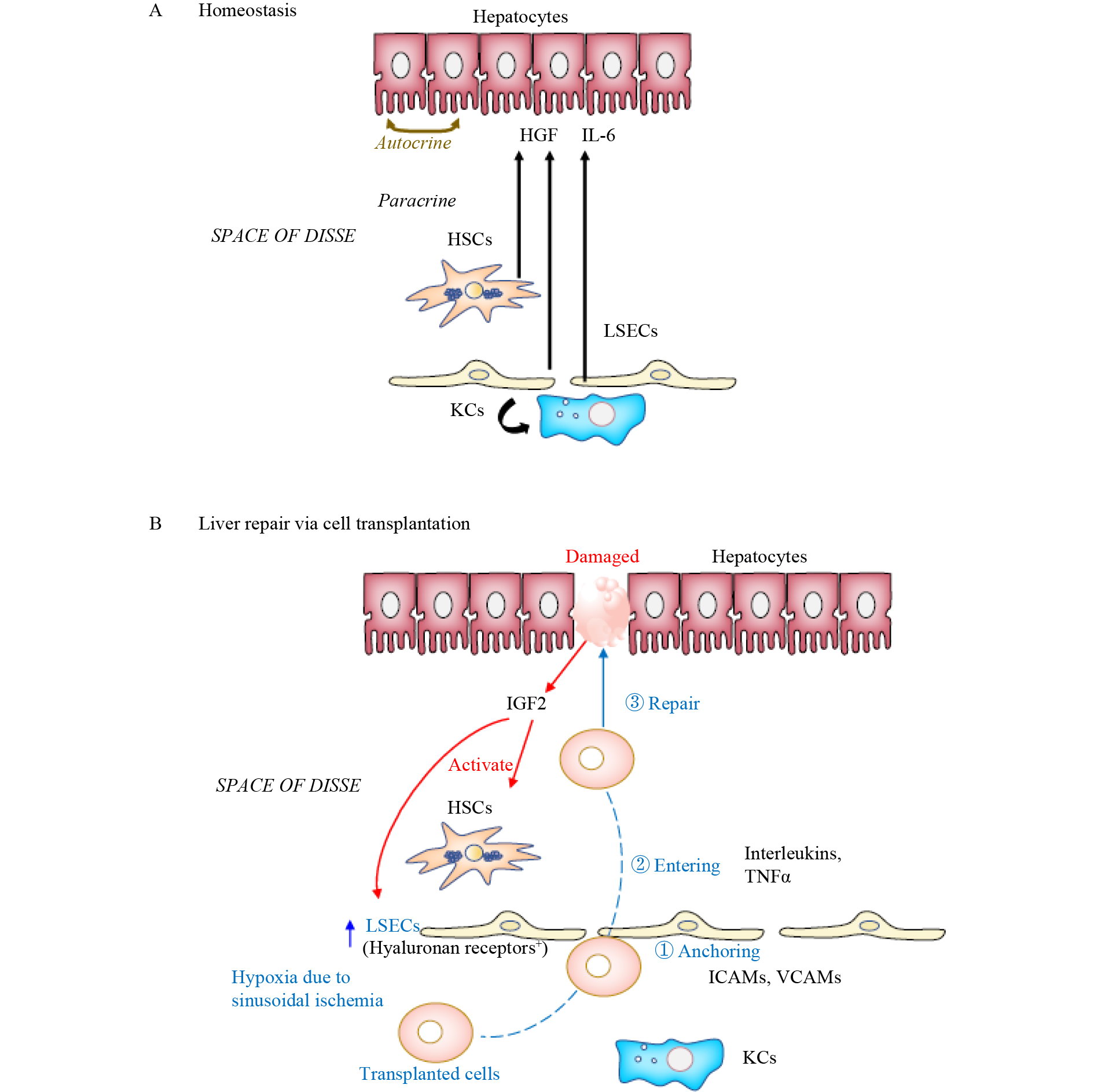
E Maslak, A Gregorius, S Chlopicki. Liver sinusoidal endothelial cells (LSECs) function and NAFLD; NO-based therapy targeted to the liver. Pharmacol Rep 2015; 67(4): 689–694
https://doi.org/10.1016/j.pharep.2015.04.010
|
| 134 |
J Poisson, S Lemoinne, C Boulanger, F Durand, R Moreau, D Valla, PE Rautou. Liver sinusoidal endothelial cells: physiology and role in liver diseases. J Hepatol 2017; 66(1): 212–227
https://doi.org/10.1016/j.jhep.2016.07.009
|
| 135 |
J Rodriguez-Vita, M Morales-Ruiz. Down the liver sinusoidal endothelial cell (LSEC) hole. Is there a role for lipid rafts in LSEC fenestration? Hepatology 2013; 57(3): 1272–1274
https://doi.org/10.1002/hep.26249
|
| 136 |
P Carmeliet, RK Jain. Molecular mechanisms and clinical applications of angiogenesis. Nature 2011; 473(7347): 298–307
https://doi.org/10.1038/nature10144
|
| 137 |
BS Ding, DJ Nolan, JM Butler, D James, AO Babazadeh, Z Rosenwaks, V Mittal, H Kobayashi, K Shido, D Lyden, TN Sato, SY Rabbany, S Rafii. Inductive angiocrine signals from sinusoidal endothelium are required for liver regeneration. Nature 2010; 468(7321): 310–315
https://doi.org/10.1038/nature09493
|
| 138 |
N Yadav, FL Jaber, Y Sharma, P Gupta, P Viswanathan, S Gupta. Efficient reconstitution of hepatic microvasculature by endothelin receptor antagonism in liver sinusoidal endothelial cells. Hum Gene Ther 2019; 30(3): 365–377
https://doi.org/10.1089/hum.2018.166
|
| 139 |
KK Sørensen, J Simon-Santamaria, RS McCuskey, B Smedsrød. Liver sinusoidal endothelial cells. Compr Physiol 2015; 5(4): 1751–1774
https://doi.org/10.1002/cphy.c140078
|
| 140 |
B Hansen, P Longati, K Elvevold, GI Nedredal, K Schledzewski, R Olsen, M Falkowski, J Kzhyshkowska, F Carlsson, S Johansson, B Smedsrød, S Goerdt, S Johansson, P McCourt. Stabilin-1 and stabilin-2 are both directed into the early endocytic pathway in hepatic sinusoidal endothelium via interactions with clathrin/AP-2, independent of ligand binding. Exp Cell Res 2005; 303(1): 160–173
https://doi.org/10.1016/j.yexcr.2004.09.017
|
| 141 |
H Ogasawara, A Inagaki, I Fathi, T Imura, H Yamana, Y Saitoh, M Matsumura, K Fukuoka, S Miyagi, Y Nakamura, K Ohashi, M Unno, T Kamei, M Goto. Preferable transplant site for hepatocyte transplantation in a rat model. Cell Transplant 2021; 30: 9636897211040012
https://doi.org/10.1177/09636897211040012
|
| 142 |
MJ Wang, F Chen, QG Liu, CC Liu, H Yao, B Yu, HB Zhang, HX Yan, Y Ye, T Chen, KJ Wangensteen, X Wang, YP Hu, ZY He. Insulin-like growth factor 2 is a key mitogen driving liver repopulation in mice. Cell Death Dis 2018; 9(2): 26
https://doi.org/10.1038/s41419-017-0186-1
|
| 143 |
Y Du, W Zhang, H Qiu, C Xiao, J Shi, LM Reid, Z He. Mouse models of liver parenchyma injuries and regeneration. Front Cell Dev Biol 2022; 10: 903740
https://doi.org/10.3389/fcell.2022.903740
|
| 144 |
Z He, H Zhang, X Zhang, D Xie, Y Chen, KJ Wangensteen, SC Ekker, M Firpo, C Liu, D Xiang, X Zi, L Hui, G Yang, X Ding, Y Hu, X Wang. Liver xeno-repopulation with human hepatocytes in Fah–/–Rag2–/– mice after pharmacological immunosuppression. Am J Pathol 2010; 177(3): 1311–1319
https://doi.org/10.2353/ajpath.2010.091154
|
| 145 |
B Su, C Liu, D Xiang, H Zhang, S Yuan, M Wang, F Chen, H Zhu, Z He, X Wang, Y Hu. Xeno-repopulation of Fah–/– Nod/Scid mice livers by human hepatocytes. Sci China Life Sci 2011; 54(3): 227–234
https://doi.org/10.1007/s11427-011-4140-7
|
| 146 |
F Lu, X Pan, W Zhang, X Su, Y Gu, H Qiu, S Shen, C Liu, W Liu, X Wang, Z Zhan, Z Liu, Z He. A three-dimensional imaging method for the quantification and localization of dynamic cell tracking posttransplantation. Front Cell Dev Biol 2021; 9: 698795
https://doi.org/10.3389/fcell.2021.698795
|
| 147 |
BÖ Akcora, G Storm, R Bansal. Inhibition of canonical WNT signaling pathway by β-catenin/CBP inhibitor ICG-001 ameliorates liver fibrosis in vivo through suppression of stromal CXCL12. Biochim Biophys Acta Mol Basis Dis 2018; 1864(3): 804–818
https://doi.org/10.1016/j.bbadis.2017.12.001
|
| 148 |
F Moroni, BJ Dwyer, C Graham, C Pass, L Bailey, L Ritchie, D Mitchell, A Glover, A Laurie, S Doig, E Hargreaves, AR Fraser, ML Turner, JDM Campbell, NWA McGowan, J Barry, JK Moore, PC Hayes, DJ Leeming, MJ Nielsen, K Musa, JA Fallowfield, SJ Forbes. Safety profile of autologous macrophage therapy for liver cirrhosis. Nat Med 2019; 25(10): 1560–1565
https://doi.org/10.1038/s41591-019-0599-8
|
| 149 |
SL Friedman. Hepatic stellate cells: protean, multifunctional, and enigmatic cells of the liver. Physiol Rev 2008; 88(1): 125–172
https://doi.org/10.1152/physrev.00013.2007
|
| 150 |
R Bataller, DA Brenner. Liver fibrosis. J Clin Invest 2005; 115(2): 209–218
https://doi.org/10.1172/JCI24282
|
| 151 |
D Kobold, A Grundmann, F Piscaglia, C Eisenbach, K Neubauer, J Steffgen, G Ramadori, T Knittel. Expression of reelin in hepatic stellate cells and during hepatic tissue repair: a novel marker for the differentiation of HSC from other liver myofibroblasts. J Hepatol 2002; 36(5): 607–613
https://doi.org/10.1016/S0168-8278(02)00050-8
|
| 152 |
JA Oben, T Roskams, S Yang, H Lin, N Sinelli, M Torbenson, U Smedh, TH Moran, Z Li, J Huang, SA Thomas, AM Diehl. Hepatic fibrogenesis requires sympathetic neurotransmitters. Gut 2004; 53(3): 438–445
https://doi.org/10.1136/gut.2003.026658
|
| 153 |
YA LeeS L Friedman. Stellate Cells and Fibrosis. In: Arias IM, Alter HJ, Boyer JL, Cohen DE, Shafritz DA, Thorgeirsson SS, Wolkoff AW. The Liver: Biology and Pathobiology. John Wiley & Sons Ltd. 2020. 444–454
|
| 154 |
M Guilliams, CA Dutertre, CL Scott, N McGovern, D Sichien, S Chakarov, S Van Gassen, J Chen, M Poidinger, S De Prijck, SJ Tavernier, I Low, SE Irac, CN Mattar, HR Sumatoh, GHL Low, TJK Chung, DKH Chan, KK Tan, TLK Hon, E Fossum, B Bogen, M Choolani, JKY Chan, A Larbi, H Luche, S Henri, Y Saeys, EW Newell, BN Lambrecht, B Malissen, F Ginhoux. Unsupervised high-dimensional analysis aligns dendritic cells across tissues and species. Immunity 2016; 45(3): 669–684
https://doi.org/10.1016/j.immuni.2016.08.015
|
| 155 |
BG Lopez, MS Tsai, JL Baratta, KJ Longmuir, RT Robertson. Characterization of Kupffer cells in livers of developing mice. Comp Hepatol 2011; 10(1): 2
https://doi.org/10.1186/1476-5926-10-2
|
| 156 |
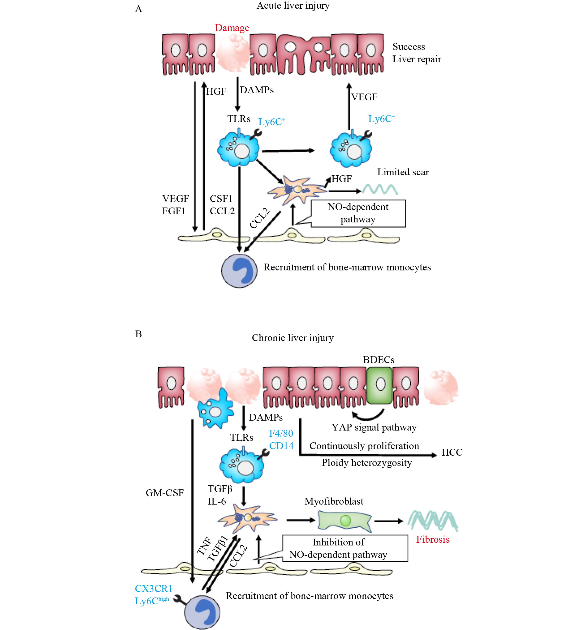
O Krenkel, F Tacke. Liver macrophages in tissue homeostasis and disease. Nat Rev Immunol 2017; 17(5): 306–321
https://doi.org/10.1038/nri.2017.11
|
| 157 |
JL Baratta, A Ngo, B Lopez, N Kasabwalla, KJ Longmuir, RT Robertson. Cellular organization of normal mouse liver: a histological, quantitative immunocytochemical, and fine structural analysis. Histochem Cell Biol 2009; 131(6): 713–726
https://doi.org/10.1007/s00418-009-0577-1
|
| 158 |
CL Scott, F Zheng, P De Baetselier, L Martens, Y Saeys, S De Prijck, S Lippens, C Abels, S Schoonooghe, G Raes, N Devoogdt, BN Lambrecht, A Beschin, M Guilliams. Bone marrow-derived monocytes give rise to self-renewing and fully differentiated Kupffer cells. Nat Commun 2016; 7(1): 10321
https://doi.org/10.1038/ncomms10321
|
| 159 |
F Moroni, BJ Dwyer, C Graham, C Pass, L Bailey, L Ritchie, D Mitchell, A Glover, A Laurie, S Doig, E Hargreaves, AR Fraser, ML Turner, JDM Campbell, NWA McGowan, J Barry, JK Moore, PC Hayes, DJ Leeming, MJ Nielsen, K Musa, JA Fallowfield, SJ Forbes. Safety profile of autologous macrophage therapy for liver cirrhosis. Nat Med 2019; 25(10): 1560–1565
https://doi.org/10.1038/s41591-019-0599-8
|
| 160 |
F Heymann, J Peusquens, I Ludwig-Portugall, M Kohlhepp, C Ergen, P Niemietz, C Martin, N van Rooijen, JC Ochando, GJ Randolph, T Luedde, F Ginhoux, C Kurts, C Trautwein, F Tacke. Liver inflammation abrogates immunological tolerance induced by Kupffer cells. Hepatology 2015; 62(1): 279–291
https://doi.org/10.1002/hep.27793
|
| 161 |
Y Wen, J Lambrecht, C Ju, F Tacke. Hepatic macrophages in liver homeostasis and diseases-diversity, plasticity and therapeutic opportunities. Cell Mol Immunol 2021; 18(1): 45–56
https://doi.org/10.1038/s41423-020-00558-8
|
| 162 |
F Heymann, L Hammerich, D Storch, M Bartneck, S Huss, V Rüsseler, N Gassler, SA Lira, T Luedde, C Trautwein, F Tacke. Hepatic macrophage migration and differentiation critical for liver fibrosis is mediated by the chemokine receptor C-C motif chemokine receptor 8 in mice. Hepatology 2012; 55(3): 898–909
https://doi.org/10.1002/hep.24764
|
| 163 |
W Bernal, J Wendon. Acute liver failure. N Engl J Med 2013; 369(26): 2525–2534
https://doi.org/10.1056/NEJMra1208937
|
| 164 |
N Nakamoto, H Ebinuma, T Kanai, PS Chu, Y Ono, Y Mikami, K Ojiro, M Lipp, PE Love, H Saito, T Hibi. CCR9+ macrophages are required for acute liver inflammation in mouse models of hepatitis. Gastroenterology 2012; 142(2): 366–376
https://doi.org/10.1053/j.gastro.2011.10.039
|
| 165 |
KR Karlmark, HW Zimmermann, C Roderburg, N Gassler, HE Wasmuth, T Luedde, C Trautwein, F Tacke. The fractalkine receptor CX3CR1 protects against liver fibrosis by controlling differentiation and survival of infiltrating hepatic monocytes. Hepatology 2010; 52(5): 1769–1782
https://doi.org/10.1002/hep.23894
|
| 166 |
E Saijou, Y Enomoto, M Matsuda, C Yuet-Yin Kok, S Akira, M Tanaka, A Miyajima. Neutrophils alleviate fibrosis in the CCl4-induced mouse chronic liver injury model. Hepatol Commun 2018; 2(6): 703–717
https://doi.org/10.1002/hep4.1178
|
| 167 |
KB Halpern, R Shenhav, O Matcovitch-Natan, B Toth, D Lemze, M Golan, EE Massasa, S Baydatch, S Landen, AE Moor, A Brandis, A Giladi, AS Avihail, E David, I Amit, S Itzkovitz. Single-cell spatial reconstruction reveals global division of labour in the mammalian liver. Nature 2017; 542(7641): 352–356
https://doi.org/10.1038/nature21065
|
| 168 |
T Chen, S Oh, S Gregory, X Shen, AM Diehl. Single-cell omics analysis reveals functional diversification of hepatocytes during liver regeneration. JCI Insight 2020; 5(22): e141024
https://doi.org/10.1172/jci.insight.141024
|
| 169 |
UV Chembazhi, S Bangru, M Hernaez, A Kalsotra. Cellular plasticity balances the metabolic and proliferation dynamics of a regenerating liver. Genome Res 2021; 31(4): 576–591
https://doi.org/10.1101/gr.267013.120
|
| 170 |
H Chen, S Tang, J Liao, M Liu, Y Lin. Therapeutic effect of human umbilical cord blood mesenchymal stem cells combined with G-CSF on rats with acute liver failure. Biochem Biophys Res Commun 2019; 517(4): 670–676
https://doi.org/10.1016/j.bbrc.2019.07.101
|
| 171 |
M Joshi, P B Patil, Z He, J Holgersson, M Olausson, S Sumitran-Holgersson. Fetal liver-derived mesenchymal stromal cells augment engraftment of transplanted hepatocytes. Cytotherapy 2012; 14(6): 657–669
https://doi.org/10.3109/14653249.2012.663526
|
|
Viewed |
|
|
|
Full text
|
|
|
|
|
Abstract
|
|
|
|
|
Cited |
|
|
|
|
| |
Shared |
|
|
|
|
| |
Discussed |
|
|
|
|